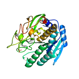
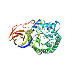
3APF
 
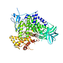 | | Crystal structure of human PI3K-gamma in complex with CH5039699 | | 分子名称: | 3-[7-(1H-benzimidazol-5-yl)-2-(morpholin-4-yl)-6,7-dihydro-5H-pyrrolo[2,3-d]pyrimidin-4-yl]phenol, Phosphatidylinositol-4,5-bisphosphate 3-kinase catalytic subunit gamma isoform, SULFATE ION | | 著者 | Nakamura, M, Fukami, T.A, Miyazaki, T, Yoshida, M. | | 登録日 | 2010-10-14 | | 公開日 | 2011-04-13 | | 最終更新日 | 2023-11-01 | | 実験手法 | X-RAY DIFFRACTION (2.82 Å) | | 主引用文献 | Discovery and biological activity of a novel class I PI3K inhibitor, CH5132799
Bioorg.Med.Chem.Lett., 21, 2011
|
|
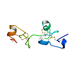
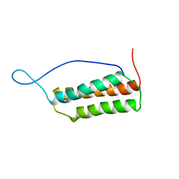
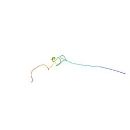
1UG8
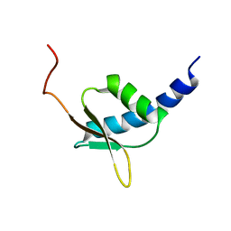 | | NMR structure of the R3H domain from Poly(A)-specific Ribonuclease | | 分子名称: | Poly(A)-specific Ribonuclease | | 著者 | Nagata, T, Muto, Y, Hayami, N, Uda, H, Shirouzu, M, Terada, T, Kigawa, T, Inoue, M, Yabuki, T, Aoki, M, Seki, E, Matsuda, T, Hirota, H, Yoshida, M, Kobayashi, N, Tanaka, A, Osanai, T, Matsuo, Y, Arakawa, T, Carninci, P, Kawai, J, Hayashizaki, Y, Yokoyama, S, RIKEN Structural Genomics/Proteomics Initiative (RSGI) | | 登録日 | 2003-06-15 | | 公開日 | 2004-08-17 | | 最終更新日 | 2023-12-27 | | 実験手法 | SOLUTION NMR | | 主引用文献 | NMR structure of the R3H domain from Poly(A)-specific Ribonuclease
To be Published
|
|
1JIE
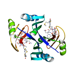 | | Crystal structure of bleomycin-binding protein from bleomycin-producing Streptomyces verticillus complexed with metal-free bleomycin | | 分子名称: | BLEOMYCIN A2, bleomycin-binding protein | | 著者 | Sugiyama, M, Kumagai, T, Hayashida, M, Maruyama, M, Matoba, Y. | | 登録日 | 2001-07-02 | | 公開日 | 2002-02-06 | | 最終更新日 | 2023-10-25 | | 実験手法 | X-RAY DIFFRACTION (1.8 Å) | | 主引用文献 | The 1.6-A crystal structure of the copper(II)-bound bleomycin complexed with the bleomycin-binding protein from bleomycin-producing Streptomyces verticillus.
J.Biol.Chem., 277, 2002
|
|
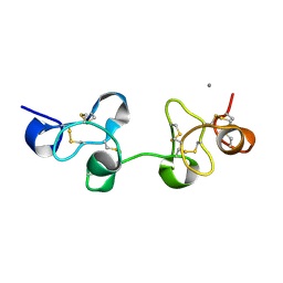
3A64
 | | Crystal structure of CcCel6C, a glycoside hydrolase family 6 enzyme, from Coprinopsis cinerea | | 分子名称: | Cellobiohydrolase, MAGNESIUM ION | | 著者 | Liu, Y, Yoshida, M, Kurakata, Y, Miyazaki, T, Nishikawa, A, Tonozuka, T. | | 登録日 | 2009-08-21 | | 公開日 | 2009-09-01 | | 最終更新日 | 2023-11-01 | | 実験手法 | X-RAY DIFFRACTION (1.6 Å) | | 主引用文献 | Crystal structure of a glycoside hydrolase family 6 enzyme, CcCel6C, a cellulase constitutively produced by Coprinopsis cinerea
Febs J., 277, 2010
|
|
1UHA
 | | Crystal Structure of Pokeweed Lectin-D2 | | 分子名称: | CALCIUM ION, lectin-D2 | | 著者 | Fujii, T, Hayashida, M, Hamasu, M, Ishiguro, M, Hata, Y. | | 登録日 | 2003-06-27 | | 公開日 | 2004-04-13 | | 最終更新日 | 2023-12-27 | | 実験手法 | X-RAY DIFFRACTION (1.5 Å) | | 主引用文献 | Structures of two lectins from the roots of pokeweed (Phytolacca americana).
Acta Crystallogr.,Sect.D, 60, 2004
|
|
1ULN
 | | Crystal Structure of Pokeweed Lectin-D1 | | 分子名称: | lectin-D | | 著者 | Fujii, T, Hayashida, M, Hamasu, M, Ishiguro, M, Hata, Y. | | 登録日 | 2003-09-16 | | 公開日 | 2004-04-13 | | 最終更新日 | 2023-12-27 | | 実験手法 | X-RAY DIFFRACTION (1.65 Å) | | 主引用文献 | Structures of two lectins from the roots of pokeweed (Phytolacca americana).
Acta Crystallogr.,Sect.D, 60, 2004
|
|
1M1N
 | | Nitrogenase MoFe protein from Azotobacter vinelandii | | 分子名称: | 3-HYDROXY-3-CARBOXY-ADIPIC ACID, CALCIUM ION, FE(7)-MO-S(9)-N CLUSTER, ... | | 著者 | Einsle, O, Tezcan, F.A, Andrade, S.L.A, Schmid, B, Yoshida, M, Howard, J.B, Rees, D.C. | | 登録日 | 2002-06-19 | | 公開日 | 2002-09-11 | | 最終更新日 | 2024-02-14 | | 実験手法 | X-RAY DIFFRACTION (1.16 Å) | | 主引用文献 | Nitrogenase MoFe-protein at 1.16 A resolution: a central ligand in the FeMo-cofactor.
Science, 297, 2002
|
|
1WI1
 | | Solution structure of the PH domain of human calcium-dependent activator protein for secretion (CAPS) | | 分子名称: | calcium-dependent activator protein for secretion, CAPS | | 著者 | Yoneyama, M, Izumi, K, Yoshida, M, Tochio, N, Koshiba, S, Inoue, M, Kigawa, T, Yokoyama, S, RIKEN Structural Genomics/Proteomics Initiative (RSGI) | | 登録日 | 2004-05-28 | | 公開日 | 2004-11-28 | | 最終更新日 | 2024-05-29 | | 実験手法 | SOLUTION NMR | | 主引用文献 | Solution structure of the PH domain of human calcium-dependent activator protein for secretion (CAPS)
To be Published
|
|
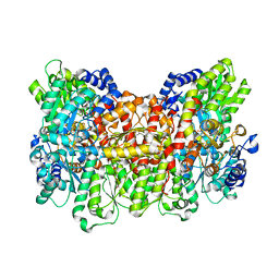
1IOK
 | | CRYSTAL STRUCTURE OF CHAPERONIN-60 FROM PARACOCCUS DENITRIFICANS | | 分子名称: | CHAPERONIN 60 | | 著者 | Fukami, T.A, Yohda, M, Taguchi, H, Yoshida, M, Miki, K. | | 登録日 | 2001-03-16 | | 公開日 | 2001-10-03 | | 最終更新日 | 2023-12-27 | | 実験手法 | X-RAY DIFFRACTION (3.2 Å) | | 主引用文献 | Crystal structure of chaperonin-60 from Paracoccus denitrificans.
J.Mol.Biol., 312, 2001
|
|
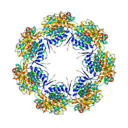
2D00
 | | Subunit F of V-type ATPase/synthase | | 分子名称: | CALCIUM ION, V-type ATP synthase subunit F | | 著者 | Makyio, H, Iino, R, Ikeda, C, Imamura, H, Tamakoshi, M, Iwata, M, Stock, D, Bernal, R.A, Carpenter, E.P, Yoshida, M, Yokoyama, K, Iwata, S. | | 登録日 | 2005-07-21 | | 公開日 | 2005-12-06 | | 最終更新日 | 2024-03-13 | | 実験手法 | X-RAY DIFFRACTION (2.2 Å) | | 主引用文献 | Structure of a central stalk subunit F of prokaryotic V-type ATPase/synthase from Thermus thermophilus
Embo J., 24, 2005
|
|
2D9S
 | | Solution structure of RSGI RUH-049, a UBA domain from mouse cDNA | | 分子名称: | CBL E3 ubiquitin protein ligase | | 著者 | Hamada, T, Hirota, H, Lin, Y.-J, Guntert, P, Kurosaki, C, Izumi, K, Yoshida, M, Koshiba, S, Kigawa, T, Yokoyama, S, RIKEN Structural Genomics/Proteomics Initiative (RSGI) | | 登録日 | 2005-12-13 | | 公開日 | 2007-01-23 | | 最終更新日 | 2024-05-29 | | 実験手法 | SOLUTION NMR | | 主引用文献 | Solution structure of RSGI RUH-049, a UBA domain from mouse cDNA
To be Published
|
|
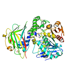
1SIU
 | | KUMAMOLISIN-AS E78H MUTANT | | 分子名称: | CALCIUM ION, SULFATE ION, kumamolisin-As | | 著者 | Li, M, Wlodawer, A, Gustchina, A, Tsuruoka, N, Ashida, M, Minakata, H, Oyama, H, Oda, K, Nishino, T, Nakayama, T. | | 登録日 | 2004-03-01 | | 公開日 | 2004-03-30 | | 最終更新日 | 2024-04-03 | | 実験手法 | X-RAY DIFFRACTION (2.31 Å) | | 主引用文献 | Crystallographic and biochemical investigations of kumamolisin-As, a serine-carboxyl peptidase with collagenase activity
J.Biol.Chem., 279, 2004
|
|
3A9B
 | | CcCel6C, a glycoside hydrolase family 6 enzyme, complexed with cellobiose | | 分子名称: | Cellobiohydrolase, MAGNESIUM ION, beta-D-glucopyranose, ... | | 著者 | Liu, Y, Yoshida, M, Kurakata, Y, Miyazaki, T, Nishikawa, A, Tonozuka, T. | | 登録日 | 2009-10-22 | | 公開日 | 2009-11-03 | | 最終更新日 | 2023-11-01 | | 実験手法 | X-RAY DIFFRACTION (1.2 Å) | | 主引用文献 | Crystal structure of a glycoside hydrolase family 6 enzyme, CcCel6C, a cellulase constitutively produced by Coprinopsis cinerea
Febs J., 277, 2010
|
|
3ABX
 | | CcCel6C, a glycoside hydrolase family 6 enzyme, complexed with p-nitrophenyl beta-D-cellotrioside | | 分子名称: | 4-nitrophenyl beta-D-glucopyranosyl-(1->4)-beta-D-glucopyranosyl-(1->4)-beta-D-glucopyranoside, Cellobiohydrolase, MAGNESIUM ION | | 著者 | Liu, Y, Yoshida, M, Kurakata, Y, Miyazaki, T, Nishikawa, A, Tonozuka, T. | | 登録日 | 2009-12-24 | | 公開日 | 2010-01-05 | | 最終更新日 | 2023-11-01 | | 実験手法 | X-RAY DIFFRACTION (1.4 Å) | | 主引用文献 | Crystal structure of a glycoside hydrolase family 6 enzyme, CcCel6C, a cellulase constitutively produced by Coprinopsis cinerea
Febs J., 277, 2010
|
|
1UK5
 | | Solution structure of the Murine BAG domain of Bcl2-associated athanogene 3 | | 分子名称: | BAG-family molecular chaperone regulator-3 | | 著者 | Hatta, R, Yoshida, M, Hayashi, F, Yokoyama, S, RIKEN Structural Genomics/Proteomics Initiative (RSGI) | | 登録日 | 2003-08-19 | | 公開日 | 2004-02-19 | | 最終更新日 | 2023-12-27 | | 実験手法 | SOLUTION NMR | | 主引用文献 | The C-terminal BAG domain of BAG5 induces conformational changes of the Hsp70 nucleotide-binding domain for ADP-ATP exchange
Structure, 18, 2010
|
|
1WPX
 | | Crystal structure of carboxypeptidase Y inhibitor complexed with the cognate proteinase | | 分子名称: | 2-acetamido-2-deoxy-beta-D-glucopyranose, Carboxypeptidase Y, Carboxypeptidase Y inhibitor, ... | | 著者 | Mima, J, Hayashida, M, Fujii, T, Narita, Y, Hayashi, R, Ueda, M, Hata, Y. | | 登録日 | 2004-09-14 | | 公開日 | 2005-03-01 | | 最終更新日 | 2020-07-29 | | 実験手法 | X-RAY DIFFRACTION (2.7 Å) | | 主引用文献 | Structure of the carboxypeptidase y inhibitor i(c) in complex with the cognate proteinase reveals a novel mode of the proteinase-protein inhibitor interaction
J.Mol.Biol., 346, 2005
|
|
2EDF
 | |
1WFF
 | | Solution structure of the zf-AN1 domain from mouse RIKEN cDNA 2810002D23 protein | | 分子名称: | RIKEN cDNA 2810002D23 protein, ZINC ION | | 著者 | Tomizawa, T, Kigawa, T, Izumi, K, Yoshida, M, Yamazaki, T, Inoue, M, Yokoyama, S, RIKEN Structural Genomics/Proteomics Initiative (RSGI) | | 登録日 | 2004-05-26 | | 公開日 | 2004-11-26 | | 最終更新日 | 2024-05-29 | | 実験手法 | SOLUTION NMR | | 主引用文献 | Solution structure of the zf-AN1 domain from mouse RIKEN cDNA 2810002D23 protein
To be Published
|
|
3VNY
 | | Crystal structure of beta-glucuronidase from Acidobacterium capsulatum | | 分子名称: | GLYCEROL, PHOSPHATE ION, beta-GLUCURONIDASE | | 著者 | Momma, M, Fujimoto, Z, Michikawa, M, Ichinose, H, Yoshida, M, Kotake, Y, Biely, P, Tsumuraya, Y, Kaneko, S. | | 登録日 | 2012-01-18 | | 公開日 | 2012-02-22 | | 最終更新日 | 2024-03-20 | | 実験手法 | X-RAY DIFFRACTION (1.5 Å) | | 主引用文献 | Structural and biochemical characterization of glycoside hydrolase family 79 beta-glucuronidase from Acidobacterium capsulatum
J.Biol.Chem., 287, 2012
|
|
3VO0
 | | Crystal structure of beta-glucuronidase from Acidobacterium capsulatum covalent-bonded with 2-deoxy-2-fluoro-D-glucuronic acid | | 分子名称: | 2,4-DINITROPHENOL, 2-deoxy-2-fluoro-alpha-D-glucopyranuronic acid, 2-deoxy-2-fluoro-beta-D-glucopyranuronic acid, ... | | 著者 | Momma, M, Fujimoto, Z, Michikawa, M, Ichinose, H, Jongkees, S, Yoshida, M, Kotake, Y, Biely, P, Tsumuraya, Y, Withers, S, Kaneko, S. | | 登録日 | 2012-01-18 | | 公開日 | 2012-02-22 | | 最終更新日 | 2023-11-08 | | 実験手法 | X-RAY DIFFRACTION (1.9 Å) | | 主引用文献 | Structural and biochemical characterization of glycoside hydrolase family 79 beta-glucuronidase from Acidobacterium capsulatum
J.Biol.Chem., 287, 2012
|
|
3VNZ
 | | Crystal structure of beta-glucuronidase from Acidobacterium capsulatum in complex with D-glucuronic acid | | 分子名称: | GLYCEROL, PHOSPHATE ION, beta-D-glucopyranuronic acid, ... | | 著者 | Momma, M, Fujimoto, Z, Michikawa, M, Ichinose, H, Yoshida, M, Kotake, Y, Biely, P, Tsumuraya, Y, Kaneko, S. | | 登録日 | 2012-01-18 | | 公開日 | 2012-02-22 | | 最終更新日 | 2023-11-08 | | 実験手法 | X-RAY DIFFRACTION (1.8 Å) | | 主引用文献 | Structural and biochemical characterization of glycoside hydrolase family 79 beta-glucuronidase from Acidobacterium capsulatum
J.Biol.Chem., 287, 2012
|
|
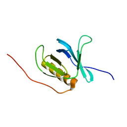
2DNC
 | | Solution Structure of RSGI RUH-054, a lipoyl domain from human 2-oxoacid dehydrogenase | | 分子名称: | Pyruvate dehydrogenase protein X component | | 著者 | Ruhul Momen, A.Z.M, Hirota, H, Hayashi, F, Kurosaki, C, Yoshida, M, Yokoyama, S, RIKEN Structural Genomics/Proteomics Initiative (RSGI) | | 登録日 | 2006-04-25 | | 公開日 | 2006-10-25 | | 最終更新日 | 2024-05-29 | | 実験手法 | SOLUTION NMR | | 主引用文献 | Solution Structure of RSGI RUH-054, a lipoyl domain from human 2-oxoacid dehydrogenase
To be published
|
|
2ENM
 | | Solution structure of the SH3 domain from mouse sorting nexin-9 | | 分子名称: | Sorting nexin-9 | | 著者 | Wakabayashi, M, Kurosaki, C, Yoshida, M, Hayashi, F, Yokoyama, S, RIKEN Structural Genomics/Proteomics Initiative (RSGI) | | 登録日 | 2007-03-28 | | 公開日 | 2007-10-02 | | 最終更新日 | 2024-05-29 | | 実験手法 | SOLUTION NMR | | 主引用文献 | Solution structure of the SH3 domain from mouse sorting nexin-9
To be Published
|
|
2ENS
 | | Solution structure of the third ig-like domain from human Advanced glycosylation end product-specific receptor | | 分子名称: | Advanced glycosylation end product-specific receptor | | 著者 | Wakabayashi, M, Kurosaki, C, Yoshida, M, Hayashi, F, Yokoyama, S, RIKEN Structural Genomics/Proteomics Initiative (RSGI) | | 登録日 | 2007-03-28 | | 公開日 | 2008-04-01 | | 最終更新日 | 2022-03-09 | | 実験手法 | SOLUTION NMR | | 主引用文献 | Solution structure of the third ig-like domain from human Advanced glycosylation end product-specific receptor
To be Published
|
|
2DNT
 | | Solution Structure of RSGI RUH-064, a Chromo Domain from Human cDNA | | 分子名称: | Chromodomain protein, Y chromosome-like, isoform b | | 著者 | Ruhul Momen, A.Z.M, Hirota, H, Izumi, K, Yoshida, M, Yokoyama, S, RIKEN Structural Genomics/Proteomics Initiative (RSGI) | | 登録日 | 2006-04-26 | | 公開日 | 2006-10-26 | | 最終更新日 | 2024-05-29 | | 実験手法 | SOLUTION NMR | | 主引用文献 | Solution Structure of RSGI RUH-064, a Chromo Domain from Human cDNA
to be published
|
|


