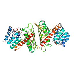
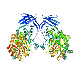
7NE2
 
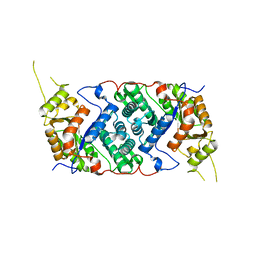
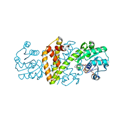
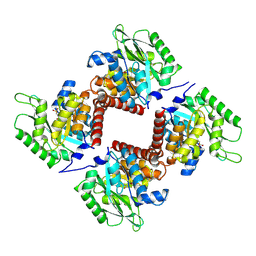 | | Crystal structure of class I SFP aldolase YihT from Salmonella enterica with SFP/ DHAP (Schiff base complex with active site Lys193) | | 分子名称: | (2~{S},3~{S},4~{R})-2,3,4,5-tetrakis(oxidanyl)-6-phosphonooxy-hexane-1-sulfonic acid, Sulfofructosephosphate aldolase, [(~{E})-2,3-bis(oxidanyl)prop-1-enyl] dihydrogen phosphate | | 著者 | Sharma, M, Davies, G.J. | | 登録日 | 2021-02-03 | | 公開日 | 2021-04-14 | | 最終更新日 | 2024-01-31 | | 実験手法 | X-RAY DIFFRACTION (1.5 Å) | | 主引用文献 | Molecular Basis of Sulfosugar Selectivity in Sulfoglycolysis.
Acs Cent.Sci., 7, 2021
|
|
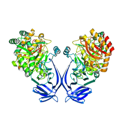
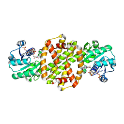
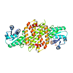
8C54
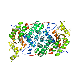
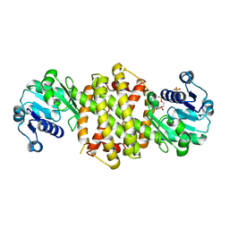
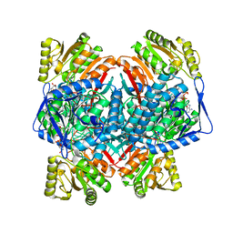 | | Cryo-EM structure of NADH bound SLA dehydrogenase RlGabD from Rhizobium leguminosarum bv. trifolii SRD1565 | | 分子名称: | 1,4-DIHYDRONICOTINAMIDE ADENINE DINUCLEOTIDE, Succinate semialdehyde dehydrogenase | | 著者 | Sharma, M, Meek, R.W, Armstrong, Z, Blaza, J.N, Alhifthi, A, Li, J, Goddard-Borger, E.D, Williams, S.J, Davies, G.J. | | 登録日 | 2023-01-06 | | 公開日 | 2023-09-20 | | 最終更新日 | 2024-04-10 | | 実験手法 | ELECTRON MICROSCOPY (2.52 Å) | | 主引用文献 | Molecular basis of sulfolactate synthesis by sulfolactaldehyde dehydrogenase from Rhizobium leguminosarum.
Chem Sci, 14, 2023
|
|
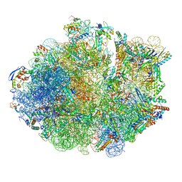
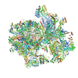
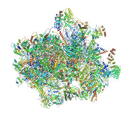
6DZP
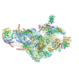
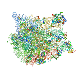
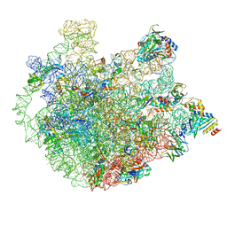 | | Cryo-EM Structure of Mycobacterium smegmatis C(minus) 50S ribosomal subunit | | 分子名称: | 23S rRNA, 50S ribosomal protein L10, 50S ribosomal protein L11, ... | | 著者 | Sharma, M.R, Li, Y, Korripella, R, Yang, Y, Kaushal, P.S, Lin, Q, Wade, J.T, Gray, A.G, Derbyshire, K.M, Agrawal, R.K, Ojha, A. | | 登録日 | 2018-07-05 | | 公開日 | 2018-10-03 | | 最終更新日 | 2024-03-13 | | 実験手法 | ELECTRON MICROSCOPY (3.42 Å) | | 主引用文献 | Zinc depletion induces ribosome hibernation in mycobacteria.
Proc. Natl. Acad. Sci. U.S.A., 115, 2018
|
|
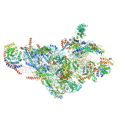
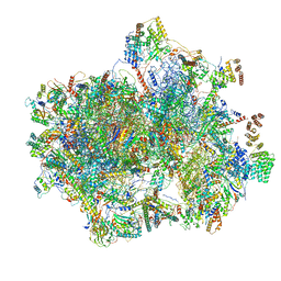
6DZK
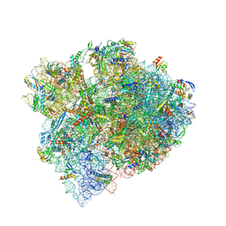
 | | Cryo-EM Structure of Mycobacterium smegmatis C(minus) 30S ribosomal subunit with MPY | | 分子名称: | 16S rRNA, 30S ribosomal protein S10, 30S ribosomal protein S11, ... | | 著者 | Sharma, M.R, Li, Y, Korripella, R, Yang, Y, Kaushal, P.S, Lin, Q, Wade, J.T, Gray, A.G, Derbyshire, K.M, Agrawal, R.K, Ojha, A. | | 登録日 | 2018-07-05 | | 公開日 | 2018-09-19 | | 最終更新日 | 2024-03-13 | | 実験手法 | ELECTRON MICROSCOPY (3.6 Å) | | 主引用文献 | Zinc depletion induces ribosome hibernation in mycobacteria.
Proc. Natl. Acad. Sci. U.S.A., 115, 2018
|
|
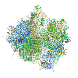
6DZI
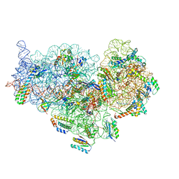
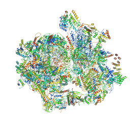
 | | Cryo-EM Structure of Mycobacterium smegmatis 70S C(minus) ribosome 70S-MPY complex | | 分子名称: | 16S rRNA, 23 S rRNA (3119-MER), 30S ribosomal protein S10, ... | | 著者 | Sharma, M.R, Li, Y, Korripella, R, Yang, Y, Kaushal, P.S, Lin, Q, Wade, J.T, Gray, A.G, Derbyshire, K.M, Agrawal, R.K, Ojha, A. | | 登録日 | 2018-07-05 | | 公開日 | 2018-09-26 | | 最終更新日 | 2024-03-13 | | 実験手法 | ELECTRON MICROSCOPY (3.46 Å) | | 主引用文献 | Zinc depletion induces ribosome hibernation in mycobacteria.
Proc. Natl. Acad. Sci. U.S.A., 115, 2018
|
|
4V61
 | | Homology model for the Spinach chloroplast 30S subunit fitted to 9.4A cryo-EM map of the 70S chlororibosome. | | 分子名称: | 16S rRNA, 23S rRNA, 4.8S rRNA, ... | | 著者 | Sharma, M.R, Wilson, D.N, Datta, P.P, Barat, C, Schluenzen, F, Fucini, P, Agrawal, R.K. | | 登録日 | 2007-11-09 | | 公開日 | 2014-07-09 | | 最終更新日 | 2024-02-28 | | 実験手法 | ELECTRON MICROSCOPY (9.4 Å) | | 主引用文献 | Cryo-EM study of the spinach chloroplast ribosome reveals the structural and functional roles of plastid-specific ribosomal proteins
Proc.Natl.Acad.Sci.Usa, 104, 2007
|
|
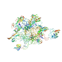
7OFX
 | | Crystal structure of a GH31 family sulfoquinovosidase mutant D455N from Agrobacterium tumefaciens in complex with sulfoquinovosyl glycerol (SQGro) | | 分子名称: | Alpha-glucosidase yihQ, [(2S,3S,4S,5R,6S)-6-[(2R)-2,3-bis(oxidanyl)propoxy]-3,4,5-tris(oxidanyl)oxan-2-yl]methanesulfonic acid | | 著者 | Sharma, M, Davies, G.J. | | 登録日 | 2021-05-05 | | 公開日 | 2022-01-19 | | 最終更新日 | 2024-01-31 | | 実験手法 | X-RAY DIFFRACTION (2.15 Å) | | 主引用文献 | Oxidative desulfurization pathway for complete catabolism of sulfoquinovose by bacteria.
Proc.Natl.Acad.Sci.USA, 119, 2022
|
|
6NF8
 | | Structure of human mitochondrial translation initiation factor 3 bound to the small ribosomal subunit -Class I | | 分子名称: | 28S ribosomal RNA, mitochondria, 28S ribosomal protein S10, ... | | 著者 | Sharma, M, Koripella, R, Agrawal, R. | | 登録日 | 2018-12-19 | | 公開日 | 2019-02-27 | | 最終更新日 | 2019-12-11 | | 実験手法 | ELECTRON MICROSCOPY (3.48 Å) | | 主引用文献 | Structure of Human Mitochondrial Translation Initiation Factor 3 Bound to the Small Ribosomal Subunit.
iScience, 12, 2019
|
|
6NEQ
 | | Structure of human mitochondrial translation initiation factor 3 bound to the small ribosomal subunit-Class-II | | 分子名称: | 28S ribosomal RNA, mitochondrial, 28S ribosomal protein S10, ... | | 著者 | Sharma, M, Koripella, R, Agrawal, R. | | 登録日 | 2018-12-18 | | 公開日 | 2019-02-27 | | 最終更新日 | 2019-12-11 | | 実験手法 | ELECTRON MICROSCOPY (3.32 Å) | | 主引用文献 | Structure of Human Mitochondrial Translation Initiation Factor 3 Bound to the Small Ribosomal Subunit.
iScience, 12, 2019
|
|
7OLF
 | |
7OH2
 | |
6NU3
 | | Structural insights into unique features of the human mitochondrial ribosome recycling | | 分子名称: | 12S rRNA, 16S rRNA, 28S ribosomal protein S10, ... | | 著者 | Sharma, M.R, Koripella, R.K, Agrawal, R.K. | | 登録日 | 2019-01-30 | | 公開日 | 2019-04-17 | | 最終更新日 | 2019-12-11 | | 実験手法 | ELECTRON MICROSCOPY (4.4 Å) | | 主引用文献 | Structural insights into unique features of the human mitochondrial ribosome recycling.
Proc.Natl.Acad.Sci.USA, 116, 2019
|
|
6NU2
 | | Structural insights into unique features of the human mitochondrial ribosome recycling | | 分子名称: | 12S rRNA, 16S rRNA, 28S ribosomal protein S10, ... | | 著者 | Sharma, M.R, Koripella, R.K, Agrawal, R.K. | | 登録日 | 2019-01-30 | | 公開日 | 2019-04-17 | | 最終更新日 | 2019-12-11 | | 実験手法 | ELECTRON MICROSCOPY (3.9 Å) | | 主引用文献 | Structural insights into unique features of the human mitochondrial ribosome recycling.
Proc.Natl.Acad.Sci.USA, 116, 2019
|
|
8P2J
 | |
1DQ7
 | | THREE-DIMENSIONAL STRUCTURE OF A NEUROTOXIN FROM RED SCORPION (BUTHUS TAMULUS) AT 2.2A RESOLUTION. | | 分子名称: | NEUROTOXIN | | 著者 | Sharma, M, Yadav, S, Karthikeyan, S, Kumar, S, Paramasivam, M, Srinivasan, A, Singh, T.P. | | 登録日 | 1999-12-30 | | 公開日 | 2000-12-30 | | 最終更新日 | 2011-07-13 | | 実験手法 | X-RAY DIFFRACTION (2.2 Å) | | 主引用文献 | Three-dimensional Structure of a Neurotoxin from Red Scorpion (Buthus tamulus) at 2.2A Resolution
To be Published
|
|
6EOD
 | |
7X2Y
 | | Crystal Structure of cis-4,5-dihydrodiol phthalate dehydrogenase in complex with NAD+ and 3-Hydroxybenzoate | | 分子名称: | 3-HYDROXYBENZOIC ACID, 4,5-dihydroxyphthalate dehydrogenase, DI(HYDROXYETHYL)ETHER, ... | | 著者 | Sharma, M, Mahto, J.K, Kumar, P. | | 登録日 | 2022-02-26 | | 公開日 | 2022-09-14 | | 最終更新日 | 2023-11-29 | | 実験手法 | X-RAY DIFFRACTION (2.48 Å) | | 主引用文献 | Conformational flexibility enables catalysis of phthalate cis-4,5-dihydrodiol dehydrogenase.
Arch.Biochem.Biophys., 727, 2022
|
|
8OZV
 | | Imine Reductase from Ajellomyces dermatitidis in complex with 2,2-difluoroacetophenone | | 分子名称: | 2,2-bis(fluoranyl)-1-phenyl-ethanone, NADPH DIHYDRO-NICOTINAMIDE-ADENINE-DINUCLEOTIDE PHOSPHATE, Oxidoreductase | | 著者 | Sharma, M, Grogan, G. | | 登録日 | 2023-05-09 | | 公開日 | 2023-08-30 | | 最終更新日 | 2023-09-13 | | 実験手法 | X-RAY DIFFRACTION (1.52 Å) | | 主引用文献 | Structure of the imine reductase from Ajellomyces dermatitidis in three crystal forms.
Acta Crystallogr.,Sect.F, 79, 2023
|
|
8OZW
 | | Imine Reductase from Ajellomyces dermatitidis in complex NADPH4 | | 分子名称: | 1,2-ETHANEDIOL, 1,4,5,6-TETRAHYDRONICOTINAMIDE ADENINE DINUCLEOTIDE PHOSPHATE, Oxidoreductase | | 著者 | Sharma, M, Grogan, G. | | 登録日 | 2023-05-09 | | 公開日 | 2023-08-30 | | 最終更新日 | 2023-09-13 | | 実験手法 | X-RAY DIFFRACTION (2.01 Å) | | 主引用文献 | Structure of the imine reductase from Ajellomyces dermatitidis in three crystal forms.
Acta Crystallogr.,Sect.F, 79, 2023
|
|
8FN2
 | | The structure of a 50S ribosomal subunit in the Lyme disease pathogen Borreliella burgdorferi | | 分子名称: | 23S ribosomal RNA, 50S ribosomal protein L10, 50S ribosomal protein L11, ... | | 著者 | Sharma, M.R, Manjari, S.R, Agrawal, E.K, Keshavan, P, Koripella, R.K, Majumdar, S, Marcinkiewicz, A.L, Lin, Y.P, Agrawal, R.K, Banavali, N.K. | | 登録日 | 2022-12-26 | | 公開日 | 2023-11-08 | | 実験手法 | ELECTRON MICROSCOPY (3.4 Å) | | 主引用文献 | The structure of a hibernating ribosome in a Lyme disease pathogen.
Nat Commun, 14, 2023
|
|
8FMW
 | | The structure of a hibernating ribosome in the Lyme disease pathogen | | 分子名称: | 16S ribosomal RNA, 23S ribosomal RNA, 30S ribosomal protein S10, ... | | 著者 | Sharma, M.R, Manjari, S.R, Agrawal, E.K, Keshavan, P, Koripella, R.K, Majumdar, S, Marcinkiewicz, A.L, Lin, Y.P, Agrawal, R.K, Banavali, N.K. | | 登録日 | 2022-12-25 | | 公開日 | 2023-11-08 | | 実験手法 | ELECTRON MICROSCOPY (2.86 Å) | | 主引用文献 | The structure of a hibernating ribosome in a Lyme disease pathogen.
Nat Commun, 14, 2023
|
|
6VMI
 | | Structure of the human mitochondrial ribosome-EF-G1 complex (ClassIII) | | 分子名称: | 12s rRNA, 16s rRNA, 28S ribosomal protein S10, ... | | 著者 | Sharma, M.R, Koripella, R.K, Agrawal, R.K. | | 登録日 | 2020-01-28 | | 公開日 | 2020-08-05 | | 最終更新日 | 2021-02-17 | | 実験手法 | ELECTRON MICROSCOPY (2.96 Å) | | 主引用文献 | Structures of the human mitochondrial ribosome bound to EF-G1 reveal distinct features of mitochondrial translation elongation.
Nat Commun, 11, 2020
|
|
6VLZ
 | | Structure of the human mitochondrial ribosome-EF-G1 complex (ClassI) | | 分子名称: | 12s rRNA, 16s rRNA, 28S ribosomal protein S10, ... | | 著者 | Sharma, M.R, Koripella, R.K, Agrawal, R.K. | | 登録日 | 2020-01-27 | | 公開日 | 2020-08-05 | | 最終更新日 | 2024-03-06 | | 実験手法 | ELECTRON MICROSCOPY (2.97 Å) | | 主引用文献 | Structures of the human mitochondrial ribosome bound to EF-G1 reveal distinct features of mitochondrial translation elongation.
Nat Commun, 11, 2020
|
|
3IY9
 | | Leishmania Tarentolae Mitochondrial Large Ribosomal Subunit Model | | 分子名称: | 39S ribosomal protein L11, mitochondrial, 39S ribosomal protein L16, ... | | 著者 | Sharma, M.R, Booth, T.M, Simpson, L, Maslov, D.A, Agrawal, R.K. | | 登録日 | 2009-04-20 | | 公開日 | 2009-07-07 | | 最終更新日 | 2024-02-21 | | 実験手法 | ELECTRON MICROSCOPY (14.1 Å) | | 主引用文献 | Structure of a mitochondrial ribosome with minimal RNA
Proc.Natl.Acad.Sci.USA, 106, 2009
|
|
8R56
 | |


