1UWM
 
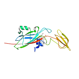
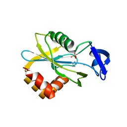
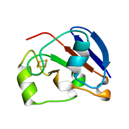 | | reduced ferredoxin 6 from Rhodobacter capsulatus | | 分子名称: | FE2/S2 (INORGANIC) CLUSTER, FERREDOXIN VI | | 著者 | Sainz, G, Jakoncic, J, Sieker, L.C, Stojanoff, V, Sanishvili, N, Asso, M, Bertrand, P, Armengaud, J, Jouanneau, Y. | | 登録日 | 2004-02-05 | | 公開日 | 2006-01-18 | | 最終更新日 | 2023-12-13 | | 実験手法 | X-RAY DIFFRACTION (2 Å) | | 主引用文献 | Structure of a [2Fe-2S] Ferredoxin from Rhodobacter Capsulatus Likely Involved in Fe-S Cluster Biogenesis and Conformational Changes Observed Upon Reduction.
J.Biol.Inorg.Chem., 11, 2006
|
|
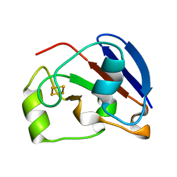
1DXH
 | | Catabolic ornithine carbamoyltransferase from Pseudomonas aeruginosa | | 分子名称: | ORNITHINE CARBAMOYLTRANSFERASE, SULFATE ION | | 著者 | Sainz, G, Vicat, J, Kahn, R, Duee, E, Tricot, C, Stalon, V, Dideberg, O. | | 登録日 | 2000-01-05 | | 公開日 | 2001-01-12 | | 最終更新日 | 2023-12-06 | | 実験手法 | X-RAY DIFFRACTION (2.5 Å) | | 主引用文献 | Crystal Structure of the Allosteric Active Form of Catabolic Ornithine Carbamoyltransferase from Pseudomonas Aeruginosa
To be Published
|
|
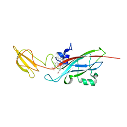
1HIZ
 | | Xylanase T6 (Xt6) from Bacillus Stearothermophilus | | 分子名称: | ENDO-1,4-BETA-XYLANASE, SULFATE ION, alpha-D-galactopyranose, ... | | 著者 | Sainz, G, Tepplitsky, A, Stojanoff, V, Thompson, A, Shoham, Y, Shoham, G. | | 登録日 | 2001-01-05 | | 公開日 | 2002-01-04 | | 最終更新日 | 2024-05-08 | | 実験手法 | X-RAY DIFFRACTION (2.4 Å) | | 主引用文献 | Structure Determination of the Extracellular Xylanase from Geobacillus Stearothermophilus by Selenomethionyl MAD Phasing
Acta Crystallogr.,Sect.D, 60, 2004
|
|
1EWH
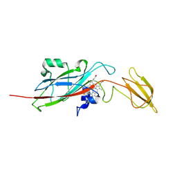 | | STRUCTURE OF CYTOCHROME F FROM CHLAMYDOMONAS REINHARDTII | | 分子名称: | ACETATE ION, CYTOCHROME F, HEME C | | 著者 | Sainz, G, Carrell, C.J, Ponamarev, M.V, Soriano, G.M, Cramer, W.A, Smith, J.L. | | 登録日 | 2000-04-25 | | 公開日 | 2000-08-09 | | 最終更新日 | 2021-03-03 | | 実験手法 | X-RAY DIFFRACTION (2.35 Å) | | 主引用文献 | Interruption of the internal water chain of cytochrome f impairs photosynthetic function.
Biochemistry, 39, 2000
|
|
1E2V
 | | N153Q mutant of cytochrome f from Chlamydomonas reinhardtii | | 分子名称: | ACETATE ION, CYTOCHROME F, HEME C | | 著者 | Sainz, G, Carrell, C.J, Ponamarev, M.V, Soriano, G.M, Cramer, W.A, Smith, J.L. | | 登録日 | 2000-05-29 | | 公開日 | 2000-08-04 | | 最終更新日 | 2023-12-06 | | 実験手法 | X-RAY DIFFRACTION (1.85 Å) | | 主引用文献 | Interruption of the Internal Water Chain of Cytochrome F Impairs Photosynthetic Function
Biochemistry, 39, 2000
|
|
1E2Z
 | | Q158L mutant of cytochrome f from Chlamydomonas reinhardtii | | 分子名称: | CYTOCHROME F, HEME C | | 著者 | Sainz, G, Carrell, C.J, Ponamarev, M.V, Soriano, G.M, Cramer, W.A, Smith, J.L. | | 登録日 | 2000-05-30 | | 公開日 | 2000-08-04 | | 最終更新日 | 2023-12-06 | | 実験手法 | X-RAY DIFFRACTION (2.5 Å) | | 主引用文献 | Interruption of the Internal Water Chain of Cytochrome F Impairs Photosynthetic Function
Biochemistry, 39, 2000
|
|
1E2W
 | | N168F mutant of cytochrome f from Chlamydomonas reinhardtii | | 分子名称: | CYTOCHROME F, HEME C | | 著者 | Sainz, G, Carrell, C.J, Ponamarev, M.V, Soriano, G.M, Cramer, W.A, Smith, J.L. | | 登録日 | 2000-05-30 | | 公開日 | 2000-08-04 | | 最終更新日 | 2023-12-06 | | 実験手法 | X-RAY DIFFRACTION (1.6 Å) | | 主引用文献 | Interruption of the Internal Water Chain of Cytochrome F Impairs Photosynthetic Function
Biochemistry, 39, 2000
|
|
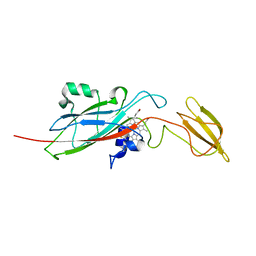
1E9M
 | | Ferredoxin VI from Rhodobacter Capsulatus | | 分子名称: | FE2/S2 (INORGANIC) CLUSTER, FERREDOXIN VI | | 著者 | Sainz, G, Armengaud, J, Stojanoff, V, Sanishvili, N, Jouanneau, Y, Larry, S. | | 登録日 | 2000-10-24 | | 公開日 | 2001-04-09 | | 最終更新日 | 2024-05-08 | | 実験手法 | X-RAY DIFFRACTION (2.07 Å) | | 主引用文献 | Crystallization and Preliminary X-Ray Diffraction Analysis of a [2Fe-2S] Ferredoxin (Fdvi) from Rhodobacter Capsulatus
Acta Crystallogr.,Sect.D, 57, 2001
|
|
1MQF
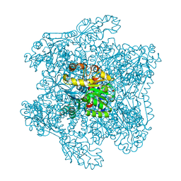
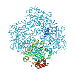
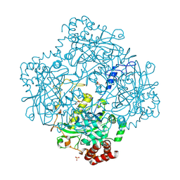 | | Compound I from Proteus mirabilis catalase | | 分子名称: | Catalase, GLYCEROL, OXYGEN ATOM, ... | | 著者 | Andreoletti, P, Pernoud, A, Sainz, G, Gouet, P, Jouve, H.M. | | 登録日 | 2002-09-16 | | 公開日 | 2002-10-02 | | 最終更新日 | 2011-07-13 | | 実験手法 | X-RAY DIFFRACTION (2.5 Å) | | 主引用文献 | Structural studies of Proteus mirabilis catalase in its ground state, oxidized state and in complex with formic acid.
Acta Crystallogr.,Sect.D, 59, 2003
|
|
1ZLX
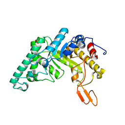
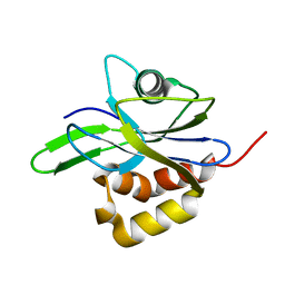
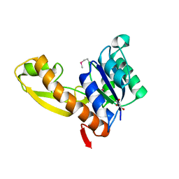 | | The apo structure of human glycinamide ribonucleotide transformylase | | 分子名称: | GLYCEROL, Phosphoribosylglycinamide formyltransferase | | 著者 | Dahms, T.E, Sainz, G, Giroux, E.L, Caperelli, C.A, Smith, J.L. | | 登録日 | 2005-05-09 | | 公開日 | 2005-08-23 | | 最終更新日 | 2023-11-15 | | 実験手法 | X-RAY DIFFRACTION (2.2 Å) | | 主引用文献 | The apo and ternary complex structures of a chemotherapeutic target: human glycinamide ribonucleotide transformylase.
Biochemistry, 44, 2005
|
|
1ZLY
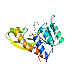 | | The structure of human glycinamide ribonucleotide transformylase in complex with alpha,beta-N-(hydroxyacetyl)-D-ribofuranosylamine and 10-formyl-5,8,dideazafolate | | 分子名称: | 4-[(4-{[(2-AMINO-4-OXO-3,4-DIHYDROQUINAZOLIN-6-YL)METHYL]AMINO}BENZOYL)AMINO]BUTANOIC ACID, 5-O-phosphono-beta-D-ribofuranosylamine, Phosphoribosylglycinamide formyltransferase | | 著者 | Dahms, T.E.S, Sainz, G, Giroux, E.L, Caperelli, C.A, Smith, J.L. | | 登録日 | 2005-05-09 | | 公開日 | 2005-08-23 | | 最終更新日 | 2023-08-23 | | 実験手法 | X-RAY DIFFRACTION (2.07 Å) | | 主引用文献 | The apo and ternary complex structures of a chemotherapeutic target: human glycinamide ribonucleotide transformylase.
Biochemistry, 44, 2005
|
|
1H7K
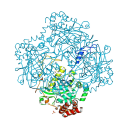
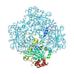 | | Formation of a tyrosyl radical intermediate in Proteus mirabilis catalase by directed mutagenesis and consequences for nucleotide reactivity | | 分子名称: | ACETATE ION, CATALASE, PROTOPORPHYRIN IX CONTAINING FE, ... | | 著者 | Andreoletti, P, Gambarelli, S, Gaillard, J, Sainz, G, Stojanoff, V, Jouve, H.M. | | 登録日 | 2001-07-08 | | 公開日 | 2004-02-25 | | 最終更新日 | 2023-12-13 | | 実験手法 | X-RAY DIFFRACTION (2.4 Å) | | 主引用文献 | Formation of a Tyrosyl Radical Intermediate in Proteus Mirabilis Catalase by Directed Mutagenesis and Consequences for Nucleotide Reactivity.
Biochemistry, 40, 2001
|
|
1H6N
 | | Formation of a tyrosyl radical intermediate in Proteus mirabilis catalase by directed mutagenesis and consequences for nucleotide reactivity | | 分子名称: | ACETATE ION, CATALASE, PROTOPORPHYRIN IX CONTAINING FE, ... | | 著者 | Andreoletti, P, Sainz, G, Jaquinod, M, Gagnon, J, Jouve, H.M. | | 登録日 | 2001-06-20 | | 公開日 | 2003-10-03 | | 最終更新日 | 2023-12-13 | | 実験手法 | X-RAY DIFFRACTION (2.11 Å) | | 主引用文献 | High Resolution Structure and Biochemical Properties of a Recombinant Proteus Mirabilis Catalase Depleted in Iron.
Proteins: Struct.,Funct., Genet., 50, 2003
|
|
1HX3
 | | CRYSTAL STRUCTURE OF E.COLI ISOPENTENYL DIPHOSPHATE:DIMETHYLALLYL DIPHOSPHATE ISOMERASE | | 分子名称: | IMIDAZOLE, ISOPENTENYL DIPHOSPHATE DELTA-ISOMERASE, MANGANESE (II) ION, ... | | 著者 | Durbecq, V, Sainz, G, Oudjama, Y, Clantin, B, Bompard-Gilles, C, Tricot, C, Caillet, J, Stalon, V, Droogmans, L, Villeret, V. | | 登録日 | 2001-01-11 | | 公開日 | 2001-07-11 | | 最終更新日 | 2024-05-29 | | 実験手法 | X-RAY DIFFRACTION (2.1 Å) | | 主引用文献 | Crystal structure of isopentenyl diphosphate:dimethylallyl diphosphate isomerase.
Embo J., 20, 2001
|
|
1E93
 | | High resolution structure and biochemical properties of a recombinant catalase depleted in iron | | 分子名称: | ACETATE ION, CATALASE, PROTOPORPHYRIN IX CONTAINING FE, ... | | 著者 | Andreoletti, P, Sainz, G, Jaquinod, M, Gagnon, J, Jouve, H.M. | | 登録日 | 2000-10-05 | | 公開日 | 2000-10-13 | | 最終更新日 | 2023-12-13 | | 実験手法 | X-RAY DIFFRACTION (2 Å) | | 主引用文献 | High-Resolution Structure and Biochemical Properties of a Recombinant Proteus Mirabilis Catalase Depleted in Iron.
Proteins: Struct.,Funct., Genet., 50, 2003
|
|
1HZT
 | | CRYSTAL STRUCTURE OF METAL-FREE ISOPENTENYL DIPHOSPHATE:DIMETHYLALLYL DIPHOSPHATE ISOMERASE | | 分子名称: | ISOPENTENYL DIPHOSPHATE DELTA-ISOMERASE | | 著者 | Durbecq, V, Sainz, G, Oudjama, Y, Clantin, B, Bompard-Gilles, C, Tricot, C, Caillet, J, Stalon, V, Droogmans, L, Villeret, V. | | 登録日 | 2001-01-26 | | 公開日 | 2001-07-26 | | 最終更新日 | 2024-03-13 | | 実験手法 | X-RAY DIFFRACTION (1.45 Å) | | 主引用文献 | Crystal structure of isopentenyl diphosphate:dimethylallyl diphosphate isomerase.
Embo J., 20, 2001
|
|


