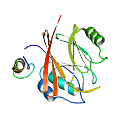
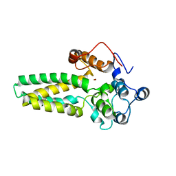
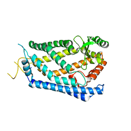
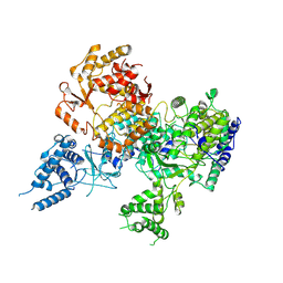
5MUZ
 
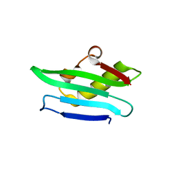 | | Structure of a C-terminal domain of a reptarenavirus L protein | | 分子名称: | L protein | | 著者 | Rosenthal, M, Gogrefe, N, Reguera, J, Vogel, D, Rauschenberger, B, Cusack, S, Gunther, S, Reindl, S. | | 登録日 | 2017-01-14 | | 公開日 | 2017-05-17 | | 最終更新日 | 2024-01-17 | | 実験手法 | X-RAY DIFFRACTION (1.776 Å) | | 主引用文献 | Structural insights into reptarenavirus cap-snatching machinery.
PLoS Pathog., 13, 2017
|
|
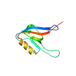
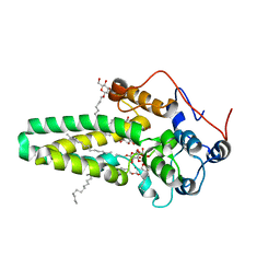
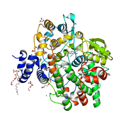
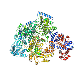
5MUY
 | | Structure of a C-terminal domain of a reptarenavirus L protein with m7GTP | | 分子名称: | 7N-METHYL-8-HYDROGUANOSINE-5'-TRIPHOSPHATE, L protein | | 著者 | Rosenthal, M, Gogrefe, N, Reguera, J, Vogel, D, Rauschenberger, B, Cusack, S, Gunther, S, Reindl, S. | | 登録日 | 2017-01-14 | | 公開日 | 2017-05-17 | | 最終更新日 | 2024-01-17 | | 実験手法 | X-RAY DIFFRACTION (1.99 Å) | | 主引用文献 | Structural insights into reptarenavirus cap-snatching machinery.
PLoS Pathog., 13, 2017
|
|
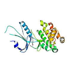
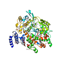
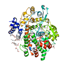
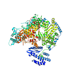
5MV0
 | | Structure of an N-terminal domain of a reptarenavirus L protein | | 分子名称: | L protein, PHOSPHATE ION | | 著者 | Rosenthal, M, Gogrefe, N, Reguera, J, Vogel, D, Rauschenberger, B, Cusack, S, Gunther, S, Reindl, S. | | 登録日 | 2017-01-14 | | 公開日 | 2017-05-17 | | 最終更新日 | 2024-05-08 | | 実験手法 | X-RAY DIFFRACTION (1.93 Å) | | 主引用文献 | Structural insights into reptarenavirus cap-snatching machinery.
PLoS Pathog., 13, 2017
|
|
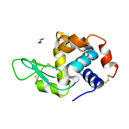
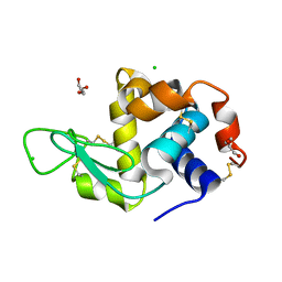
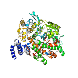
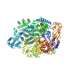
5MUS
 | | Structure of the C-terminal domain of a reptarenavirus L protein | | 分子名称: | CHLORIDE ION, GLYCEROL, L protein | | 著者 | Rosenthal, M, Gogrefe, N, Reguera, J, Vogel, D, Rauschenberger, B, Cusack, S, Gunther, S, Reindl, S. | | 登録日 | 2017-01-14 | | 公開日 | 2017-05-17 | | 最終更新日 | 2017-05-24 | | 実験手法 | X-RAY DIFFRACTION (2.009 Å) | | 主引用文献 | Structural insights into reptarenavirus cap-snatching machinery.
PLoS Pathog., 13, 2017
|
|
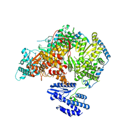
5GTU
 | |
2JRE
 | | C60-1, a PDZ domain designed using statistical coupling analysis | | 分子名称: | C60-1 PDZ domain peptide | | 著者 | Larson, C, Stiffler, M, Li, P, Rosen, M, MacBeath, G, Ranganathan, R. | | 登録日 | 2007-06-25 | | 公開日 | 2008-07-01 | | 最終更新日 | 2023-12-20 | | 実験手法 | SOLUTION NMR | | 主引用文献 | C60-1, a PDZ domain designed using statistical coupling analysis
To be Published
|
|
7MF0
 | | Co-crystal structure of PERK with inhibitor (R)-2-amino-N-cyclopropyl-5-(4-(2-(3,5-difluorophenyl)-2-hydroxyacetamido)-2-methylphenyl)nicotinamide | | 分子名称: | 2-amino-N-cyclopropyl-5-(4-{[(2R)-2-(3,5-difluorophenyl)-2-hydroxyacetyl]amino}-2-methylphenyl)pyridine-3-carboxamide, Eukaryotic translation initiation factor 2-alpha kinase 3,Eukaryotic translation initiation factor 2-alpha kinase 3 | | 著者 | Wiens, B, Koszelak-Rosenblum, M, Surman, M.D, Zhu, G, Mulvihill, M.J. | | 登録日 | 2021-04-08 | | 公開日 | 2021-05-19 | | 最終更新日 | 2023-10-18 | | 実験手法 | X-RAY DIFFRACTION (2.809 Å) | | 主引用文献 | Discovery of 2-amino-3-amido-5-aryl-pyridines as highly potent, orally bioavailable, and efficacious PERK kinase inhibitors.
Bioorg.Med.Chem.Lett., 43, 2021
|
|
6Q88
 | | RT structure of HEWL at 5 kGy | | 分子名称: | CHLORIDE ION, DI(HYDROXYETHYL)ETHER, Lysozyme C | | 著者 | de la Mora, E, Coquelle, N, Bury, C.S, Rosenthal, M, Garman, E.F, Burghammer, M, Colletier, J.P, Weik, M. | | 登録日 | 2018-12-14 | | 公開日 | 2020-01-15 | | 最終更新日 | 2024-01-24 | | 実験手法 | X-RAY DIFFRACTION (1.74007058 Å) | | 主引用文献 | Radiation damage and dose limits in serial synchrotron crystallography at cryo- and room temperatures.
Proc.Natl.Acad.Sci.USA, 117, 2020
|
|
6Q8T
 | | Cryo structure of HEWL at 81 kGy | | 分子名称: | CHLORIDE ION, DI(HYDROXYETHYL)ETHER, Lysozyme C | | 著者 | de la Mora, E, Coquelle, N, Bury, C.S, Rosenthal, M, Garman, E.F, Burghammer, M, Colletier, J.P, Weik, M. | | 登録日 | 2018-12-16 | | 公開日 | 2020-01-15 | | 最終更新日 | 2024-01-24 | | 実験手法 | X-RAY DIFFRACTION (1.74008667 Å) | | 主引用文献 | Radiation damage and dose limits in serial synchrotron crystallography at cryo- and room temperatures.
Proc.Natl.Acad.Sci.USA, 117, 2020
|
|
4ZR1
 | | Hydroxylase domain of scs7p | | 分子名称: | Ceramide very long chain fatty acid hydroxylase SCS7, TRIDECANE, ZINC ION, ... | | 著者 | Zhu, G, Koszelak-Rosenblum, M, Malkowski, M.G, Membrane Protein Structural Biology Consortium (MPSBC) | | 登録日 | 2015-05-11 | | 公開日 | 2015-07-29 | | 最終更新日 | 2024-03-06 | | 実験手法 | X-RAY DIFFRACTION (2.6008 Å) | | 主引用文献 | The Crystal Structure of an Integral Membrane Fatty Acid alpha-Hydroxylase.
J.Biol.Chem., 290, 2015
|
|
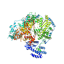
4ZR0
 | |
6WO0
 | | human Artemis/SNM1C catalytic domain, crystal form 1 | | 分子名称: | GLYCEROL, Protein artemis, ZINC ION | | 著者 | Karim, F, Liu, S, Laciak, A.R, Volk, L, Rosenblum, M, Curtis, R, Huang, N, Carr, G, Zhu, G. | | 登録日 | 2020-04-23 | | 公開日 | 2020-07-01 | | 最終更新日 | 2024-04-03 | | 実験手法 | X-RAY DIFFRACTION (1.97 Å) | | 主引用文献 | Structural analysis of the catalytic domain of Artemis endonuclease/SNM1C reveals distinct structural features.
J.Biol.Chem., 295, 2020
|
|
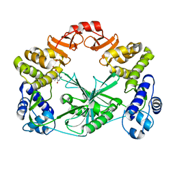
4HHR
 | | Crystal Structure of fatty acid alpha-dioxygenase (Arabidopsis thaliana) | | 分子名称: | Alpha-dioxygenase, CALCIUM ION, CHLORIDE ION, ... | | 著者 | Goulah, C.C, Zhu, G, Koszelak-Rosenblum, M, Malkowski, M.G. | | 登録日 | 2012-10-10 | | 公開日 | 2013-02-20 | | 最終更新日 | 2024-02-28 | | 実験手法 | X-RAY DIFFRACTION (1.51 Å) | | 主引用文献 | The crystal structure of alpha-Dioxygenase provides insight into diversity in the cyclooxygenase-peroxidase superfamily.
Biochemistry, 52, 2013
|
|
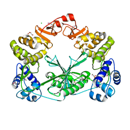
4HHS
 | | Crystal Structure of fatty acid alpha-dioxygenase (Arabidopsis thaliana) | | 分子名称: | Alpha-dioxygenase, CALCIUM ION, CHLORIDE ION, ... | | 著者 | Goulah, C.C, Zhu, G, Koszelak-Rosenblum, M, Malkowski, M.G. | | 登録日 | 2012-10-10 | | 公開日 | 2013-02-20 | | 最終更新日 | 2024-02-28 | | 実験手法 | X-RAY DIFFRACTION (1.7 Å) | | 主引用文献 | The crystal structure of alpha-Dioxygenase provides insight into diversity in the cyclooxygenase-peroxidase superfamily.
Biochemistry, 52, 2013
|
|
4IL3
 | | Crystal Structure of S. mikatae Ste24p | | 分子名称: | Ste24p, ZINC ION | | 著者 | Pryor Jr, E.E, Horanyi, P.S, Clark, K, Fedoriw, N, Connelly, S.M, Koszelak-Rosenblum, M, Zhu, G, Malkowski, M.G, Dumont, M.E, Wiener, M.C, Membrane Protein Structural Biology Consortium (MPSBC) | | 登録日 | 2012-12-28 | | 公開日 | 2013-02-20 | | 最終更新日 | 2023-09-20 | | 実験手法 | X-RAY DIFFRACTION (3.102 Å) | | 主引用文献 | Structure of the integral membrane protein CAAX protease Ste24p.
Science, 339, 2013
|
|
4KVJ
 | | Crystal structure of Oryza sativa fatty acid alpha-dioxygenase with hydrogen peroxide | | 分子名称: | 3,6,9,12,15,18-HEXAOXAICOSANE-1,20-DIOL, CALCIUM ION, CHLORIDE ION, ... | | 著者 | Zhu, G, Koszelak-Rosenblum, M, Malkowski, M.G. | | 登録日 | 2013-05-22 | | 公開日 | 2013-08-21 | | 最終更新日 | 2024-02-28 | | 実験手法 | X-RAY DIFFRACTION (2.12 Å) | | 主引用文献 | Crystal structures of alpha-dioxygenase from Oryza sativa: Insights into substrate binding and activation by hydrogen peroxide.
Protein Sci., 22, 2013
|
|
4KVL
 | | Crystal structure of Oryza sativa fatty acid alpha-dioxygenase Y379F with palmitic acid | | 分子名称: | CALCIUM ION, CHLORIDE ION, Fatty acid alpha-oxidase, ... | | 著者 | Zhu, G, Koszelak-Rosenblum, M, Malkowski, M.G. | | 登録日 | 2013-05-22 | | 公開日 | 2013-08-21 | | 最終更新日 | 2024-02-28 | | 実験手法 | X-RAY DIFFRACTION (1.96 Å) | | 主引用文献 | Crystal structures of alpha-dioxygenase from Oryza sativa: Insights into substrate binding and activation by hydrogen peroxide.
Protein Sci., 22, 2013
|
|
7OE7
 | | Apo-structure of Lassa virus L protein (well-resolved alpha ribbon) [APO-RIBBON] | | 分子名称: | MAGNESIUM ION, RNA-directed RNA polymerase L, ZINC ION | | 著者 | Kouba, T, Vogel, D, Thorkelsson, S, Quemin, E, Williams, H.M, Milewski, M, Busch, C, Gunther, S, Grunewald, K, Rosenthal, M, Cusack, S. | | 登録日 | 2021-05-01 | | 公開日 | 2021-12-01 | | 最終更新日 | 2024-05-01 | | 実験手法 | ELECTRON MICROSCOPY (3.73 Å) | | 主引用文献 | Conformational changes in Lassa virus L protein associated with promoter binding and RNA synthesis activity.
Nat Commun, 12, 2021
|
|
7OJK
 | | Lassa virus L protein bound to the distal promoter duplex [DISTAL-PROMOTER] | | 分子名称: | 3' RNA, 5' RNA, RNA-directed RNA polymerase L, ... | | 著者 | Kouba, T, Vogel, D, Thorkelsson, S, Quemin, E, Williams, H.M, Milewski, M, Busch, C, Gunther, S, Grunewald, K, Rosenthal, M, Cusack, S. | | 登録日 | 2021-05-16 | | 公開日 | 2021-12-01 | | 最終更新日 | 2024-05-01 | | 実験手法 | ELECTRON MICROSCOPY (3.89 Å) | | 主引用文献 | Conformational changes in Lassa virus L protein associated with promoter binding and RNA synthesis activity.
Nat Commun, 12, 2021
|
|
7OEA
 | | Lassa virus L protein bound to 3' promoter RNA (well-resolved polymerase core) [3END-CORE] | | 分子名称: | 3' vRNA, MAGNESIUM ION, RNA-directed RNA polymerase L, ... | | 著者 | Kouba, T, Vogel, D, Thorkelsson, S, Quemin, E, Williams, H.M, Milewski, M, Busch, C, Gunther, S, Grunewald, K, Rosenthal, M, Cusack, S. | | 登録日 | 2021-05-02 | | 公開日 | 2021-12-01 | | 最終更新日 | 2024-05-01 | | 実験手法 | ELECTRON MICROSCOPY (2.7 Å) | | 主引用文献 | Conformational changes in Lassa virus L protein associated with promoter binding and RNA synthesis activity.
Nat Commun, 12, 2021
|
|
7OCH
 | | Apo-structure of Lassa virus L protein (well-resolved polymerase core) [APO-CORE] | | 分子名称: | MAGNESIUM ION, RNA-directed RNA polymerase L, ZINC ION | | 著者 | Kouba, T, Vogel, D, Thorkelsson, S, Quemin, E, Williams, H.M, Milewski, M, Busch, C, Gunther, S, Grunewald, K, Rosenthal, M, Cusack, S. | | 登録日 | 2021-04-26 | | 公開日 | 2021-12-01 | | 最終更新日 | 2024-05-01 | | 実験手法 | ELECTRON MICROSCOPY (3.14 Å) | | 主引用文献 | Conformational changes in Lassa virus L protein associated with promoter binding and RNA synthesis activity.
Nat Commun, 12, 2021
|
|
7OJN
 | | Lassa virus L protein in an elongation conformation [ELONGATION] | | 分子名称: | 3' RNA, 5' RNA, 5'-O-[(S)-hydroxy{[(S)-hydroxy(phosphonooxy)phosphoryl]amino}phosphoryl]uridine, ... | | 著者 | Kouba, T, Vogel, D, Thorkelsson, S, Quemin, E, Williams, H.M, Milewski, M, Busch, C, Gunther, S, Grunewald, K, Rosenthal, M, Cusack, S. | | 登録日 | 2021-05-16 | | 公開日 | 2021-12-01 | | 最終更新日 | 2024-05-01 | | 実験手法 | ELECTRON MICROSCOPY (2.92 Å) | | 主引用文献 | Conformational changes in Lassa virus L protein associated with promoter binding and RNA synthesis activity.
Nat Commun, 12, 2021
|
|
7OEB
 | | Lassa virus L protein bound to 3' promoter RNA (well-resolved endonuclease) [3END-ENDO] | | 分子名称: | 3' vRNA, MAGNESIUM ION, RNA-directed RNA polymerase L, ... | | 著者 | Kouba, T, Vogel, D, Thorkelsson, S, Quemin, E, Williams, H.M, Milewski, M, Busch, C, Gunther, S, Grunewald, K, Rosenthal, M, Cusack, S. | | 登録日 | 2021-05-02 | | 公開日 | 2021-12-01 | | 最終更新日 | 2024-05-01 | | 実験手法 | ELECTRON MICROSCOPY (3.04 Å) | | 主引用文献 | Conformational changes in Lassa virus L protein associated with promoter binding and RNA synthesis activity.
Nat Commun, 12, 2021
|
|
7OE3
 | | Apo-structure of Lassa virus L protein (well-resolved endonuclease) [APO-ENDO] | | 分子名称: | MAGNESIUM ION, RNA-directed RNA polymerase L, ZINC ION | | 著者 | Kouba, T, Vogel, D, Thorkelsson, S, Quemin, E, Williams, H.M, Milewski, M, Busch, C, Gunther, S, Grunewald, K, Rosenthal, M, Cusack, S. | | 登録日 | 2021-05-01 | | 公開日 | 2021-12-01 | | 最終更新日 | 2024-05-01 | | 実験手法 | ELECTRON MICROSCOPY (3.35 Å) | | 主引用文献 | Conformational changes in Lassa virus L protein associated with promoter binding and RNA synthesis activity.
Nat Commun, 12, 2021
|
|
7OJL
 | | Lassa virus L protein in a pre-initiation conformation [PREINITIATION] | | 分子名称: | 3' RNA, 5' RNA, MANGANESE (II) ION, ... | | 著者 | Kouba, T, Vogel, D, Thorkelsson, S, Quemin, E, Williams, H.M, Milewski, M, Busch, C, Gunther, S, Grunewald, K, Rosenthal, M, Cusack, S. | | 登録日 | 2021-05-16 | | 公開日 | 2021-12-01 | | 最終更新日 | 2024-05-01 | | 実験手法 | ELECTRON MICROSCOPY (3.3 Å) | | 主引用文献 | Conformational changes in Lassa virus L protein associated with promoter binding and RNA synthesis activity.
Nat Commun, 12, 2021
|
|


