4G5G
 
 | |
4ZJX
 | |
2AFA
 | |
1YT8
 | |
1CQV
 | |
1Y9Q
 | |
1YAV
 | |
1YBD
 | |
1Y9I
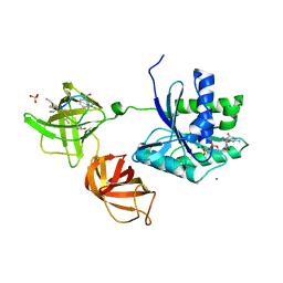
 | | Crystal structure of low temperature requirement C protein from Listeria monocytogenes | | 分子名称: | CALCIUM ION, GLYCEROL, MAGNESIUM ION, ... | | 著者 | Kumaran, D, Swaminathan, S, Burley, S.K, New York SGX Research Center for Structural Genomics (NYSGXRC) | | 登録日 | 2004-12-15 | | 公開日 | 2004-12-28 | | 最終更新日 | 2021-02-03 | | 実験手法 | X-RAY DIFFRACTION (1.8 Å) | | 主引用文献 | Crystal structure of phosphatidylglycerophosphatase (PGPase), a putative membrane-bound lipid phosphatase, reveals a novel binuclear metal binding site and two "proton wires".
Proteins, 64, 2006
|
|
6FCE
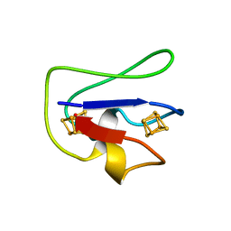
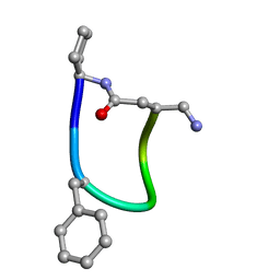 | | NMR ensemble of Macrocyclic Peptidomimetic Containing Constrained a,a-dialkylated Amino Acids with Potent and Selective Activity at Human Melanocortin Receptors | | 分子名称: | ACP-HIS-DPHE-ARG-TRP-ASP-NH2 | | 著者 | Brancaccio, D, Carotenuto, A, Grieco, P, Merlino, F, Zhou, Y, Cai, M, Yousif, A.M, Di Maro, S, Novellino, E, Hruby, V.J. | | 登録日 | 2017-12-20 | | 公開日 | 2018-04-25 | | 最終更新日 | 2018-05-23 | | 実験手法 | SOLUTION NMR | | 主引用文献 | Development of Macrocyclic Peptidomimetics Containing Constrained alpha , alpha-Dialkylated Amino Acids with Potent and Selective Activity at Human Melanocortin Receptors.
J. Med. Chem., 61, 2018
|
|
1RRM
 | |
1F1M
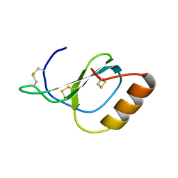
 | | CRYSTAL STRUCTURE OF OUTER SURFACE PROTEIN C (OSPC) | | 分子名称: | OUTER SURFACE PROTEIN C, ZINC ION | | 著者 | Kumaran, D, Eswaramoorthy, S, Dunn, J.J, Swaminathan, S. | | 登録日 | 2000-05-19 | | 公開日 | 2001-04-04 | | 最終更新日 | 2024-02-07 | | 実験手法 | X-RAY DIFFRACTION (1.8 Å) | | 主引用文献 | Crystal structure of outer surface protein C (OspC) from the Lyme disease spirochete, Borrelia burgdorferi.
EMBO J., 20, 2001
|
|
3SQS
 | |
1I4P
 | |
1I4R
 | |
3URH
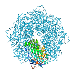
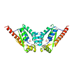
 | | Crystal structure of a dihydrolipoamide dehydrogenase from Sinorhizobium meliloti 1021 | | 分子名称: | 1,2-ETHANEDIOL, Dihydrolipoyl dehydrogenase, FLAVIN-ADENINE DINUCLEOTIDE, ... | | 著者 | Kumaran, D, Chamala, S, Evans, B, Foti, R, Gizzi, A, Hillerich, B, Kar, A, LaFleur, J, Seidel, R, Villigas, G, Zencheck, W, Almo, S.C, Swaminathan, S, New York Structural Genomics Research Consortium (NYSGRC) | | 登録日 | 2011-11-22 | | 公開日 | 2011-12-14 | | 実験手法 | X-RAY DIFFRACTION (1.9 Å) | | 主引用文献 | Crystal structure of a dihydrolipoamide dehydrogenase from Sinorhizobium meliloti 1021
TO BE PUBLISHED
|
|
1I4Q
 | |
4HED
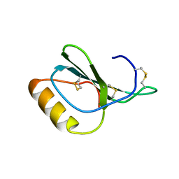 | | Zebrafish chemokine CXL1 | | 分子名称: | Uncharacterized protein | | 著者 | Rajasekaran, D, Fan, C, Meng, W, Pflugrath, J.W, Lolis, E.J. | | 登録日 | 2012-10-03 | | 公開日 | 2013-08-21 | | 最終更新日 | 2014-04-23 | | 実験手法 | X-RAY DIFFRACTION (1.62 Å) | | 主引用文献 | Structural insight into the evolution of a new chemokine family from zebrafish.
Proteins, 82, 2014
|
|
1F89
 | | Crystal structure of Saccharomyces cerevisiae Nit3, a member of branch 10 of the nitrilase superfamily | | 分子名称: | 32.5 KDA PROTEIN YLR351C | | 著者 | Kumaran, D, Eswaramoorthy, S, Studier, F.W, Swaminathan, S, Burley, S.K, New York SGX Research Center for Structural Genomics (NYSGXRC) | | 登録日 | 2000-06-29 | | 公開日 | 2001-10-04 | | 最終更新日 | 2024-02-07 | | 実験手法 | X-RAY DIFFRACTION (2.4 Å) | | 主引用文献 | Crystal structure of a putative CN hydrolase from yeast
Proteins, 52, 2003
|
|
4HCS
 | | Structure of Novel subfamily CX chemokine solved by sulfur SAD | | 分子名称: | Uncharacterized protein | | 著者 | Rajasekaran, D, Fan, C, Meng, W, Pflugrath, J.W, Lolis, E.J. | | 登録日 | 2012-10-01 | | 公開日 | 2013-10-16 | | 最終更新日 | 2014-04-23 | | 実験手法 | X-RAY DIFFRACTION (1.28 Å) | | 主引用文献 | Structural insight into the evolution of a new chemokine family from zebrafish.
Proteins, 82, 2014
|
|
1FCA
 | |
3V8B
 | | Crystal Structure of a 3-ketoacyl-ACP reductase from Sinorhizobium meliloti 1021 | | 分子名称: | Putative dehydrogenase, possibly 3-oxoacyl-[acyl-carrier protein] reductase | | 著者 | Kumaran, D, Chamala, S, Evans, B, Foti, R, Gizzi, A, Hillerich, B, Kar, A, Lafleur, J, Seidel, R, Villigas, G, Zencheck, W, Almo, S.C, Swaminathan, S, New York Structural Genomics Research Consortium (NYSGRC) | | 登録日 | 2011-12-22 | | 公開日 | 2012-01-11 | | 最終更新日 | 2023-12-06 | | 実験手法 | X-RAY DIFFRACTION (2.7 Å) | | 主引用文献 | Crystal Structure of a 3-ketoacyl-ACP reductase from Sinorhizobium meliloti 1021
To be Published
|
|
3US8
 | | Crystal Structure of an isocitrate dehydrogenase from Sinorhizobium meliloti 1021 | | 分子名称: | Isocitrate dehydrogenase [NADP], SULFATE ION | | 著者 | Kumaran, D, Chamala, S, Evans, B, Foti, R, Gizzi, A, Hillerich, B, Kar, A, LaFleur, J, Seidel, R, Villigas, G, Zencheck, W, Almo, S.C, Swaminathan, S, New York Structural Genomics Research Consortium (NYSGRC) | | 登録日 | 2011-11-23 | | 公開日 | 2011-12-14 | | 実験手法 | X-RAY DIFFRACTION (2.25 Å) | | 主引用文献 | Crystal Structure of an isocitrate dehydrogenase from Sinorhizobium meliloti 1021
To be Published
|
|
4E3Z
 | | Crystal Structure of a oxidoreductase from Rhizobium etli CFN 42 | | 分子名称: | Putative oxidoreductase protein | | 著者 | Kumaran, D, Chamala, S, Evans, B, Foti, R, Gizzi, A, Hillerich, B, Kar, A, Lafleur, J, Seidel, R, Villigas, G, Zencheck, W, Almo, S.C, Swaminathan, S, New York Structural Genomics Research Consortium (NYSGRC) | | 登録日 | 2012-03-11 | | 公開日 | 2012-03-21 | | 実験手法 | X-RAY DIFFRACTION (2 Å) | | 主引用文献 | Crystal Structure of a oxidoreductase from Rhizobium etli CFN 42
To be Published
|
|
3T61
 | | Crystal Structure of a gluconokinase from Sinorhizobium meliloti 1021 | | 分子名称: | Gluconokinase, PHOSPHATE ION | | 著者 | Kumaran, D, Chamala, S, Evans, B, Foti, R, Gizzi, A, Hillerich, B, Kar, A, LaFleur, J, Seidel, R, Villigas, G, Zencheck, W, Almo, S.C, Swaminathan, S, New York Structural Genomics Research Consortium (NYSGRC) | | 登録日 | 2011-07-28 | | 公開日 | 2011-08-17 | | 最終更新日 | 2012-03-21 | | 実験手法 | X-RAY DIFFRACTION (2.2 Å) | | 主引用文献 | Crystal Structure of a gluconokinase from Sinorhizobium meliloti 1021
To be Published
|
|


