Loading
PDBj
MenuPDBj@FacebookPDBj@TwitterPDBj@YouTubewwPDB FoundationwwPDB
RCSB PDBPDBeBMRBAdv. SearchSearch help
PDB: 43 results

2OGR
DownloadVisualize
BU of 2ogr by Molmil
Crystal Structure of Yellow Fluorescent Protein from Zoanthus sp. at 1.8 A Resolution
Descriptor: FLUORESCENT PROTEIN FP538
Authors:Pletneva, N.V, Pletnev, S.V, Tikhonova, T.V, Pletnev, V.Z.
Deposit date:2007-01-08
Release date:2007-09-25
Last modified:2023-12-27
Method:X-RAY DIFFRACTION (1.8 Å)
Cite:Three-dimensional structure of yellow fluorescent protein zYFP538 from Zoanthus sp. at the resolution 1.8 angstrom
Bioorg.Khim., 33
4ZFS
DownloadVisualize
BU of 4zfs by Molmil
Phototoxic Fluorescent Protein KillerOrange
Descriptor: KillerOrange
Authors:Pletneva, N.V, Pletnev, V.Z, Pletnev, S.
Deposit date:2015-04-21
Release date:2015-12-23
Last modified:2024-07-10
Method:X-RAY DIFFRACTION (2.01 Å)
Cite:Crystal Structure of Phototoxic Orange Fluorescent Proteins with a Tryptophan-Based Chromophore.
Plos One, 10, 2015
4EDS
DownloadVisualize
BU of 4eds by Molmil
Crystal structure of far-red fluorescent protein eqFP670
Descriptor: far-red fluorescent protein eqFP650
Authors:Pletnev, S, Pletnev, V.Z, Pletneva, N.V.
Deposit date:2012-03-27
Release date:2012-09-05
Last modified:2013-03-27
Method:X-RAY DIFFRACTION (1.6 Å)
Cite:Structural basis for bathochromic shift of fluorescence in far-red fluorescent proteins eqFP650 and eqFP670.
Acta Crystallogr.,Sect.D, 68, 2012
4EDO
DownloadVisualize
BU of 4edo by Molmil
Crystal structure of far-red fluorescent protein eqFP650
Descriptor: far-red fluorescent protein eqFP650
Authors:Pletnev, S, Pletnev, V.Z, Pletneva, N.V.
Deposit date:2012-03-27
Release date:2012-09-05
Last modified:2013-03-27
Method:X-RAY DIFFRACTION (1.8 Å)
Cite:Structural basis for bathochromic shift of fluorescence in far-red fluorescent proteins eqFP650 and eqFP670.
Acta Crystallogr.,Sect.D, 68, 2012
3PJ5
DownloadVisualize
BU of 3pj5 by Molmil
Crystal structure of far-red fluorescent protein Katushka crystallized at pH 5.0
Descriptor: Red fluorescent protein eqFP578, SULFATE ION
Authors:Pletnev, S, Pletneva, N.V, Pletnev, V.Z.
Deposit date:2010-11-08
Release date:2011-05-25
Last modified:2012-02-22
Method:X-RAY DIFFRACTION (1.6 Å)
Cite:Crystallographic study of red fluorescent protein eqFP578 and its far-red variant Katushka reveals opposite pH-induced isomerization of chromophore.
Protein Sci., 20, 2011
3PJB
DownloadVisualize
BU of 3pjb by Molmil
Crystal structure of red fluorescent protein eqFP578 crystallized at pH 4.0
Descriptor: GLYCEROL, Red fluorescent protein eqFP578
Authors:Pletnev, S, Pletneva, N.V, Pletnev, V.Z.
Deposit date:2010-11-09
Release date:2011-05-25
Last modified:2018-01-24
Method:X-RAY DIFFRACTION (1.75 Å)
Cite:Crystallographic study of red fluorescent protein eqFP578 and its far-red variant Katushka reveals opposite pH-induced isomerization of chromophore.
Protein Sci., 20, 2011
4HE4
DownloadVisualize
BU of 4he4 by Molmil
Crystal structure of the yellow fluorescent protein phiYFP (Phialidium sp.)
Descriptor: Yellow fluorescent protein
Authors:Pletneva, N, Pletnev, S, Pletnev, V.Z.
Deposit date:2012-10-03
Release date:2013-05-29
Last modified:2023-12-06
Method:X-RAY DIFFRACTION (2.05 Å)
Cite:Yellow fluorescent protein phiYFPv (Phialidium): structure and structure-based mutagenesis.
Acta Crystallogr.,Sect.D, 69, 2013
4N3D
DownloadVisualize
BU of 4n3d by Molmil
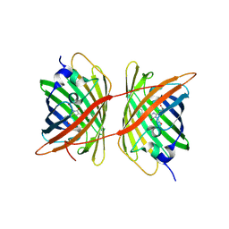
Crystal structure of the dimeric variant EGFP-K162Q in P61 space group
Descriptor: GLYCEROL, Green fluorescent protein, PHOSPHATE ION, ...
Authors:Pletneva, N.V, Pletnev, V.Z, Pletnev, S.V.
Deposit date:2013-10-07
Release date:2014-08-27
Method:X-RAY DIFFRACTION (1.34 Å)
Cite:Three dimensional structure of the dimeric gene-engineered variant of green fluorescent protein egfp-K162Q in P61 crystal space group
Rus.J.Bioorg.Chem., 40, 2014
3PIB
DownloadVisualize
BU of 3pib by Molmil
Crystal structure of red fluorescent protein eqFP578 crystallized at pH 5.5
Descriptor: GLYCEROL, eqFP578 fluorescent protein
Authors:Pletnev, S, Pletneva, N.V, Pletnev, V.Z.
Deposit date:2010-11-05
Release date:2011-05-25
Last modified:2012-02-22
Method:X-RAY DIFFRACTION (1.154 Å)
Cite:Crystallographic study of red fluorescent protein eqFP578 and its far-red variant Katushka reveals opposite pH-induced isomerization of chromophore.
Protein Sci., 20, 2011
3PJ7
DownloadVisualize
BU of 3pj7 by Molmil
Crystal structure of far-red fluorescent protein Katushka crystallized at pH 8.5
Descriptor: Red fluorescent protein eqFP578
Authors:Pletnev, S, Pletneva, N.V, Pletnev, V.Z.
Deposit date:2010-11-08
Release date:2011-05-25
Last modified:2012-02-22
Method:X-RAY DIFFRACTION (1.85 Å)
Cite:Crystallographic study of red fluorescent protein eqFP578 and its far-red variant Katushka reveals opposite pH-induced isomerization of chromophore.
Protein Sci., 20, 2011
3LVD
DownloadVisualize
BU of 3lvd by Molmil
Crystal structure of GFP-like protein aceGFP_G222E (A. coerulescens). UV-photoconverted green form.
Descriptor: GLYCEROL, Green fluorescent protein
Authors:Pletneva, N.V, Pletnev, V.Z, Pletnev, S.V.
Deposit date:2010-02-19
Release date:2010-03-09
Last modified:2023-11-15
Method:X-RAY DIFFRACTION (1.751 Å)
Cite:Structural evidence for a dehydrated intermediate in green fluorescent protein chromophore biosynthesis.
J.Biol.Chem., 285, 2010
3LVC
DownloadVisualize
BU of 3lvc by Molmil
Crystal structure of GFP-like protein aceGFP_G222E (A. coerulescens). Colorless form.
Descriptor: GLYCEROL, Green fluorescent protein
Authors:Pletneva, N.V, Pletnev, V.Z, Pletnev, S.V.
Deposit date:2010-02-19
Release date:2010-03-09
Last modified:2023-11-15
Method:X-RAY DIFFRACTION (1.14 Å)
Cite:Structural evidence for a dehydrated intermediate in green fluorescent protein chromophore biosynthesis.
J.Biol.Chem., 285, 2010
3LVA
DownloadVisualize
BU of 3lva by Molmil
Crystal structure of colorless GFP-like protein from Aequorea coerulescens
Descriptor: GLYCEROL, Green fluorescent protein, SULFATE ION
Authors:Pletneva, N.V, Pletnev, V.Z, Pletnev, S.V.
Deposit date:2010-02-19
Release date:2010-03-09
Last modified:2023-11-15
Method:X-RAY DIFFRACTION (1.5 Å)
Cite:Structural evidence for a dehydrated intermediate in green fluorescent protein chromophore biosynthesis.
J.Biol.Chem., 285, 2010
3GL4
DownloadVisualize
BU of 3gl4 by Molmil
X-ray structure of photobleached killerred
Descriptor: KillerRed
Authors:Pletnev, S, Pletneva, N.V, Pletnev, V.Z.
Deposit date:2009-03-11
Release date:2009-09-08
Last modified:2023-11-15
Method:X-RAY DIFFRACTION (2.15 Å)
Cite:Structural basis for phototoxicity of the genetically encoded photosensitizer KillerRed.
J.Biol.Chem., 284, 2009
3GB3
DownloadVisualize
BU of 3gb3 by Molmil
X-ray structure of genetically encoded photosensitizer KillerRed in native form
Descriptor: KillerRed, SULFATE ION
Authors:Pletnev, S, Pletneva, N.V, Pletnev, V.Z.
Deposit date:2009-02-18
Release date:2009-09-08
Last modified:2023-11-15
Method:X-RAY DIFFRACTION (1.75 Å)
Cite:Structural basis for phototoxicity of the genetically encoded photosensitizer KillerRed.
J.Biol.Chem., 284, 2009
2BQP
DownloadVisualize
BU of 2bqp by Molmil
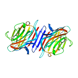
THE STRUCTURE OF THE PEA LECTIN-D-GLUCOPYRANOSE COMPLEX
Descriptor: CALCIUM ION, MANGANESE (II) ION, PROTEIN (PEA LECTIN), ...
Authors:Ruzeinikov, S.N, Mikhailova Yu, I, Tsygannik, I.N, Pangborn, W, Duax, W, Pletnev, V.Z.
Deposit date:1998-12-08
Release date:1998-12-16
Last modified:2023-08-23
Method:X-RAY DIFFRACTION (1.9 Å)
Cite:The Structure of Pea Lectin-D-Glucopyranose Complex at a 1.9 A Resolution
RUSS.J.BIOORGANIC CHEM., 23, 1997
1F90
DownloadVisualize
BU of 1f90 by Molmil
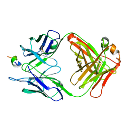
FAB FRAGMENT OF MONOCLONAL ANTIBODY (LNKB-2) AGAINST HUMAN INTERLEUKIN-2 IN COMPLEX WITH ANTIGENIC PEPTIDE
Descriptor: ANTIGENIC NONAPEPTIDE, FAB FRAGMENT OF MONOCLONAL ANTIBODY
Authors:Afonin, P.V, Fokin, A.V, Tsigannik, I.N, Mikhailova, I.Y, Onoprienko, L.V, Mikhaleva, I.I, Ivanov, V.T, Mareeva, T.Y, Nesmeyanov, V.A, Li, N, Duax, W.L, Pletnev, V.Z.
Deposit date:2000-07-06
Release date:2001-07-11
Last modified:2018-04-04
Method:X-RAY DIFFRACTION (2.6 Å)
Cite:Crystal structure of an anti-interleukin-2 monoclonal antibody Fab complexed with an antigenic nonapeptide.
Protein Sci., 10, 2001
1BQP
DownloadVisualize
BU of 1bqp by Molmil
THE STRUCTURE OF THE PEA LECTIN-D-MANNOPYRANOSE COMPLEX
Descriptor: CALCIUM ION, MANGANESE (II) ION, PROTEIN (LECTIN), ...
Authors:Ruzeinikov, S.N, Mikhailova, I.Y, Tsygannik, I.N, Pangborn, W, Duax, W, Pletnev, V.Z.
Deposit date:1998-08-17
Release date:1998-08-26
Last modified:2023-08-09
Method:X-RAY DIFFRACTION (2.1 Å)
Cite:The Structure of the Pea Lectin-D-Mannopyranose Complex at a 2.1 A Resolution
RUSS.J.BIOORGANIC CHEM., 24, 1998
<12


224201

PDB entries from 2024-08-28

PDB statisticsPDBj update infoContact PDBjnumon