Loading
PDBj
MenuPDBj@FacebookPDBj@TwitterPDBj@YouTubewwPDB FoundationwwPDB
RCSB PDBPDBeBMRBAdv. SearchSearch help
PDB: 85 results

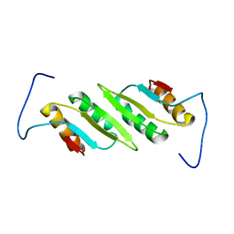
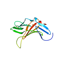
2LSS
DownloadVisualize
BU of 2lss by Molmil
Solution structure of the R. rickettsii cold shock-like protein
Descriptor: Cold shock-like protein
Authors:Veldkamp, C.T, Peterson, F.C, Gerarden, K.P, Fuchs, A.M, Koch, J.M, Mueller, M.M.
Deposit date:2012-05-04
Release date:2012-05-16
Last modified:2024-05-15
Method:SOLUTION NMR
Cite:Solution structure of the cold-shock-like protein from Rickettsia rickettsii.
Acta Crystallogr.,Sect.F, 68, 2012
2L48
DownloadVisualize
BU of 2l48 by Molmil
Solution structure of the PlyG cell wall binding domain
Descriptor: N-acetylmuramoyl-L-alanine amidase
Authors:Volkman, B.F, Dias, J.S, Peterson, F.C.
Deposit date:2010-10-01
Release date:2011-10-12
Last modified:2024-05-01
Method:SOLUTION NMR
Cite:Tbd
To be Published
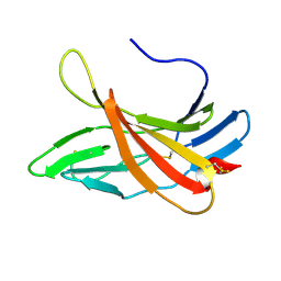
2KVB
DownloadVisualize
BU of 2kvb by Molmil
Solution structure of CI-MPR domain 5 bound to N-acetylglucosaminyl 6-phosphomethylmannoside
Descriptor: Cation-independent mannose-6-phosphate receptor
Authors:Olson, L.J, Peterson, F.C, Volkman, B.F, Dahms, N.M.
Deposit date:2010-03-10
Release date:2010-07-07
Last modified:2023-06-14
Method:SOLUTION NMR
Cite:Structural basis for recognition of phosphodiester-containing lysosomal enzymes by the cation-independent mannose 6-phosphate receptor.
Proc.Natl.Acad.Sci.USA, 107, 2010
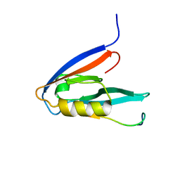
2KOH
DownloadVisualize
BU of 2koh by Molmil
NMR structure of mouse Par3-PDZ3 in complex with VE-Cadherin C-terminus
Descriptor: Cadherin-5, Partitioning defective 3 homolog
Authors:Volkman, B.F, Tyler, R.C, Peterson, F.C.
Deposit date:2009-09-22
Release date:2010-02-09
Last modified:2024-05-22
Method:SOLUTION NMR
Cite:Distal interactions within the par3-VE-cadherin complex.
Biochemistry, 49, 2010
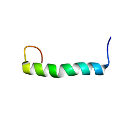
2KY5
DownloadVisualize
BU of 2ky5 by Molmil
Solution structure of the PECAM-1 cytoplasmic tail with DPC
Descriptor: Platelet endothelial cell adhesion molecule
Authors:Lytle, B.L, Peterson, F.C, Volkman, B.F, Paddock, C, Newman, D.K, Center for Eukaryotic Structural Genomics (CESG)
Deposit date:2010-05-14
Release date:2010-05-26
Last modified:2024-05-15
Method:SOLUTION NMR
Cite:Residues within a lipid-associated segment of the PECAM-1 cytoplasmic domain are susceptible to inducible, sequential phosphorylation.
Blood, 117, 2011
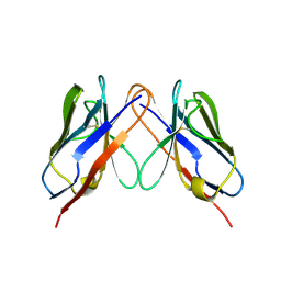
2KQN
DownloadVisualize
BU of 2kqn by Molmil
Solution structure of the AL-09 H87Y immunoglobulin light chain variable domain
Descriptor: Ig kappa chain V-I region AU
Authors:Volkman, B.F, Peterson, F.C, Ramirez-Alvarado, M, Baden, E.M.
Deposit date:2009-11-11
Release date:2010-03-16
Last modified:2022-03-16
Method:SOLUTION NMR
Cite:A single mutation promotes amyloidogenicity through a highly promiscuous dimer interface.
Structure, 18, 2010
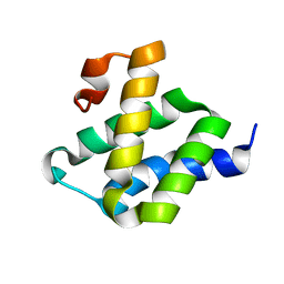
2LWX
DownloadVisualize
BU of 2lwx by Molmil
Solution structure of the C-terminal Pdr1-activating domain of the J-protein Zuo1
Descriptor: Zuotin
Authors:Volkman, B.F, Ducett, J.K, Peterson, F.C, Craig, E.A.
Deposit date:2012-08-08
Release date:2012-10-03
Last modified:2024-05-15
Method:SOLUTION NMR
Cite:Unfolding of the C-terminal domain of the j-protein zuo1 releases autoinhibition and activates pdr1-dependent transcription.
J.Mol.Biol., 425, 2013
2N55
DownloadVisualize
BU of 2n55 by Molmil
Structure of constitutively monomeric CXCL12 in complex with the CXCR4 N-terminus
Descriptor: C-X-C chemokine receptor type 4, Stromal cell-derived factor 1
Authors:Ziarek, J.J, Peterson, F.C, Volkman, B.F.
Deposit date:2015-07-07
Release date:2016-04-27
Last modified:2024-10-09
Method:SOLUTION NMR
Cite:Structural basis for chemokine recognition by a G protein-coupled receptor and implications for receptor activation.
Sci Signal, 10, 2017
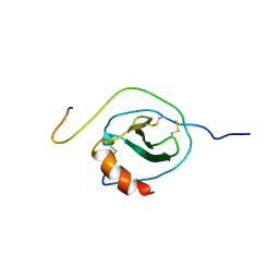
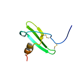
2KOL
DownloadVisualize
BU of 2kol by Molmil
Solution structure of human SDF1-alpha H25R
Descriptor: Stromal cell-derived factor 1
Authors:Volkman, B.F, Ziarek, J.J, Peterson, F.C, Veldkamp, C.T.
Deposit date:2009-09-24
Release date:2010-10-06
Last modified:2023-06-14
Method:SOLUTION NMR
Cite:Solution structure of human SDF1-alpha H25R
To be Published
2KVA
DownloadVisualize
BU of 2kva by Molmil
SOLUTION STRUCTURE OF CI-MPR ligand-free domain 5
Descriptor: Cation-independent mannose-6-phosphate receptor
Authors:Olson, L.J, Peterson, F.C, Volkman, B.F, Dahms, N.M.
Deposit date:2010-03-10
Release date:2010-07-07
Last modified:2024-10-16
Method:SOLUTION NMR
Cite:Structural basis for recognition of phosphodiester-containing lysosomal enzymes by the cation-independent mannose 6-phosphate receptor.
Proc.Natl.Acad.Sci.USA, 107, 2010
<1234


226707

PDB entries from 2024-10-30

PDB statisticsPDBj update infoContact PDBjnumon