Loading
PDBj
MenuPDBj@FacebookPDBj@TwitterPDBj@YouTubewwPDB FoundationwwPDB
RCSB PDBPDBeBMRBAdv. SearchSearch help
PDB: 531 results

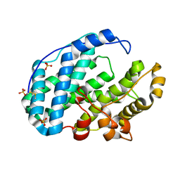
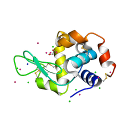
5MYY
DownloadVisualize
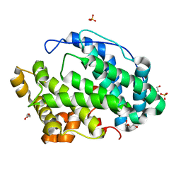
BU of 5myy by Molmil
Hen Egg-White Lysozyme (HEWL) cocrystallized in the presence of Cadmium sulphate
Descriptor: 1,2-ETHANEDIOL, CADMIUM ION, CHLORIDE ION, ...
Authors:Panneerselvam, S, Burkhardt, A, Meents, A.
Deposit date:2017-01-30
Release date:2017-07-12
Last modified:2019-10-16
Method:X-RAY DIFFRACTION (1.1 Å)
Cite:Rapid cadmium SAD phasing at the standard wavelength (1 angstrom ).
Acta Crystallogr D Struct Biol, 73, 2017
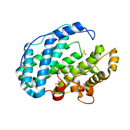
2WZJ
DownloadVisualize
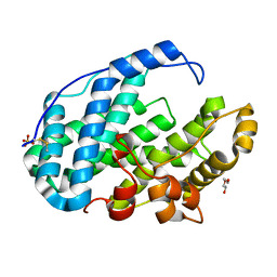
BU of 2wzj by Molmil
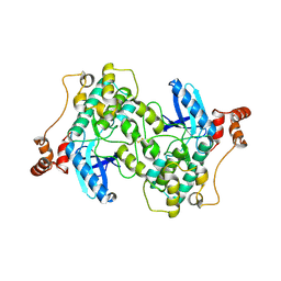
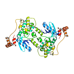
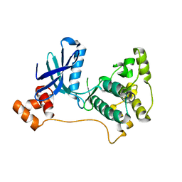
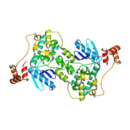
Catalytic and UBA domain of kinase MARK2/(Par-1) K82R, T208E double mutant
Descriptor: SERINE/THREONINE-PROTEIN KINASE MARK2
Authors:Panneerselvam, S, Marx, A, Mandelkow, E.-M, Mandelkow, E.
Deposit date:2009-11-30
Release date:2009-12-22
Last modified:2023-12-20
Method:X-RAY DIFFRACTION (2.786 Å)
Cite:Structure and Function of Polarity-Inducing Kinase Family Mark/Par-1 within the Branch of Ampk/Snf1-Related Kinases.
Faseb J., 24, 2010
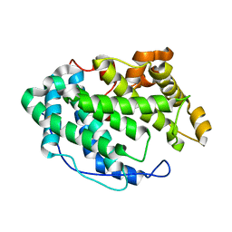
5JW8
DownloadVisualize
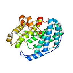
BU of 5jw8 by Molmil
Crystal structure of the Type IV pilin subunit PilE from Neisseria meningitidis
Descriptor: Major pilin PilE
Authors:Kolappan, S, Craig, L.
Deposit date:2016-05-11
Release date:2016-07-06
Last modified:2023-09-27
Method:X-RAY DIFFRACTION (1.439 Å)
Cite:Structure of the Neisseria meningitidis Type IV pilus.
Nat Commun, 7, 2016
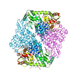
7FHZ
DownloadVisualize
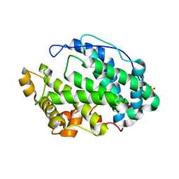
BU of 7fhz by Molmil
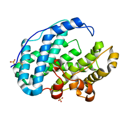
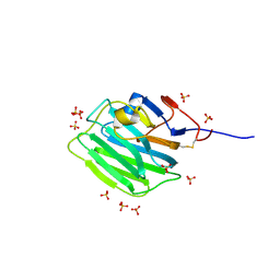
Crystal structure of Multi-functional Polysaccharide lyase Smlt1473 (WT) from Stenotrophomonas maltophilia (strain K279a) in apo form at pH 9.0
Descriptor: Polysaccharide lyase, SULFATE ION
Authors:Pandey, S, Berger, B.W, Acharya, R.
Deposit date:2021-07-30
Release date:2021-10-27
Last modified:2023-11-29
Method:X-RAY DIFFRACTION (2.5 Å)
Cite:Structural insights into the mechanism of pH-selective substrate specificity of the polysaccharide lyase Smlt1473.
J.Biol.Chem., 297, 2021
7FI1
DownloadVisualize
BU of 7fi1 by Molmil
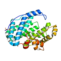
Crystal structure of Multi-functional Polysaccharide lyase Smlt1473 (WT) from Stenotrophomonas maltophilia (strain K279a) in ManA bound form at pH-7.0
Descriptor: Polysaccharide lyase, SULFATE ION, beta-D-mannopyranuronic acid-(1-4)-beta-D-mannopyranuronic acid-(1-4)-beta-D-mannopyranuronic acid
Authors:Pandey, S, Berger, B.W, Acharya, R.
Deposit date:2021-07-30
Release date:2021-10-27
Last modified:2023-11-29
Method:X-RAY DIFFRACTION (2.43 Å)
Cite:Structural insights into the mechanism of pH-selective substrate specificity of the polysaccharide lyase Smlt1473.
J.Biol.Chem., 297, 2021
7FI2
DownloadVisualize
BU of 7fi2 by Molmil
Crystal structure of Multi-functional Polysaccharide lyase Smlt1473-H168A from Stenotrophomonas maltophilia (strain K279a) at pH-5.0
Descriptor: DI(HYDROXYETHYL)ETHER, Polysaccharide lyase, SULFATE ION
Authors:Pandey, S, Berger, B.W, Acharya, R.
Deposit date:2021-07-30
Release date:2021-10-27
Last modified:2023-11-29
Method:X-RAY DIFFRACTION (2.6 Å)
Cite:Structural insights into the mechanism of pH-selective substrate specificity of the polysaccharide lyase Smlt1473.
J.Biol.Chem., 297, 2021
7FI0
DownloadVisualize
BU of 7fi0 by Molmil
Crystal structure of Multi-functional Polysaccharide lyase Smlt1473 (WT) from Stenotrophomonas maltophilia (strain K279a) in ManA bound form at pH-5.0
Descriptor: DI(HYDROXYETHYL)ETHER, Polysaccharide lyase, SULFATE ION, ...
Authors:Pandey, S, Berger, B.W, Acharya, R.
Deposit date:2021-07-30
Release date:2021-10-27
Last modified:2023-11-29
Method:X-RAY DIFFRACTION (2.31 Å)
Cite:Structural insights into the mechanism of pH-selective substrate specificity of the polysaccharide lyase Smlt1473.
J.Biol.Chem., 297, 2021
7FHY
DownloadVisualize
BU of 7fhy by Molmil
Crystal structure of Multi-functional Polysaccharide lyase Smlt1473 (WT) from Stenotrophomonas maltophilia (strain K279a) in apo form at pH 7.0
Descriptor: Polysaccharide lyase, SULFATE ION
Authors:Pandey, S, Berger, B.W, Acharya, R.
Deposit date:2021-07-30
Release date:2021-10-27
Last modified:2023-11-29
Method:X-RAY DIFFRACTION (2.2 Å)
Cite:Structural insights into the mechanism of pH-selective substrate specificity of the polysaccharide lyase Smlt1473.
J.Biol.Chem., 297, 2021
7FHX
DownloadVisualize
BU of 7fhx by Molmil
Crystal structure of Multi-functional Polysaccharide lyase Smlt1473 (WT) from Stenotrophomonas maltophilia (strain K279a) in apo form at pH 5.0
Descriptor: DI(HYDROXYETHYL)ETHER, Polysaccharide lyase, SULFATE ION
Authors:Pandey, S, Berger, B.W, Acharya, R.
Deposit date:2021-07-30
Release date:2021-10-27
Last modified:2023-11-29
Method:X-RAY DIFFRACTION (2.63 Å)
Cite:Structural insights into the mechanism of pH-selective substrate specificity of the polysaccharide lyase Smlt1473.
J.Biol.Chem., 297, 2021
7FHU
DownloadVisualize
BU of 7fhu by Molmil
Crystal structure of Multi-functional Polysaccharide lyase Smlt1473 from Stenotrophomonas maltophilia (strain K279a) in apo form at pH 8.5
Descriptor: Polysaccharide lyase, SULFATE ION
Authors:Pandey, S, Berger, B.W, Acharya, R.
Deposit date:2021-07-30
Release date:2021-10-27
Last modified:2023-11-29
Method:X-RAY DIFFRACTION (2.43 Å)
Cite:Structural insights into the mechanism of pH-selective substrate specificity of the polysaccharide lyase Smlt1473.
J.Biol.Chem., 297, 2021
7FHV
DownloadVisualize
BU of 7fhv by Molmil
Crystal structure of Multi-functional Polysaccharide lyase Smlt1473 (WT) from Stenotrophomonas maltophilia (strain K279a) in apo form at pH 6.5
Descriptor: DI(HYDROXYETHYL)ETHER, Polysaccharide lyase, SULFATE ION
Authors:Pandey, S, Berger, B.W, Acharya, R.
Deposit date:2021-07-30
Release date:2021-10-27
Last modified:2023-11-29
Method:X-RAY DIFFRACTION (2.28 Å)
Cite:Structural insights into the mechanism of pH-selective substrate specificity of the polysaccharide lyase Smlt1473.
J.Biol.Chem., 297, 2021
7FHW
DownloadVisualize
BU of 7fhw by Molmil
Crystal structure of Multi-functional Polysaccharide lyase Smlt1473 from Stenotrophomonas maltophilia (strain K279a) in apo form at pH 5.5
Descriptor: Polysaccharide lyase
Authors:Pandey, S, Berger, B.W, Acharya, R.
Deposit date:2021-07-30
Release date:2021-10-27
Last modified:2023-11-29
Method:X-RAY DIFFRACTION (2.061 Å)
Cite:Structural insights into the mechanism of pH-selective substrate specificity of the polysaccharide lyase Smlt1473.
J.Biol.Chem., 297, 2021
1OZG
DownloadVisualize
BU of 1ozg by Molmil
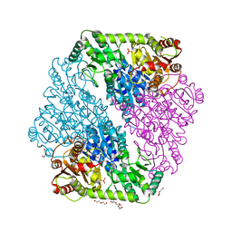
The crystal structure of Klebsiella pneumoniae acetolactate synthase with enzyme-bound cofactor and with an unusual intermediate
Descriptor: 2-HYDROXYETHYL DIHYDROTHIACHROME DIPHOSPHATE, Acetolactate synthase, catabolic, ...
Authors:Pang, S.S, Duggleby, R.G, Schowen, R.L, Guddat, L.W.
Deposit date:2003-04-09
Release date:2003-11-04
Last modified:2023-08-16
Method:X-RAY DIFFRACTION (2.3 Å)
Cite:The Crystal Structures of Klebsiella pneumoniae Acetolactate Synthase with Enzyme-bound Cofactor and with an Unusual Intermediate.
J.Biol.Chem., 279, 2004
7ZC9
DownloadVisualize
BU of 7zc9 by Molmil
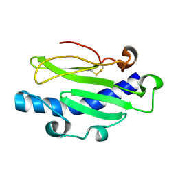
Human Pikachurin/EGFLAM C-terminal Laminin-G domain (LG3)
Descriptor: 2-acetamido-2-deoxy-beta-D-glucopyranose, Pikachurin, SULFATE ION
Authors:Pantalone, S, Forneris, F.
Deposit date:2022-03-26
Release date:2023-07-19
Last modified:2024-02-07
Method:X-RAY DIFFRACTION (2.1 Å)
Cite:Structure of the photoreceptor synaptic assembly of the extracellular matrix protein pikachurin with the orphan receptor GPR179.
Sci.Signal., 16, 2023
7ZCB
DownloadVisualize
BU of 7zcb by Molmil
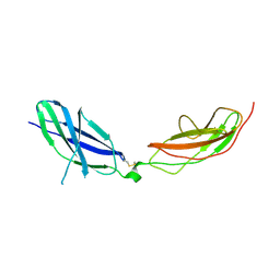
Human Pikachurin/EGFLAM N-terminal Fibronectin-III (1-2) domains
Descriptor: CHLORIDE ION, Pikachurin, beta-D-mannopyranose-(1-4)-2-acetamido-2-deoxy-beta-D-glucopyranose-(1-4)-2-acetamido-2-deoxy-beta-D-glucopyranose
Authors:Pantalone, S, Savino, S, Viti, L.V, Forneris, F.
Deposit date:2022-03-26
Release date:2023-07-19
Last modified:2024-02-07
Method:X-RAY DIFFRACTION (2.5 Å)
Cite:Structure of the photoreceptor synaptic assembly of the extracellular matrix protein pikachurin with the orphan receptor GPR179.
Sci.Signal., 16, 2023
1OZF
DownloadVisualize
BU of 1ozf by Molmil
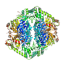
The crystal structure of Klebsiella pneumoniae acetolactate synthase with enzyme-bound cofactors
Descriptor: Acetolactate synthase, catabolic, DI(HYDROXYETHYL)ETHER, ...
Authors:Pang, S.S, Duggleby, R.G, Schowen, R.L, Guddat, L.W.
Deposit date:2003-04-09
Release date:2003-11-11
Last modified:2023-08-16
Method:X-RAY DIFFRACTION (2.3 Å)
Cite:The Crystal Structures of Klebsiella pneumoniae Acetolactate Synthase with Enzyme-bound Cofactor and with an Unusual Intermediate.
J.Biol.Chem., 279, 2004
1OZH
DownloadVisualize
BU of 1ozh by Molmil
The crystal structure of Klebsiella pneumoniae acetolactate synthase with enzyme-bound cofactor and with an unusual intermediate.
Descriptor: 2-HYDROXYETHYL DIHYDROTHIACHROME DIPHOSPHATE, Acetolactate synthase, catabolic, ...
Authors:Pang, S.S, Duggleby, R.G, Schowen, R.L, Guddat, L.W.
Deposit date:2003-04-09
Release date:2003-11-04
Last modified:2023-08-16
Method:X-RAY DIFFRACTION (2 Å)
Cite:The Crystal Structures of Klebsiella pneumoniae Acetolactate Synthase with Enzyme-bound Cofactor and with an Unusual Intermediate.
J.Biol.Chem., 279, 2004
1ZMV
DownloadVisualize
BU of 1zmv by Molmil
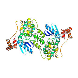
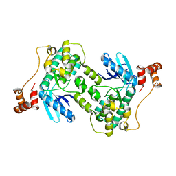
Catalytic and ubiqutin-associated domains of MARK2/PAR-1: K82R mutant
Descriptor: MAP/Microtubule affinity regulating kinase 2
Authors:Panneerselvam, S, Marx, A, Mandelkow, E.-M, Mandelkow, E.
Deposit date:2005-05-11
Release date:2006-02-14
Last modified:2023-08-23
Method:X-RAY DIFFRACTION (3.105 Å)
Cite:Structure of the catalytic and ubiquitin-associated domains of the protein kinase MARK/Par-1.
Structure, 14, 2006
2R0I
DownloadVisualize
BU of 2r0i by Molmil
Crystal structure of a kinase MARK2/Par-1 mutant
Descriptor: Serine/threonine-protein kinase MARK2
Authors:Panneerselvam, S, Marx, A, Mandelkow, E.-M, Mandelkow, E.
Deposit date:2007-08-20
Release date:2008-08-26
Last modified:2023-08-30
Method:X-RAY DIFFRACTION (2.202 Å)
Cite:Crystal structure of a kinase MARK2/Par-1 mutant
To be Published
1Y8G
DownloadVisualize
BU of 1y8g by Molmil
Catalytic and ubiqutin-associated domains of MARK2/PAR-1: Inactive double mutant with selenomethionine
Descriptor: MAP/Microtubule affinity-regulating kinase 2
Authors:Panneerselvam, S, Marx, A, Mandelkow, E.-M, Mandelkow, E.
Deposit date:2004-12-12
Release date:2006-02-14
Last modified:2024-04-03
Method:X-RAY DIFFRACTION (2.501 Å)
Cite:Structure of the catalytic and ubiquitin-associated domains of the protein kinase MARK/Par-1.
Structure, 14, 2006
2BD0
DownloadVisualize
BU of 2bd0 by Molmil
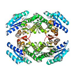
Chlorobium tepidum Sepiapterin Reductase complexed with NADP and Sepiapterin
Descriptor: BIOPTERIN, NADP NICOTINAMIDE-ADENINE-DINUCLEOTIDE PHOSPHATE, sepiapterin reductase
Authors:Supangat, S, Seo, K.H, Choi, Y.K, Park, Y.S, Lee, K.H.
Deposit date:2005-10-19
Release date:2005-11-29
Last modified:2024-03-13
Method:X-RAY DIFFRACTION (1.7 Å)
Cite:Structure of Chlorobium tepidum sepiapterin reductase complex reveals the novel substrate binding mode for stereospecific production of L-threo-tetrahydrobiopterin
J.Biol.Chem., 281, 2006
1ZMW
DownloadVisualize
BU of 1zmw by Molmil
Catalytic and ubiqutin-associated domains of MARK2/PAR-1: T208A/S212A inactive double mutant
Descriptor: MAP/Microtubule affinity regulating kinase 2
Authors:Panneerselvam, S, Marx, A, Mandelkow, E.-M, Mandelkow, E.
Deposit date:2005-05-11
Release date:2006-02-14
Last modified:2023-08-23
Method:X-RAY DIFFRACTION (2.802 Å)
Cite:Structure of the catalytic and ubiquitin-associated domains of the protein kinase MARK/Par-1.
Structure, 14, 2006
1ZMU
DownloadVisualize
BU of 1zmu by Molmil
Catalytic and ubiqutin-associated domains of MARK2/PAR-1: Wild type
Descriptor: MAP/Microtubule affinity regulating kinase 2
Authors:Panneerselvam, S, Marx, A, Mandelkow, E.-M, Mandelkow, E.
Deposit date:2005-05-11
Release date:2006-02-14
Last modified:2023-08-23
Method:X-RAY DIFFRACTION (2.9 Å)
Cite:Structure of the catalytic and ubiquitin-associated domains of the protein kinase MARK/Par-1.
Structure, 14, 2006
7ZF2
DownloadVisualize
BU of 7zf2 by Molmil
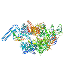
Protomeric substructure from an octameric assembly of M. tuberculosis RNA polymerase in complex with sigma-b initiation factor
Descriptor: DNA-directed RNA polymerase subunit alpha, DNA-directed RNA polymerase subunit beta, DNA-directed RNA polymerase subunit beta', ...
Authors:Trapani, S, Bron, P, Lai Kee Him, J, Brodolin, K, Morichaud, Z, Vishwakarma, R.
Deposit date:2022-03-31
Release date:2023-02-08
Last modified:2024-03-27
Method:ELECTRON MICROSCOPY (3.86 Å)
Cite:Structural basis of the mycobacterial stress-response RNA polymerase auto-inhibition via oligomerization.
Nat Commun, 14, 2023
1L1G
DownloadVisualize
BU of 1l1g by Molmil
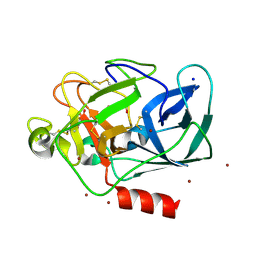
The Structure of Porcine Pancreatic Elastase Complexed with Xenon and Bromide, Cryoprotected with Glycerol
Descriptor: BROMIDE ION, ELASTASE 1, GLYCEROL, ...
Authors:Panjikar, S, Tucker, P.A.
Deposit date:2002-02-16
Release date:2002-08-28
Last modified:2023-08-16
Method:X-RAY DIFFRACTION (1.5 Å)
Cite:Xenon derivatization of halide-soaked protein crystals.
Acta Crystallogr.,Sect.D, 58, 2002

222926

건을2024-07-24부터공개중

PDB statisticsPDBj update infoContact PDBjnumon