2MGO
 
 | |
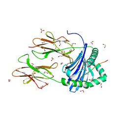
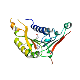
8CMF
 | | Human Leukocyte Antigen class II allotype DR1 presenting SARS-CoV-2 nsp3 epitope (orf1ab)1350-1364 | | 分子名称: | 1,2-ETHANEDIOL, HLA class II histocompatibility antigen, DR alpha chain, ... | | 著者 | MacLachlan, B.J, Mason, G.H, Sourfield, D.O, Godkin, A.J, Rizkallah, P.J. | | 登録日 | 2023-02-19 | | 公開日 | 2023-07-26 | | 最終更新日 | 2023-08-02 | | 実験手法 | X-RAY DIFFRACTION (2.2 Å) | | 主引用文献 | Structural definition of HLA class II-presented SARS-CoV-2 epitopes reveals a mechanism to escape pre-existing CD4 + T cell immunity.
Cell Rep, 42, 2023
|
|
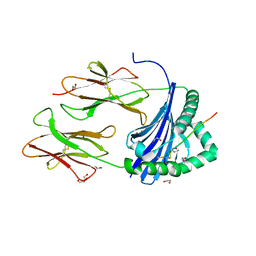
8CMI
 | | Human Leukocyte Antigen class II allotype DR1 presenting SARS-CoV-2 Omicron (BA.1) Spike peptide S761-775 | | 分子名称: | 1,2-ETHANEDIOL, HLA class II histocompatibility antigen, DR alpha chain, ... | | 著者 | MacLachlan, B.J, Mason, G.H, Sourfield, D.O, Godkin, A.J, Rizkallah, P.J. | | 登録日 | 2023-02-19 | | 公開日 | 2023-07-26 | | 最終更新日 | 2023-08-02 | | 実験手法 | X-RAY DIFFRACTION (2.6 Å) | | 主引用文献 | Structural definition of HLA class II-presented SARS-CoV-2 epitopes reveals a mechanism to escape pre-existing CD4 + T cell immunity.
Cell Rep, 42, 2023
|
|
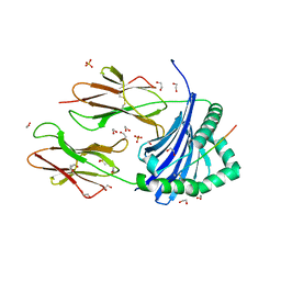
8CMD
 | | Human Leukocyte Antigen class II allotype DR1 presenting SARS-CoV-2 Spike peptide S761-775 | | 分子名称: | 1,2-ETHANEDIOL, HLA class II histocompatibility antigen, DR alpha chain, ... | | 著者 | MacLachlan, B.J, Mason, G.H, Sourfield, D.O, Godkin, A.J, Rizkallah, P.J. | | 登録日 | 2023-02-19 | | 公開日 | 2023-07-26 | | 最終更新日 | 2023-08-02 | | 実験手法 | X-RAY DIFFRACTION (2.54 Å) | | 主引用文献 | Structural definition of HLA class II-presented SARS-CoV-2 epitopes reveals a mechanism to escape pre-existing CD4 + T cell immunity.
Cell Rep, 42, 2023
|
|
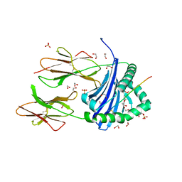
8CME
 | | Human Leukocyte Antigen class II allotype DR1 presenting SARS-CoV-2 Membrane peptide M176-190 | | 分子名称: | 1,2-ETHANEDIOL, 2-(N-MORPHOLINO)-ETHANESULFONIC ACID, HLA class II histocompatibility antigen, ... | | 著者 | MacLachlan, B.J, Mason, G.H, Sourfield, D.O, Godkin, A.J, Rizkallah, P.J. | | 登録日 | 2023-02-19 | | 公開日 | 2023-07-26 | | 最終更新日 | 2023-08-02 | | 実験手法 | X-RAY DIFFRACTION (2.26 Å) | | 主引用文献 | Structural definition of HLA class II-presented SARS-CoV-2 epitopes reveals a mechanism to escape pre-existing CD4 + T cell immunity.
Cell Rep, 42, 2023
|
|
8CMH
 | | Human Leukocyte Antigen class II allotype DR1 presenting SARS-CoV-2 Omicron (BA.1) Spike peptide S486-505 | | 分子名称: | 1,2-ETHANEDIOL, 2-AMINO-ETHANETHIOL, HLA class II histocompatibility antigen, ... | | 著者 | MacLachlan, B.J, Mason, G.H, Sourfield, D.O, Godkin, A.J, Rizkallah, P.J. | | 登録日 | 2023-02-19 | | 公開日 | 2023-07-26 | | 最終更新日 | 2024-04-03 | | 実験手法 | X-RAY DIFFRACTION (1.64 Å) | | 主引用文献 | Structural definition of HLA class II-presented SARS-CoV-2 epitopes reveals a mechanism to escape pre-existing CD4 + T cell immunity.
Cell Rep, 42, 2023
|
|
8CMB
 | | Human Leukocyte Antigen class II allotype DR1 presenting SARS-CoV-2 Spike peptide S486-505 | | 分子名称: | 1,2-ETHANEDIOL, 2-AMINO-ETHANETHIOL, HLA class II histocompatibility antigen, ... | | 著者 | MacLachlan, B.J, Mason, G.H, Godkin, A.J, Rizkallah, P.J. | | 登録日 | 2023-02-19 | | 公開日 | 2023-07-26 | | 最終更新日 | 2024-04-03 | | 実験手法 | X-RAY DIFFRACTION (1.84 Å) | | 主引用文献 | Structural definition of HLA class II-presented SARS-CoV-2 epitopes reveals a mechanism to escape pre-existing CD4 + T cell immunity.
Cell Rep, 42, 2023
|
|
8CMC
 | | Human Leukocyte Antigen class II allotype DR1 presenting SARS-CoV-2 Spike peptide S511-530 | | 分子名称: | 1,2-ETHANEDIOL, 2-(N-MORPHOLINO)-ETHANESULFONIC ACID, HLA class II histocompatibility antigen, ... | | 著者 | MacLachlan, B.J, Mason, G.H, Sourfield, D.O, Godkin, A.J, Rizkallah, P.J. | | 登録日 | 2023-02-19 | | 公開日 | 2023-07-26 | | 最終更新日 | 2023-08-02 | | 実験手法 | X-RAY DIFFRACTION (1.42 Å) | | 主引用文献 | Structural definition of HLA class II-presented SARS-CoV-2 epitopes reveals a mechanism to escape pre-existing CD4 + T cell immunity.
Cell Rep, 42, 2023
|
|
8CMG
 | | Human Leukocyte Antigen class II allotype DR1 presenting SARS-CoV-2 nsp14 peptide (orf1ab)6420-6434 | | 分子名称: | 1,2-ETHANEDIOL, HLA class II histocompatibility antigen, DR alpha chain, ... | | 著者 | MacLachlan, B.J, Mason, G.H, Sourfield, D.O, Godkin, A.J, Rizkallah, P.J. | | 登録日 | 2023-02-19 | | 公開日 | 2023-07-26 | | 最終更新日 | 2023-08-02 | | 実験手法 | X-RAY DIFFRACTION (1.64 Å) | | 主引用文献 | Structural definition of HLA class II-presented SARS-CoV-2 epitopes reveals a mechanism to escape pre-existing CD4 + T cell immunity.
Cell Rep, 42, 2023
|
|
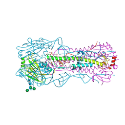
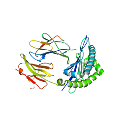
4W8N
 | | The crystal structure of hemagglutinin from a swine influenza virus (A/swine/Missouri/2124514/2006) | | 分子名称: | 2-acetamido-2-deoxy-beta-D-glucopyranose, Hemagglutinin | | 著者 | Yang, H, Carney, P.J, Tumpey, T.M, Stevens, J. | | 登録日 | 2014-08-25 | | 公開日 | 2015-02-11 | | 最終更新日 | 2023-12-27 | | 実験手法 | X-RAY DIFFRACTION (2.9 Å) | | 主引用文献 | Assessment of transmission, pathogenesis and adaptation of H2 subtype influenza viruses in ferrets.
Virology, 477C, 2015
|
|
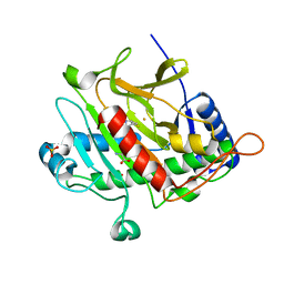
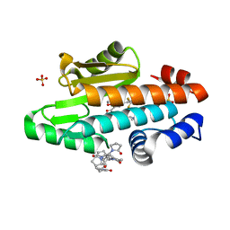
1J98
 | | The 1.2 Angstrom Structure of Bacillus subtilis LuxS | | 分子名称: | AUTOINDUCER-2 PRODUCTION PROTEIN LUXS, ZINC ION | | 著者 | Ruzheinikov, S.N, Das, S.K, Sedelnikova, S.E, Hartley, A, Foster, S.J, Horsburgh, M.J, Cox, A.G, McCleod, C.W, Mekhalfia, A, Blackburn, G.M, Rice, D.W, Baker, P.J. | | 登録日 | 2001-05-24 | | 公開日 | 2001-06-06 | | 最終更新日 | 2021-10-27 | | 実験手法 | X-RAY DIFFRACTION (1.2 Å) | | 主引用文献 | The 1.2 A Structure of a Novel Quorum-Sensing Protein, Bacillus subtilis LuxS
J.Mol.Biol., 313, 2001
|
|
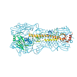
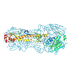
4WE4
 | | The crystal structure of hemagglutinin from 1968 H3N2 influenza virus | | 分子名称: | 2-acetamido-2-deoxy-beta-D-glucopyranose, 2-acetamido-2-deoxy-beta-D-glucopyranose-(1-2)-alpha-D-mannopyranose-(1-6)-[alpha-D-mannopyranose-(1-3)]beta-D-mannopyranose-(1-4)-2-acetamido-2-deoxy-beta-D-glucopyranose-(1-4)-2-acetamido-2-deoxy-beta-D-glucopyranose, 2-acetamido-2-deoxy-beta-D-glucopyranose-(1-4)-2-acetamido-2-deoxy-beta-D-glucopyranose, ... | | 著者 | Yang, H, Carney, P.J, Chang, J.C, Guo, Z, Villanueva, J.M, Stevens, J. | | 登録日 | 2014-09-09 | | 公開日 | 2015-02-11 | | 最終更新日 | 2023-12-27 | | 実験手法 | X-RAY DIFFRACTION (2.351 Å) | | 主引用文献 | Structure and receptor binding preferences of recombinant human A(H3N2) virus hemagglutinins.
Virology, 477C, 2015
|
|
4WE9
 | | The crystal structure of hemagglutinin from influenza virus A/Victoria/361/2011 in complex with 3'SLN | | 分子名称: | 2-acetamido-2-deoxy-beta-D-glucopyranose, 2-acetamido-2-deoxy-beta-D-glucopyranose-(1-4)-2-acetamido-2-deoxy-beta-D-glucopyranose, Hemagglutinin | | 著者 | Yang, H, Carney, P.J, Chang, J.C, Guo, Z, Villanueva, J.M, Stevens, J. | | 登録日 | 2014-09-09 | | 公開日 | 2015-02-11 | | 最終更新日 | 2023-12-27 | | 実験手法 | X-RAY DIFFRACTION (2.2 Å) | | 主引用文献 | Structure and receptor binding preferences of recombinant human A(H3N2) virus hemagglutinins.
Virology, 477C, 2015
|
|
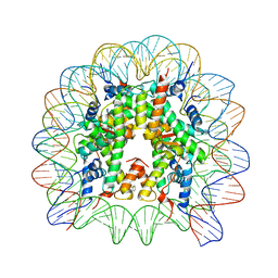
8Q3M
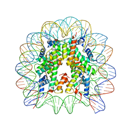 | | Structure of Nucleosome Core with a Bound Kaposi Sarcoma Associated Herpesvirus LANA Peptide Having a Methionine to Ornithine Substitution | | 分子名称: | DNA (145-MER), Histone H2A type 1-B/E, Histone H2B type 1-K, ... | | 著者 | De Falco, L, Batchelor, L.K, Dyson, P.J, Davey, C.A. | | 登録日 | 2023-08-04 | | 公開日 | 2024-03-27 | | 実験手法 | X-RAY DIFFRACTION (2.503 Å) | | 主引用文献 | Viral peptide conjugates for metal-warhead delivery to chromatin.
Rsc Adv, 14, 2024
|
|
8Q36
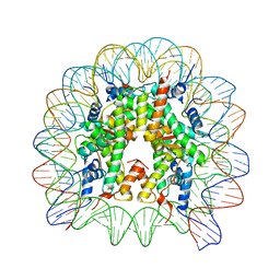 | | Structure of Nucleosome Core with a Bound Metallopeptide Conjugate (Foamy Virus GAG Peptide-Au[I] Compound) | | 分子名称: | DNA (145-MER), GAG structural protein, Histone H2A type 1-B/E, ... | | 著者 | De Falco, L, Batchelor, L.K, Dyson, P.J, Davey, C.A. | | 登録日 | 2023-08-03 | | 公開日 | 2024-03-27 | | 実験手法 | X-RAY DIFFRACTION (2.604 Å) | | 主引用文献 | Viral peptide conjugates for metal-warhead delivery to chromatin.
Rsc Adv, 14, 2024
|
|
4XI1
 | | Crystal structure of U-box 2 of LubX / LegU2 / Lpp2887 from Legionella pneumophila str. Paris, wild-type | | 分子名称: | CHLORIDE ION, E3 ubiquitin-protein ligase LubX, GLYCEROL, ... | | 著者 | Stogios, P.J, Quaile, T, Skarina, T, Cuff, M, Di Leo, R, Yim, V, Savchenko, A, Joachimiak, A, Midwest Center for Structural Genomics (MCSG) | | 登録日 | 2015-01-06 | | 公開日 | 2015-01-21 | | 最終更新日 | 2023-09-27 | | 実験手法 | X-RAY DIFFRACTION (2.983 Å) | | 主引用文献 | Molecular Characterization of LubX: Functional Divergence of the U-Box Fold by Legionella pneumophila.
Structure, 23, 2015
|
|
8Q3X
 | | Structure of Nucleosome Core with a Bound Metallopeptide Conjugate (Kaposi Sarcoma Associated Herpesvirus LANA Peptide-Au[I] Compound) | | 分子名称: | 4-diphenylphosphanylbenzoic acid, DNA (145-MER), GOLD ION, ... | | 著者 | De Falco, L, Batchelor, L.K, Dyson, P.J, Davey, C.A. | | 登録日 | 2023-08-04 | | 公開日 | 2024-03-27 | | 実験手法 | X-RAY DIFFRACTION (2.301 Å) | | 主引用文献 | Viral peptide conjugates for metal-warhead delivery to chromatin.
Rsc Adv, 14, 2024
|
|
2IVJ
 | | Isopenicillin N Synthase From Aspergillus Nidulans (Anaerobic Ac- cyclopropylglycine Fe Complex) | | 分子名称: | D-(L-A-AMINOADIPOYL)-L-CYSTEINYL-D-CYCLOPROPYLGLYCINE, FE (II) ION, ISOPENICILLIN N SYNTHETASE, ... | | 著者 | Howard-Jones, A.R, Elkins, J.M, Clifton, I.J, Roach, P.L, Adlington, R.M, Baldwin, J.E, Rutledge, P.J. | | 登録日 | 2006-06-13 | | 公開日 | 2007-04-10 | | 最終更新日 | 2024-05-08 | | 実験手法 | X-RAY DIFFRACTION (1.46 Å) | | 主引用文献 | Interactions of Isopenicillin N Synthase with Cyclopropyl-Containing Substrate Analogues Reveal New Mechanistic Insight.
Biochemistry, 46, 2007
|
|
2LYE
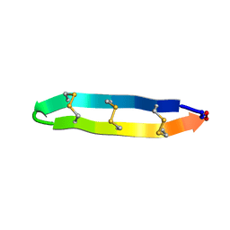 | | High resolution NMR solution structure of a symmetrical theta-defensin, BTD-2 | | 分子名称: | BTD-2 | | 著者 | Conibear, A.C, Rosengren, K, Harvey, P.J, Craik, D.J. | | 登録日 | 2012-09-18 | | 公開日 | 2012-11-28 | | 最終更新日 | 2023-06-14 | | 実験手法 | SOLUTION NMR | | 主引用文献 | Structural characterization of the cyclic cystine ladder motif of theta-defensins.
Biochemistry, 51, 2012
|
|
6ORQ
 | | Modified BG505 SOSIP-based immunogen RC1 in complex with the elicited V3-glycan patch antibody Ab275MUR | | 分子名称: | 2-acetamido-2-deoxy-beta-D-glucopyranose, 2-acetamido-2-deoxy-beta-D-glucopyranose-(1-4)-2-acetamido-2-deoxy-beta-D-glucopyranose, Ab275MUR antibody Fab heavy chain, ... | | 著者 | Abernathy, M.E, Gristick, H.B, Bjorkman, P.J. | | 登録日 | 2019-04-30 | | 公開日 | 2019-06-12 | | 最終更新日 | 2020-07-29 | | 実験手法 | ELECTRON MICROSCOPY (4.4 Å) | | 主引用文献 | Immunization expands B cells specific to HIV-1 V3 glycan in mice and macaques.
Nature, 570, 2019
|
|
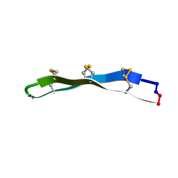
2LYF
 | |
4XCX
 | | METHYLTRANSFERASE DOMAIN OF SMALL RNA 2'-O-METHYLTRANSFERASE | | 分子名称: | S-ADENOSYL-L-HOMOCYSTEINE, Small RNA 2'-O-methyltransferase | | 著者 | Walker, J.R, Zeng, H, Dong, A, Li, Y, Wernimont, A, Bountra, C, Arrowsmith, C.H, Edwards, A.M, Brown, P.J, Wu, H, Structural Genomics Consortium (SGC) | | 登録日 | 2014-12-18 | | 公開日 | 2015-01-14 | | 最終更新日 | 2023-09-27 | | 実験手法 | X-RAY DIFFRACTION (2.84 Å) | | 主引用文献 | Crystal structure of human C1ORF59 in complex with SAH
To be published
|
|
7K77
 | | The crystal structure of the 2009 H1N1 PA endonuclease mutant I38T in complex with SJ001008025 | | 分子名称: | 5-hydroxy-N-[2-(2-methoxypyridin-4-yl)ethyl]-6-oxo-2-[2-(trifluoromethyl)phenyl]-3,6-dihydropyrimidine-4-carboxamide, Hexa Vinylpyrrolidone K15, MANGANESE (II) ION, ... | | 著者 | Cuypers, M.G, Slavish, P.J, Rankovic, Z, White, S.W. | | 登録日 | 2020-09-22 | | 公開日 | 2021-09-22 | | 最終更新日 | 2023-10-18 | | 実験手法 | X-RAY DIFFRACTION (2.78 Å) | | 主引用文献 | The crystal structure of the 2009 H1N1 PA endonuclease mutant I38T in complex with SJ001008025
To Be Published
|
|
4WDI
 | |
4WE8
 | | The crystal structure of hemagglutinin of influenza virus A/Victoria/361/2011 | | 分子名称: | 2-acetamido-2-deoxy-beta-D-glucopyranose, 2-acetamido-2-deoxy-beta-D-glucopyranose-(1-4)-2-acetamido-2-deoxy-beta-D-glucopyranose, Hemagglutinin | | 著者 | Yang, H, Carney, P.J, Chang, J.C, Guo, Z, Villanueva, J.M, Stevens, J. | | 登録日 | 2014-09-09 | | 公開日 | 2015-02-11 | | 最終更新日 | 2023-12-27 | | 実験手法 | X-RAY DIFFRACTION (2.1 Å) | | 主引用文献 | Structure and receptor binding preferences of recombinant human A(H3N2) virus hemagglutinins.
Virology, 477C, 2015
|
|


