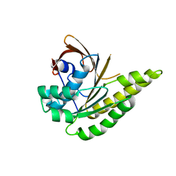
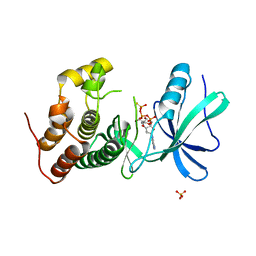
5KDD
 
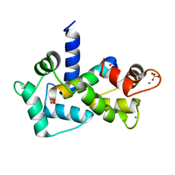
 | | Apo-structure of humanised RadA-mutant humRadA22 | | 分子名称: | DNA repair and recombination protein RadA, SULFATE ION | | 著者 | Fischer, G, Marsh, M, Moschetti, T, Sharpe, T, Scott, D, Morgan, M, Ng, H, Skidmore, J, Venkitaraman, A, Abell, C, Blundell, T.L, Hyvonen, M. | | 登録日 | 2016-06-08 | | 公開日 | 2016-10-19 | | 最終更新日 | 2024-06-19 | | 実験手法 | X-RAY DIFFRACTION (1.99 Å) | | 主引用文献 | Engineering Archeal Surrogate Systems for the Development of Protein-Protein Interaction Inhibitors against Human RAD51.
J.Mol.Biol., 428, 2016
|
|
5L8V
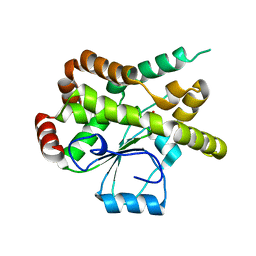
 | | Apo-structure of humanised RadA-mutant humRadA4 | | 分子名称: | DNA repair and recombination protein RadA, PHOSPHATE ION | | 著者 | Marsh, M, Fischer, G, Moschetti, T, Sharpe, T, Scott, D, Morgan, M, Ng, H, Skidmore, J, Venkitaraman, A, Abell, C, Blundell, T.L, Hyvonen, M. | | 登録日 | 2016-06-08 | | 公開日 | 2016-10-19 | | 最終更新日 | 2024-02-07 | | 実験手法 | X-RAY DIFFRACTION (1.5 Å) | | 主引用文献 | Engineering Archeal Surrogate Systems for the Development of Protein-Protein Interaction Inhibitors against Human RAD51.
J.Mol.Biol., 428, 2016
|
|
5LBI
 | | Apo-structure of humanised RadA-mutant humRadA3 | | 分子名称: | CALCIUM ION, DNA repair and recombination protein RadA | | 著者 | Marsh, M, Fischer, G, Moschetti, T, Sharpe, T, Scott, D, Morgan, M, Ng, H, Skidmore, J, Venkitaraman, A, Abell, C, Blundell, T.L, Hyvonen, M. | | 登録日 | 2016-06-16 | | 公開日 | 2016-10-26 | | 最終更新日 | 2024-02-07 | | 実験手法 | X-RAY DIFFRACTION (1.43 Å) | | 主引用文献 | Engineering Archeal Surrogate Systems for the Development of Protein-Protein Interaction Inhibitors against Human RAD51.
J.Mol.Biol., 428, 2016
|
|
5LB4
 | | Apo-structure of humanised RadA-mutant humRadA14 | | 分子名称: | DNA repair and recombination protein RadA | | 著者 | Marsh, M, Fischer, G, Moschetti, T, Sharpe, T, Scott, D, Morgan, M, Ng, H, Skidmore, J, Venkitaraman, A, Abell, C, Blundell, T.L, Hyvonen, M. | | 登録日 | 2016-06-15 | | 公開日 | 2016-10-19 | | 最終更新日 | 2024-02-07 | | 実験手法 | X-RAY DIFFRACTION (1.98 Å) | | 主引用文献 | Engineering Archeal Surrogate Systems for the Development of Protein-Protein Interaction Inhibitors against Human RAD51.
J.Mol.Biol., 428, 2016
|
|
5LB2
 | | Apo-structure of humanised RadA-mutant humRadA2 | | 分子名称: | DNA repair and recombination protein RadA | | 著者 | Marsh, M, Fischer, G, Moschetti, T, Sharpe, T, Scott, D, Morgan, M, Ng, H, Skidmore, J, Venkitaraman, A, Abell, C, Blundell, T.L, Hyvonen, M. | | 登録日 | 2016-06-15 | | 公開日 | 2016-10-19 | | 最終更新日 | 2024-02-07 | | 実験手法 | X-RAY DIFFRACTION (2.1 Å) | | 主引用文献 | Engineering Archeal Surrogate Systems for the Development of Protein-Protein Interaction Inhibitors against Human RAD51.
J.Mol.Biol., 428, 2016
|
|
4WUU
 | |
3SE1
 | | Frog M-ferritin with magnesium, R72D mutant | | 分子名称: | CHLORIDE ION, Ferritin, middle subunit, ... | | 著者 | Tosha, T, Ng, H.L, Alber, T, Theil, E.C. | | 登録日 | 2011-06-10 | | 公開日 | 2012-02-29 | | 最終更新日 | 2024-02-28 | | 実験手法 | X-RAY DIFFRACTION (1.65 Å) | | 主引用文献 | Frog M-ferritin with magnesium, R72D mutant
TO BE PUBLISHED
|
|
3SH6
 | | Frog M-ferritin, D122R mutant, with magnesium | | 分子名称: | CHLORIDE ION, Ferritin, middle subunit, ... | | 著者 | Tosha, T, Ng, H.L, Alber, T, Theil, E.C. | | 登録日 | 2011-06-16 | | 公開日 | 2012-02-29 | | 最終更新日 | 2024-02-28 | | 実験手法 | X-RAY DIFFRACTION (1.4 Å) | | 主引用文献 | Frog M-ferritin, D122R mutant, with magnesium
To be Published
|
|
3SHX
 | | Frog M-ferritin with magnesium, L134P mutant | | 分子名称: | CHLORIDE ION, Ferritin, middle subunit, ... | | 著者 | Tosha, T, Ng, H.L, Alber, T, Theil, E.C. | | 登録日 | 2011-06-17 | | 公開日 | 2012-02-29 | | 最終更新日 | 2024-02-28 | | 実験手法 | X-RAY DIFFRACTION (1.35 Å) | | 主引用文献 | Frog M-ferritin with magnesium, L134P mutant
To be Published
|
|
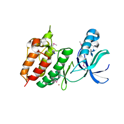
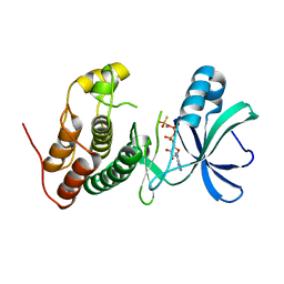
2O5G
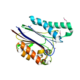
 | | Calmodulin-smooth muscle light chain kinase peptide complex | | 分子名称: | CALCIUM ION, Calmodulin, SULFATE ION, ... | | 著者 | Valentine, K.G, Ng, H.L, Schneeweis, J.K, Kranz, J.K, Frederick, K.K, Alber, T, Wand, A.J. | | 登録日 | 2006-12-05 | | 公開日 | 2007-12-25 | | 最終更新日 | 2023-12-27 | | 実験手法 | X-RAY DIFFRACTION (1.08 Å) | | 主引用文献 | Ultrahigh resolution crystal structure of calmodulin-smooth muscle light kinase peptide complex
To be Published
|
|
1YWF
 | |
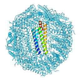
3KA4
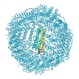
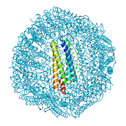
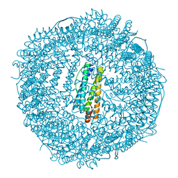
 | | Frog M-ferritin with cobalt | | 分子名称: | CHLORIDE ION, COBALT (II) ION, Ferritin, ... | | 著者 | Tosha, T, Ng, H.L, Theil, E, Alber, T, Bhattasali, O. | | 登録日 | 2009-10-18 | | 公開日 | 2010-10-06 | | 最終更新日 | 2023-09-06 | | 実験手法 | X-RAY DIFFRACTION (1.4 Å) | | 主引用文献 | Moving Metal Ions through Ferritin-Protein Nanocages from Three-Fold Pores to Catalytic Sites.
J.Am.Chem.Soc., 132, 2010
|
|
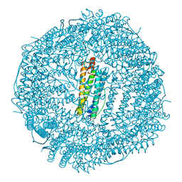
3KA6
 | | Frog M-ferritin, EED mutant, with cobalt | | 分子名称: | CHLORIDE ION, COBALT (II) ION, Ferritin, ... | | 著者 | Tosha, T, Ng, H.L, Theil, E, Alber, T, Bhattasali, O. | | 登録日 | 2009-10-18 | | 公開日 | 2010-10-06 | | 最終更新日 | 2023-09-06 | | 実験手法 | X-RAY DIFFRACTION (1.4 Å) | | 主引用文献 | Moving Metal Ions through Ferritin-Protein Nanocages from Three-Fold Pores to Catalytic Sites.
J.Am.Chem.Soc., 132, 2010
|
|
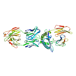
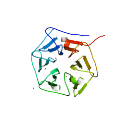
1RWI
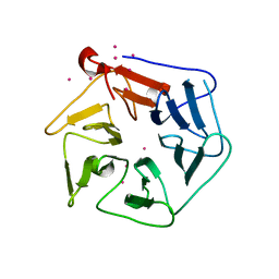 | | Extracellular domain of Mycobacterium tuberculosis PknD | | 分子名称: | CADMIUM ION, Serine/threonine-protein kinase pknD | | 著者 | Good, M.C, Greenstein, A.E, Young, T.A, Ng, H.L, Alber, T, TB Structural Genomics Consortium (TBSGC) | | 登録日 | 2003-12-16 | | 公開日 | 2004-04-27 | | 最終更新日 | 2024-02-14 | | 実験手法 | X-RAY DIFFRACTION (1.8 Å) | | 主引用文献 | Sensor Domain of the Mycobacterium tuberculosis Receptor Ser/Thr Protein Kinase, PknD, forms a Highly Symmetric beta Propeller.
J.Mol.Biol., 339, 2004
|
|
3KA8
 | | Frog M-ferritin, EQH mutant, with cobalt | | 分子名称: | CHLORIDE ION, COBALT (II) ION, Ferritin, ... | | 著者 | Tosha, T, Ng, H.L, Theil, E, Alber, T, Bhattasali, O. | | 登録日 | 2009-10-19 | | 公開日 | 2010-10-06 | | 最終更新日 | 2023-09-06 | | 実験手法 | X-RAY DIFFRACTION (1.35 Å) | | 主引用文献 | Moving Metal Ions through Ferritin-Protein Nanocages from Three-Fold Pores to Catalytic Sites.
J.Am.Chem.Soc., 132, 2010
|
|
3KA3
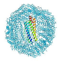
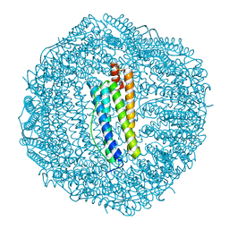 | | Frog M-ferritin with magnesium | | 分子名称: | CHLORIDE ION, Ferritin, middle subunit, ... | | 著者 | Tosha, T, Ng, H.L, Theil, E, Alber, T, Bhattasali, O. | | 登録日 | 2009-10-18 | | 公開日 | 2010-10-06 | | 最終更新日 | 2024-02-21 | | 実験手法 | X-RAY DIFFRACTION (1.4 Å) | | 主引用文献 | Moving Metal Ions through Ferritin-Protein Nanocages from Three-Fold Pores to Catalytic Sites.
J.Am.Chem.Soc., 132, 2010
|
|
1TXO
 | | Crystal structure of the Mycobacterium tuberculosis serine/threonine phosphatase PstP/Ppp at 1.95 A. | | 分子名称: | MANGANESE (II) ION, Putative Bacterial Enzyme | | 著者 | Pullen, K.E, Ng, H.L, Sung, P.Y, Good, M.C, Smith, S.M, Alber, T, TB Structural Genomics Consortium (TBSGC) | | 登録日 | 2004-07-05 | | 公開日 | 2004-11-23 | | 最終更新日 | 2017-10-11 | | 実験手法 | X-RAY DIFFRACTION (1.95 Å) | | 主引用文献 | An Alternate Conformation and a Third Metal in PstP/Ppp, the M. tuberculosis PP2C-Family Ser/Thr Protein Phosphatase.
Structure, 12, 2004
|
|
3KA9
 | | Frog M-ferritin, EEH mutant, with cobalt | | 分子名称: | CHLORIDE ION, COBALT (II) ION, Ferritin, ... | | 著者 | Tosha, T, Ng, H.L, Theil, E, Alber, T, Bhattasali, O. | | 登録日 | 2009-10-19 | | 公開日 | 2010-10-06 | | 最終更新日 | 2023-09-06 | | 実験手法 | X-RAY DIFFRACTION (1.45 Å) | | 主引用文献 | Frog M-ferritin, EEH mutant, with cobalt
To be Published
|
|
2H34
 | | Apoenzyme crystal structure of the tuberculosis serine/threonine kinase, PknE | | 分子名称: | BROMIDE ION, SODIUM ION, Serine/threonine-protein kinase pknE | | 著者 | Gay, L.M, Ng, H.L, Alber, T. | | 登録日 | 2006-05-22 | | 公開日 | 2006-07-18 | | 最終更新日 | 2017-10-18 | | 実験手法 | X-RAY DIFFRACTION (2.8 Å) | | 主引用文献 | A Conserved Dimer and Global Conformational Changes in the Structure of apo-PknE Ser/Thr Protein Kinase from Mycobacterium tuberculosis.
J.Mol.Biol., 360, 2006
|
|
1RWL
 | | Extracellular domain of Mycobacterium tuberculosis PknD | | 分子名称: | CADMIUM ION, Serine/threonine-protein kinase pknD | | 著者 | Good, M.C, Greenstein, A.E, Young, T.A, Ng, H.L, Alber, T, TB Structural Genomics Consortium (TBSGC) | | 登録日 | 2003-12-16 | | 公開日 | 2004-04-27 | | 最終更新日 | 2023-08-23 | | 実験手法 | X-RAY DIFFRACTION (1.9 Å) | | 主引用文献 | Sensor Domain of the Mycobacterium tuberculosis Receptor Ser/Thr Protein Kinase, PknD, forms a Highly Symmetric beta Propeller.
J.Mol.Biol., 339, 2004
|
|
3ORL
 | | Mycobacterium tuberculosis PknB kinase domain L33D mutant (crystal form 3) | | 分子名称: | MANGANESE (II) ION, PHOSPHOTHIOPHOSPHORIC ACID-ADENYLATE ESTER, Serine/threonine protein kinase | | 著者 | Echols, N, Lombana, T.N, Thomsen, N.D, Ng, H.-L, Alber, T, TB Structural Genomics Consortium (TBSGC) | | 登録日 | 2010-09-07 | | 公開日 | 2010-12-15 | | 最終更新日 | 2023-09-06 | | 実験手法 | X-RAY DIFFRACTION (2.9 Å) | | 主引用文献 | Allosteric activation mechanism of the Mycobacterium tuberculosis receptor Ser/Thr kinase, PknB
Structure, 18, 2010
|
|
3ORK
 | | Mycobacterium tuberculosis PknB kinase domain L33D mutant (crystal form 2) | | 分子名称: | 2-AMINO-2-HYDROXYMETHYL-PROPANE-1,3-DIOL, MANGANESE (II) ION, PHOSPHOTHIOPHOSPHORIC ACID-ADENYLATE ESTER, ... | | 著者 | Echols, N, Lombana, T.N, Thomsen, N.D, Ng, H.-L, Alber, T, TB Structural Genomics Consortium (TBSGC) | | 登録日 | 2010-09-07 | | 公開日 | 2010-12-15 | | 最終更新日 | 2023-09-06 | | 実験手法 | X-RAY DIFFRACTION (1.6 Å) | | 主引用文献 | Allosteric activation mechanism of the Mycobacterium tuberculosis receptor Ser/Thr kinase, PknB
Structure, 18, 2010
|
|
1LEY
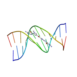 | | STRUCTURE OF A DICATIONIC MONOIMIDAZOLE LEXITROPSIN BOUND TO DNA (ORIENTATION 2) | | 分子名称: | DNA (5'-D(*CP*GP*CP*GP*AP*AP*TP*TP*CP*GP*CP*G)-3'), MONOIMIDAZOLE LEXITROPSIN | | 著者 | Goodsell, D.S, Ng, H.L, Kopka, M.L, Lown, J.W, Dickerson, R.E. | | 登録日 | 1995-10-10 | | 公開日 | 1996-04-03 | | 最終更新日 | 2024-02-14 | | 実験手法 | X-RAY DIFFRACTION (2.25 Å) | | 主引用文献 | Structure of a dicationic monoimidazole lexitropsin bound to DNA.
Biochemistry, 34, 1995
|
|
1LEX
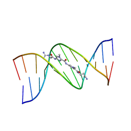 | | STRUCTURE OF A DICATIONIC MONOIMIDAZOLE LEXITROPSIN BOUND TO DNA (ORIENTATION 1) | | 分子名称: | DNA (5'-D(*CP*GP*CP*GP*AP*AP*TP*TP*CP*GP*CP*G)-3'), MONOIMIDAZOLE LEXITROPSIN | | 著者 | Goodsell, D.S, Ng, H.L, Kopka, M.L, Lown, J.W, Dickerson, R.E. | | 登録日 | 1995-10-10 | | 公開日 | 1996-04-03 | | 最終更新日 | 2024-02-14 | | 実験手法 | X-RAY DIFFRACTION (2.25 Å) | | 主引用文献 | Structure of a dicationic monoimidazole lexitropsin bound to DNA.
Biochemistry, 34, 1995
|
|
3IP2
 | | Crystal structure of red fluorescent protein Neptune at pH 7.0 | | 分子名称: | Neptune red fluorescent protein | | 著者 | Lin, M.Z, McKeown, M.R, Ng, H.L, Aguilera, T.A, Shaner, N.C, Ma, W, Adams, S.R, Campbell, R.E, Alber, T, Tsien, R.Y. | | 登録日 | 2009-08-15 | | 公開日 | 2009-12-15 | | 最終更新日 | 2023-11-22 | | 実験手法 | X-RAY DIFFRACTION (1.6 Å) | | 主引用文献 | Autofluorescent proteins with excitation in the optical window for intravital imaging in mammals.
Chem.Biol., 16, 2009
|
|


