Loading
PDBj
MenuPDBj@FacebookPDBj@TwitterPDBj@YouTubewwPDB FoundationwwPDB
RCSB PDBPDBeBMRBAdv. SearchSearch help
PDB: 171 results

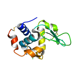
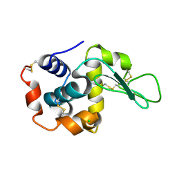
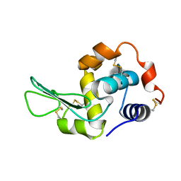
6IG6
DownloadVisualize
BU of 6ig6 by Molmil
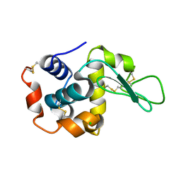
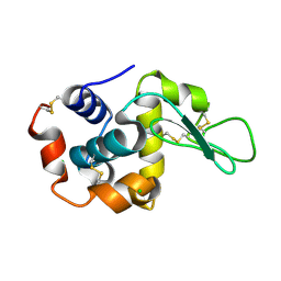
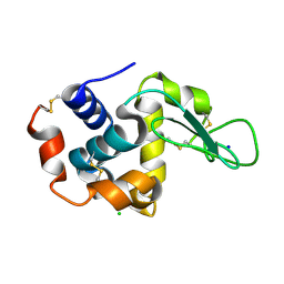
Crystal structure of lysozyme delivered in polyacrylamide using x-ray free electron laser
Descriptor: CHLORIDE ION, Lysozyme C, SODIUM ION
Authors:Nam, K.H.
Deposit date:2018-09-25
Release date:2018-11-07
Last modified:2024-10-16
Method:X-RAY DIFFRACTION (1.7 Å)
Cite:Polyacrylamide injection matrix for serial femtosecond crystallography.
Sci Rep, 9, 2019
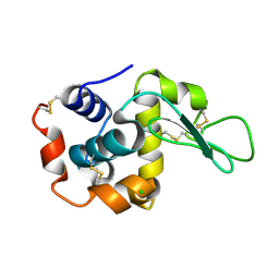
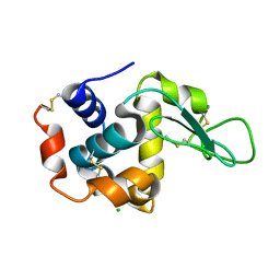
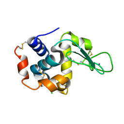
6IRJ
DownloadVisualize
BU of 6irj by Molmil
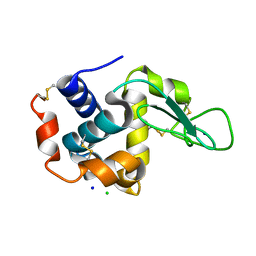
Crystal structure of lysozyme by fixed-target serial femtosecond crystallography
Descriptor: CHLORIDE ION, Lysozyme C, SODIUM ION
Authors:Nam, K.H.
Deposit date:2018-11-13
Release date:2019-05-22
Last modified:2023-11-22
Method:X-RAY DIFFRACTION (1.65 Å)
Cite:Nylon mesh-based sample holder for fixed-target serial femtosecond crystallography.
Sci Rep, 9, 2019
6IG7
DownloadVisualize
BU of 6ig7 by Molmil
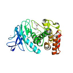
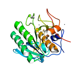
Crystal structure of thermolysin delivered in polyacrylamide using x-ray free electron laser
Descriptor: CALCIUM ION, LEUCINE, LYSINE, ...
Authors:Nam, K.H.
Deposit date:2018-09-25
Release date:2018-11-07
Last modified:2023-11-22
Method:X-RAY DIFFRACTION (1.8 Å)
Cite:Polyacrylamide injection matrix for serial femtosecond crystallography.
Sci Rep, 9, 2019
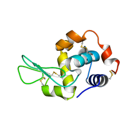
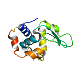
6JXQ
DownloadVisualize
BU of 6jxq by Molmil
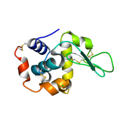
Room temperature structure of lysozyme delivered in polyacrylamide by serial millisecond crystallography
Descriptor: CHLORIDE ION, Lysozyme C
Authors:Nam, K.H.
Deposit date:2019-04-24
Release date:2019-05-15
Last modified:2023-11-22
Method:X-RAY DIFFRACTION (1.76 Å)
Cite:Sample delivery using viscous media, a syringe and a syringe pump for serial crystallography.
J.Synchrotron Radiat., 26, 2019
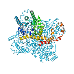
6KCC
DownloadVisualize
BU of 6kcc by Molmil
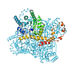
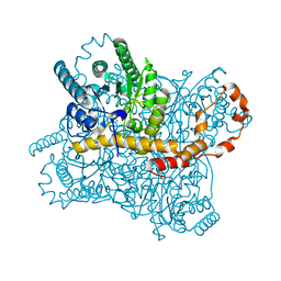
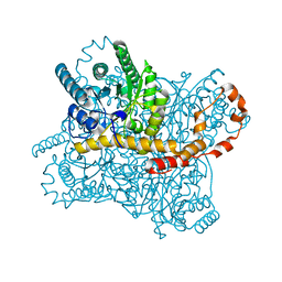
Room temperature structure of glucose isomerase delivered in shortening B by serial millisecond crystallography
Descriptor: MAGNESIUM ION, Xylose isomerase
Authors:Nam, K.H.
Deposit date:2019-06-27
Release date:2020-05-20
Last modified:2023-11-22
Method:X-RAY DIFFRACTION (2 Å)
Cite:Shortening injection matrix for serial crystallography.
Sci Rep, 10, 2020
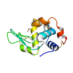
6KCB
DownloadVisualize
BU of 6kcb by Molmil
Room temperature structure of lysozyme delivered in shortening A by serial millisecond crystallography
Descriptor: CHLORIDE ION, Lysozyme C, SODIUM ION
Authors:Nam, K.H.
Deposit date:2019-06-27
Release date:2020-05-20
Last modified:2024-10-16
Method:X-RAY DIFFRACTION (1.8 Å)
Cite:Shortening injection matrix for serial crystallography.
Sci Rep, 10, 2020
6KD2
DownloadVisualize
BU of 6kd2 by Molmil
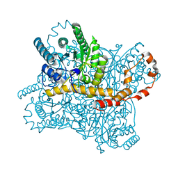
Room temperature structure of glucose isomerase delivered in gelatin by serial millisecond crystallography
Descriptor: MAGNESIUM ION, Xylose isomerase
Authors:Nam, K.H.
Deposit date:2019-06-30
Release date:2019-07-17
Last modified:2023-11-22
Method:X-RAY DIFFRACTION (1.7 Å)
Cite:Stable sample delivery in viscous media via a capillary for serial crystallography.
J.Appl.Crystallogr., 53, 2020
6KKF
DownloadVisualize
BU of 6kkf by Molmil
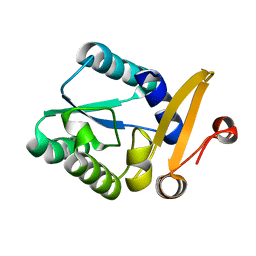
Crystal structure of proteinase K complexed with a triglycine
Descriptor: CALCIUM ION, Proteinase K, glycylglycylglycine
Authors:Nam, K.H.
Deposit date:2019-07-25
Release date:2019-09-25
Last modified:2024-10-23
Method:X-RAY DIFFRACTION (1.4 Å)
Cite:Triglycine-Based Approach for Identifying the Substrate Recognition Site of an Enzyme
Crystals, 9, 2019
6KCA
DownloadVisualize
BU of 6kca by Molmil
Room temperature structure of glucose isomerase delivered in shortening A by serial millisecond crystallography
Descriptor: MAGNESIUM ION, Xylose isomerase
Authors:Nam, K.H.
Deposit date:2019-06-27
Release date:2020-05-20
Last modified:2023-11-22
Method:X-RAY DIFFRACTION (1.9 Å)
Cite:Shortening injection matrix for serial crystallography.
Sci Rep, 10, 2020
6JXP
DownloadVisualize
BU of 6jxp by Molmil
Room temperature structure of lysozyme delivered in LCP by serial millisecond crystallography
Descriptor: CHLORIDE ION, Lysozyme C
Authors:Nam, K.H.
Deposit date:2019-04-24
Release date:2019-05-15
Last modified:2024-10-16
Method:X-RAY DIFFRACTION (1.56 Å)
Cite:Sample delivery using viscous media, a syringe and a syringe pump for serial crystallography.
J.Synchrotron Radiat., 26, 2019
6IRK
DownloadVisualize
BU of 6irk by Molmil
Crystal structure of glucose isomerase by fixed-target serial femtosecond crystallography
Descriptor: MAGNESIUM ION, Xylose isomerase
Authors:Nam, K.H.
Deposit date:2018-11-13
Release date:2019-05-22
Last modified:2023-11-22
Method:X-RAY DIFFRACTION (1.75 Å)
Cite:Nylon mesh-based sample holder for fixed-target serial femtosecond crystallography.
Sci Rep, 9, 2019
6KD1
DownloadVisualize
BU of 6kd1 by Molmil
Room temperature structure of lysozyme delivered in agarose by serial millisecond crystallography
Descriptor: CHLORIDE ION, Lysozyme C, SODIUM ION
Authors:Nam, K.H.
Deposit date:2019-06-30
Release date:2019-07-17
Last modified:2024-10-30
Method:X-RAY DIFFRACTION (1.85 Å)
Cite:Stable sample delivery in viscous media via a capillary for serial crystallography.
J.Appl.Crystallogr., 53, 2020
6K1Y
DownloadVisualize
BU of 6k1y by Molmil
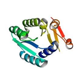
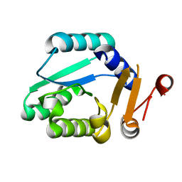
Crystal structure of Rhodothermus marinus substrate-binding protein at pH 7.5
Descriptor: ABC-type uncharacterized transport system periplasmic component-like protein
Authors:Nam, K.H.
Deposit date:2019-05-13
Release date:2019-08-21
Last modified:2023-11-22
Method:X-RAY DIFFRACTION (1.9 Å)
Cite:Crystal structure of Rhodothermus marinus substrate-binding protein at pH 7.5
To Be Published
6K1X
DownloadVisualize
BU of 6k1x by Molmil
Crystal structure of Rhodothermus marinus substrate-binding protein at pH 6.0
Descriptor: ABC-type uncharacterized transport system periplasmic component-like protein
Authors:Nam, K.H.
Deposit date:2019-05-13
Release date:2019-08-21
Last modified:2023-11-22
Method:X-RAY DIFFRACTION (1.802 Å)
Cite:Crystal structure of Rhodothermus marinus substrate-binding protein at pH 6.0
To Be Published
6KCD
DownloadVisualize
BU of 6kcd by Molmil
Room temperature structure of lysozyme delivered in shortening B by serial millisecond crystallography
Descriptor: CHLORIDE ION, Lysozyme C, SODIUM ION
Authors:Nam, K.H.
Deposit date:2019-06-27
Release date:2020-05-20
Last modified:2024-10-23
Method:X-RAY DIFFRACTION (1.5 Å)
Cite:Shortening injection matrix for serial crystallography.
Sci Rep, 10, 2020
6K1W
DownloadVisualize
BU of 6k1w by Molmil
Crystal structure of Rhodothermus marinus substrate-binding protein at pH 5.5
Descriptor: ABC-type uncharacterized transport system periplasmic component-like protein
Authors:Nam, K.H.
Deposit date:2019-05-13
Release date:2019-08-21
Last modified:2023-11-22
Method:X-RAY DIFFRACTION (1.5 Å)
Cite:Crystal structure of Rhodothermus marinus substrate-binding protein at pH 5.5
To Be Published
7WBF
DownloadVisualize
BU of 7wbf by Molmil
Crystal structure of lysozyme
Descriptor: CHLORIDE ION, Lysozyme C
Authors:Nam, K.H.
Deposit date:2021-12-16
Release date:2022-01-19
Last modified:2023-11-29
Method:X-RAY DIFFRACTION (1.6 Å)
Cite:Processing of Multicrystal Diffraction Patterns in Macromolecular Crystallography Using Serial Crystallography Programs.
Crystals, 12, 2022
7WBD
DownloadVisualize
BU of 7wbd by Molmil
Crystal structure of lysozyme (multilcrystal diffraction, CrystFEL/XGANDALF)
Descriptor: CHLORIDE ION, Lysozyme C
Authors:Nam, K.H.
Deposit date:2021-12-16
Release date:2022-01-19
Last modified:2023-11-29
Method:X-RAY DIFFRACTION (1.9 Å)
Cite:Processing of Multicrystal Diffraction Patterns in Macromolecular Crystallography Using Serial Crystallography Programs.
Crystals, 12, 2022
7WBE
DownloadVisualize
BU of 7wbe by Molmil
Crystal structure of lysozyme (multilcrystal diffraction, CrystFEL/MOSFLM)
Descriptor: CHLORIDE ION, Lysozyme C
Authors:Nam, K.H.
Deposit date:2021-12-16
Release date:2022-01-19
Last modified:2024-10-16
Method:X-RAY DIFFRACTION (1.9 Å)
Cite:Processing of Multicrystal Diffraction Patterns in Macromolecular Crystallography Using Serial Crystallography Programs.
Crystals, 12, 2022
7WKR
DownloadVisualize
BU of 7wkr by Molmil
Room temperature structure of lysozyme solved by serial synchrotron crystallography
Descriptor: CHLORIDE ION, Lysozyme C
Authors:Nam, K.H.
Deposit date:2022-01-11
Release date:2022-02-02
Last modified:2024-10-23
Method:X-RAY DIFFRACTION (1.6 Å)
Cite:Combination of an inject-and-transfer system for serial femtosecond crystallography.
J.Appl.Crystallogr., 55, 2022
7WUC
DownloadVisualize
BU of 7wuc by Molmil
Room-temperature structure of lysozyme by serial femtosecond crystallography (BITS)
Descriptor: Lysozyme C
Authors:Nam, K.H.
Deposit date:2022-02-08
Release date:2022-03-02
Last modified:2023-11-29
Method:X-RAY DIFFRACTION (2.1 Å)
Cite:Combination of an inject-and-transfer system for serial femtosecond crystallography.
J.Appl.Crystallogr., 55, 2022
7XF7
DownloadVisualize
BU of 7xf7 by Molmil
Crystal Structure of Human Lysozyme Complexed with N-Acetyl-alpha-D-Glucosamine
Descriptor: 2-acetamido-2-deoxy-alpha-D-glucopyranose, Lysozyme C
Authors:Nam, K.H.
Deposit date:2022-04-01
Release date:2022-04-13
Last modified:2024-10-30
Method:X-RAY DIFFRACTION (1.55 Å)
Cite:Crystal Structure of Human Lysozyme Complexed with N-Acetyl-alpha-d-glucosamine.
Appl Sci (Basel), 12, 2022
7XF6
DownloadVisualize
BU of 7xf6 by Molmil
Crystal Structure of Human Lysozyme
Descriptor: ACETATE ION, Lysozyme C
Authors:Nam, K.H.
Deposit date:2022-04-01
Release date:2022-04-13
Last modified:2024-10-23
Method:X-RAY DIFFRACTION (1.3 Å)
Cite:Crystal Structure of Human Lysozyme Complexed with N-Acetyl-alpha-d-glucosamine.
Appl Sci (Basel), 12, 2022
7XF8
DownloadVisualize
BU of 7xf8 by Molmil
Crystal Structure of Human Lysozyme Complexed with N-Acetyl-alpha-D-Glucosamine
Descriptor: 2-acetamido-2-deoxy-alpha-D-glucopyranose, Lysozyme C
Authors:Nam, K.H.
Deposit date:2022-04-01
Release date:2022-04-13
Last modified:2024-10-16
Method:X-RAY DIFFRACTION (1.6 Å)
Cite:Crystal Structure of Human Lysozyme Complexed with N-Acetyl-alpha-d-glucosamine.
Appl Sci (Basel), 12, 2022
5ZYC
DownloadVisualize
BU of 5zyc by Molmil
Crystal Structure of Glucose Isomerase Soaked with Mn2+
Descriptor: 1,2-ETHANEDIOL, ACETATE ION, MANGANESE (II) ION, ...
Authors:Nam, K.H.
Deposit date:2018-05-24
Release date:2018-11-28
Last modified:2023-11-22
Method:X-RAY DIFFRACTION (1.75 Å)
Cite:Structural analysis of substrate recognition by glucose isomerase in Mn2+binding mode at M2 site in S. rubiginosus
Biochem. Biophys. Res. Commun., 503, 2018

226707

PDB entries from 2024-10-30

PDB statisticsPDBj update infoContact PDBjnumon