6JXQ
 
 | |
6KD2
 | |
6KCA
 | |
6JXP
 | |
6KD1
 | |
6K1X
 | |
6KCD
 | |
6KCB
 | |
6K1W
 | |
6KCC
 | |
6K1Y
 | |
7WKR
 | |
7WUC
 | |
7WBE
 | |
7WBD
 | |
7WBF
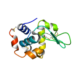
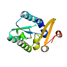
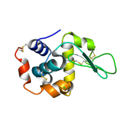
 | | Crystal structure of lysozyme | | 分子名称: | CHLORIDE ION, Lysozyme C | | 著者 | Nam, K.H. | | 登録日 | 2021-12-16 | | 公開日 | 2022-01-19 | | 最終更新日 | 2023-11-29 | | 実験手法 | X-RAY DIFFRACTION (1.6 Å) | | 主引用文献 | Processing of Multicrystal Diffraction Patterns in Macromolecular Crystallography Using Serial Crystallography Programs.
Crystals, 12, 2022
|
|
7XF7
 | |
7XF6
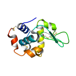
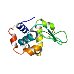
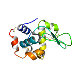
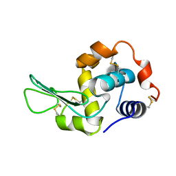
 | | Crystal Structure of Human Lysozyme | | 分子名称: | ACETATE ION, Lysozyme C | | 著者 | Nam, K.H. | | 登録日 | 2022-04-01 | | 公開日 | 2022-04-13 | | 最終更新日 | 2023-11-29 | | 実験手法 | X-RAY DIFFRACTION (1.3 Å) | | 主引用文献 | Crystal Structure of Human Lysozyme Complexed with N-Acetyl-alpha-d-glucosamine.
Appl Sci (Basel), 12, 2022
|
|
7XF8
 | |
6LNG
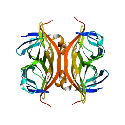 | | Rapid crystallization of streptavidin using charged peptides | | 分子名称: | GLYCEROL, Streptavidin | | 著者 | Minamihata, K, Tsukamoto, K, Adachi, M, Shimizu, R, Mishina, M, Kuroki, R, Nagamune, T. | | 登録日 | 2019-12-30 | | 公開日 | 2020-03-18 | | 最終更新日 | 2023-11-22 | | 実験手法 | X-RAY DIFFRACTION (1.8000015 Å) | | 主引用文献 | Genetically fused charged peptides induce rapid crystallization of proteins.
Chem.Commun.(Camb.), 56, 2020
|
|
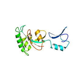
5L6Q
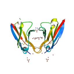 | | Refolded AL protein from cardiac amyloidosis | | 分子名称: | CARBONATE ION, CHLORIDE ION, DI(HYDROXYETHYL)ETHER, ... | | 著者 | Annamalai, K, Liberta, F, Vielberg, M.-T, Lilie, H, Guehrs, K.-H, Schierhorn, A, Koehler, R, Schmidt, A, Haupt, C, Hegenbart, O, Schoenland, S, Groll, M, Faendrich, M. | | 登録日 | 2016-05-31 | | 公開日 | 2017-05-31 | | 最終更新日 | 2024-01-10 | | 実験手法 | X-RAY DIFFRACTION (1.4 Å) | | 主引用文献 | Common Fibril Structures Imply Systemically Conserved Protein Misfolding Pathways In Vivo.
Angew. Chem. Int. Ed. Engl., 56, 2017
|
|
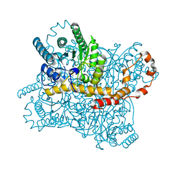
6GV1
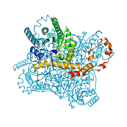
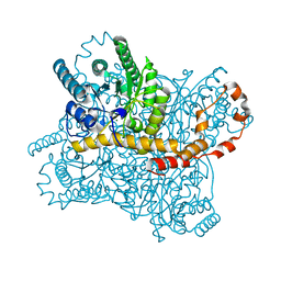
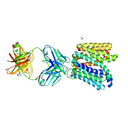 | | Crystal structure of E.coli Multidrug/H+ antiporter MdfA in outward open conformation with bound Fab fragment | | 分子名称: | Fab fragment YN1074 heavy chain, Fab fragment YN1074 light chain, Major Facilitator Superfamily multidrug/H+ antiporter MdfA from E.coli, ... | | 著者 | Nagarathinam, K, Parthier, C, Stubbs, M.T, Tanabe, M. | | 登録日 | 2018-06-20 | | 公開日 | 2018-10-03 | | 最終更新日 | 2024-01-17 | | 実験手法 | X-RAY DIFFRACTION (3.4 Å) | | 主引用文献 | Outward open conformation of a Major Facilitator Superfamily multidrug/H+antiporter provides insights into switching mechanism.
Nat Commun, 9, 2018
|
|
1G91
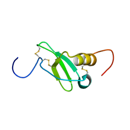 | | SOLUTION STRUCTURE OF MYELOID PROGENITOR INHIBITORY FACTOR-1 (MPIF-1) | | 分子名称: | MYELOID PROGENITOR INHIBITORY FACTOR-1 | | 著者 | Rajarathnam, K, Li, Y, Rohrer, T, Gentz, R. | | 登録日 | 2000-11-21 | | 公開日 | 2001-03-07 | | 最終更新日 | 2022-12-21 | | 実験手法 | SOLUTION NMR | | 主引用文献 | Solution structure and dynamics of myeloid progenitor inhibitory factor-1 (MPIF-1), a novel monomeric CC chemokine.
J.Biol.Chem., 276, 2001
|
|
7KKF
 | |
6IAK
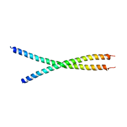 | | The crystal structure of the chicken CREB3 bZIP | | 分子名称: | Uncharacterized protein | | 著者 | Sabaratnam, K, Renner, M. | | 登録日 | 2018-11-26 | | 公開日 | 2019-12-11 | | 最終更新日 | 2020-06-24 | | 実験手法 | X-RAY DIFFRACTION (3.95 Å) | | 主引用文献 | Insights from the crystal structure of the chicken CREB3 bZIP suggest that members of the CREB3 subfamily transcription factors may be activated in response to oxidative stress.
Protein Sci., 28, 2019
|
|


