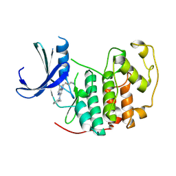
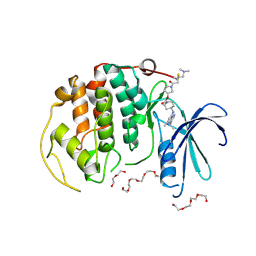
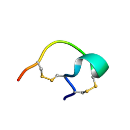
2GG1
 
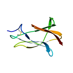 | | NMR solution structure of domain III of the E-protein of tick-borne Langat flavivirus (includes RDC restraints) | | 分子名称: | Genome polyprotein | | 著者 | Mukherjee, M, Dutta, K, White, M.A, Cowburn, D, Fox, R.O. | | 登録日 | 2006-03-23 | | 公開日 | 2006-04-25 | | 最終更新日 | 2023-11-29 | | 実験手法 | SOLUTION NMR | | 主引用文献 | NMR solution structure and backbone dynamics of domain III of the E protein of tick-borne Langat flavivirus suggests a potential site for molecular recognition.
Protein Sci., 15, 2006
|
|
6FHK
 | |
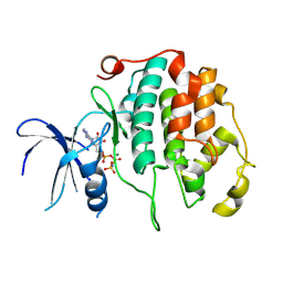
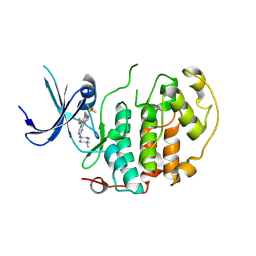
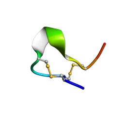
1Z66
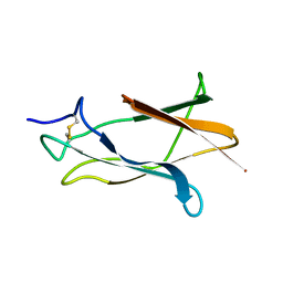 | | NMR solution structure of domain III of E-protein of tick-borne Langat flavivirus (no RDC restraints) | | 分子名称: | Major envelope protein E | | 著者 | Mukherjee, M, Dutta, K, White, M.A, Cowburn, D, Fox, R.O. | | 登録日 | 2005-03-21 | | 公開日 | 2006-03-28 | | 最終更新日 | 2022-03-02 | | 実験手法 | SOLUTION NMR | | 主引用文献 | NMR solution structure and backbone dynamics of domain III of the E protein of tick-borne Langat flavivirus suggests a potential site for molecular recognition.
Protein Sci., 15, 2006
|
|
8R9B
 | |
8RU8
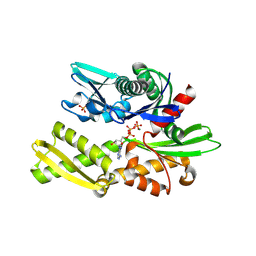
 | | A crystal form of a human CDK2-CDK7 chimera | | 分子名称: | (3R,4R)-4-[[[7-[(phenylmethyl)amino]-3-propan-2-yl-pyrazolo[1,5-a]pyrimidin-5-yl]amino]methyl]piperidin-3-ol, Cyclin-dependent kinase 2 | | 著者 | Mukherjee, M, Cleasby, A. | | 登録日 | 2024-01-30 | | 公開日 | 2024-06-26 | | 実験手法 | X-RAY DIFFRACTION (1.51 Å) | | 主引用文献 | Protein engineering enables a soakable crystal form of human CDK7 primed for high-throughput crystallography and structure-based drug design.
Structure, 2024
|
|
8R99
 | |
8R9O
 | | A soakable crystal form of human CDK7 in complex with AMP-PNP | | 分子名称: | Cyclin-dependent kinase 7, ~{N}-[(1~{S})-2-(dimethylamino)-1-phenyl-ethyl]-6,6-dimethyl-3-[[4-(propanoylamino)phenyl]carbonylamino]-1,4-dihydropyrrolo[3,4-c]pyrazole-5-carboxamide | | 著者 | Mukherjee, M, Cleasby, A. | | 登録日 | 2023-11-30 | | 公開日 | 2024-05-29 | | 最終更新日 | 2024-06-26 | | 実験手法 | X-RAY DIFFRACTION (2.22 Å) | | 主引用文献 | Protein engineering enables a soakable crystal form of human CDK7 primed for high-throughput crystallography and structure-based drug design.
Structure, 2024
|
|
8R9A
 | | A soakable crystal form of human CDK7 in complex with AMP-PNP | | 分子名称: | (3R,4R)-4-[[[7-[(phenylmethyl)amino]-3-propan-2-yl-pyrazolo[1,5-a]pyrimidin-5-yl]amino]methyl]piperidin-3-ol, Cyclin-dependent kinase 7, DI(HYDROXYETHYL)ETHER | | 著者 | Mukherjee, M, Cleasby, A. | | 登録日 | 2023-11-30 | | 公開日 | 2024-05-29 | | 最終更新日 | 2024-06-26 | | 実験手法 | X-RAY DIFFRACTION (1.71 Å) | | 主引用文献 | Protein engineering enables a soakable crystal form of human CDK7 primed for high-throughput crystallography and structure-based drug design.
Structure, 2024
|
|
8R9S
 | | A soakable crystal form of human CDK7 in complex with AMP-PNP | | 分子名称: | 3,6,9,12,15,18,21,24,27,30,33,36,39-TRIDECAOXAHENTETRACONTANE-1,41-DIOL, Cyclin-dependent kinase 7, DI(HYDROXYETHYL)ETHER, ... | | 著者 | Mukherjee, M, Cleasby, A. | | 登録日 | 2023-11-30 | | 公開日 | 2024-05-29 | | 最終更新日 | 2024-06-26 | | 実験手法 | X-RAY DIFFRACTION (2.78 Å) | | 主引用文献 | Protein engineering enables a soakable crystal form of human CDK7 primed for high-throughput crystallography and structure-based drug design.
Structure, 2024
|
|
8R9U
 | | A soakable crystal form of human CDK7 in complex with AMP-PNP | | 分子名称: | 7-dimethylphosphoryl-3-[2-[[(3~{S})-6,6-dimethylpiperidin-3-yl]amino]-5-(trifluoromethyl)pyrimidin-4-yl]-1~{H}-indole-6-carbonitrile, Cyclin-dependent kinase 7 | | 著者 | Mukherjee, M, Cleasby, A. | | 登録日 | 2023-11-30 | | 公開日 | 2024-05-29 | | 最終更新日 | 2024-06-26 | | 実験手法 | X-RAY DIFFRACTION (1.937 Å) | | 主引用文献 | Protein engineering enables a soakable crystal form of human CDK7 primed for high-throughput crystallography and structure-based drug design.
Structure, 2024
|
|
5ODS
 | |
2MQ1
 | |
2G6U
 | |
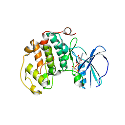
5HPK
 | | System-wide modulation of HECT E3 ligases with selective ubiquitin variant probes: NEDD4L and UbV NL.1 | | 分子名称: | E3 ubiquitin-protein ligase NEDD4-like, Ubiquitin variant NL.1 | | 著者 | Wu, K.-P, Mukherjee, M, Mercredi, P.Y, Schulman, B.A. | | 登録日 | 2016-01-20 | | 公開日 | 2016-03-16 | | 最終更新日 | 2023-09-27 | | 実験手法 | X-RAY DIFFRACTION (2.431 Å) | | 主引用文献 | System-Wide Modulation of HECT E3 Ligases with Selective Ubiquitin Variant Probes.
Mol.Cell, 62, 2016
|
|
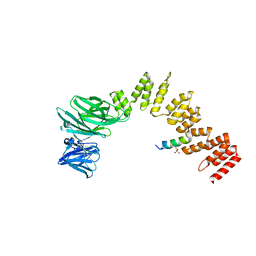
1R8T
 | | Solution structures of high affinity miniprotein ligands to Streptavidin | | 分子名称: | MP1 | | 著者 | Luo, J, Mukherjee, M, Fan, X, Yang, H, Liu, D, Khan, R, White, M, Fox, R.O. | | 登録日 | 2003-10-28 | | 公開日 | 2005-02-15 | | 最終更新日 | 2022-03-02 | | 実験手法 | SOLUTION NMR | | 主引用文献 | Structure-based design of high affinity miniprotein ligands
To be Published
|
|
3VK6
 | |


