5KAU
 
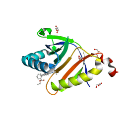 | | The structure of SAV2435 bound to RHODAMINE 6G | | 分子名称: | GLYCEROL, RHODAMINE 6G, SA2223 protein | | 著者 | Moreno, A, Wade, H. | | 登録日 | 2016-06-02 | | 公開日 | 2016-08-24 | | 最終更新日 | 2023-09-27 | | 実験手法 | X-RAY DIFFRACTION (1.95 Å) | | 主引用文献 | Solution Binding and Structural Analyses Reveal Potential Multidrug Resistance Functions for SAV2435 and CTR107 and Other GyrI-like Proteins.
Biochemistry, 55, 2016
|
|
5KCB
 
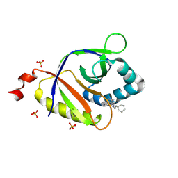 | | The structure of SAV2435 bound to ethidium bromide | | 分子名称: | ETHIDIUM, SA2223 protein, SULFATE ION | | 著者 | Moreno, A, Wade, H. | | 登録日 | 2016-06-06 | | 公開日 | 2016-08-24 | | 最終更新日 | 2024-10-30 | | 実験手法 | X-RAY DIFFRACTION (2.101 Å) | | 主引用文献 | Solution Binding and Structural Analyses Reveal Potential Multidrug Resistance Functions for SAV2435 and CTR107 and Other GyrI-like Proteins.
Biochemistry, 55, 2016
|
|
5KAX
 
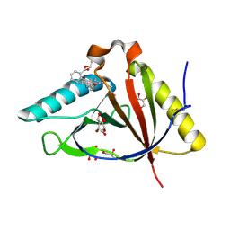 | | The structure of CTR107 protein bound to RHODAMINE 6G | | 分子名称: | CTR107 protein, GLYCEROL, RHODAMINE 6G | | 著者 | Moreno, A, Wade, H. | | 登録日 | 2016-06-02 | | 公開日 | 2016-08-24 | | 最終更新日 | 2024-10-09 | | 実験手法 | X-RAY DIFFRACTION (2 Å) | | 主引用文献 | Solution Binding and Structural Analyses Reveal Potential Multidrug Resistance Functions for SAV2435 and CTR107 and Other GyrI-like Proteins.
Biochemistry, 55, 2016
|
|
5KAW
 
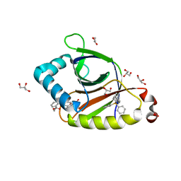 | |
5KAV
 
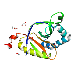 | | The structure of SAV2435 | | 分子名称: | GLYCEROL, SA2223 protein | | 著者 | Moreno, A, Wade, H. | | 登録日 | 2016-06-02 | | 公開日 | 2016-08-24 | | 最終更新日 | 2024-02-28 | | 実験手法 | X-RAY DIFFRACTION (2 Å) | | 主引用文献 | Solution Binding and Structural Analyses Reveal Potential Multidrug Resistance Functions for SAV2435 and CTR107 and Other GyrI-like Proteins.
Biochemistry, 55, 2016
|
|
5KAT
 
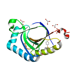 | | The structure of SAV2435 bound to TETRAPHENYLPHOSPHONIUM | | 分子名称: | GLYCEROL, SA2223 protein, TETRAPHENYLPHOSPHONIUM | | 著者 | Moreno, A, Wade, H. | | 登録日 | 2016-06-02 | | 公開日 | 2016-08-24 | | 最終更新日 | 2024-02-28 | | 実験手法 | X-RAY DIFFRACTION (2.101 Å) | | 主引用文献 | Solution Binding and Structural Analyses Reveal Potential Multidrug Resistance Functions for SAV2435 and CTR107 and Other GyrI-like Proteins.
Biochemistry, 55, 2016
|
|
7Q1L
 
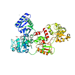 | | Glycosilated Human Serum Apo-tranferrin | | 分子名称: | 1,2-ETHANEDIOL, 2-acetamido-2-deoxy-beta-D-glucopyranose-(1-4)-2-acetamido-2-deoxy-beta-D-glucopyranose, GLYCEROL, ... | | 著者 | Gavira, J.A, Moreno, A, Campos-Escamilla, C, Gonzalez-Ramirez, L.A, Siliqi, D. | | 登録日 | 2021-10-20 | | 公開日 | 2022-03-16 | | 最終更新日 | 2024-10-16 | | 実験手法 | X-RAY DIFFRACTION (3 Å) | | 主引用文献 | X-ray Characterization of Conformational Changes of Human Apo- and Holo-Transferrin.
Int J Mol Sci, 22, 2021
|
|
4UWW
 
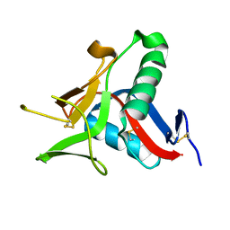 | | Crystallographic Structure of the Intramineral Protein Struthicalcin from Struthio camelus Eggshell | | 分子名称: | STRUTHIOCALCIN-1 | | 著者 | Ruiz, R.R, Moreno, A, Romero, A. | | 登録日 | 2014-08-14 | | 公開日 | 2015-04-08 | | 最終更新日 | 2024-10-23 | | 実験手法 | X-RAY DIFFRACTION (1.44 Å) | | 主引用文献 | Crystal Structure of Struthiocalcin-1, an Intramineral Protein from Struthio Camelus Eggshell, in Two Different Crystal Forms.
Acta Crystallogr.,Sect.D, 71, 2015
|
|
4UXM
 
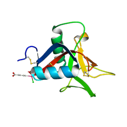 | | Crystal Structure of Struthiocalcin-1, a different crystal form. | | 分子名称: | (4-CARBOXYPHENYL)(CHLORO)MERCURY, STRUTHIOCALCIN-1 | | 著者 | Ruiz-Arellano, R.R, Moreno, A, Romero, A. | | 登録日 | 2014-08-26 | | 公開日 | 2015-04-08 | | 最終更新日 | 2024-10-16 | | 実験手法 | X-RAY DIFFRACTION (1.5 Å) | | 主引用文献 | Crystal Structure of Struthiocalcin-1, an Intramineral Protein from Struthio Camelus Eggshell, in Two Different Crystal Forms.
Acta Crystallogr.,Sect.D, 71, 2015
|
|
5AJL
 
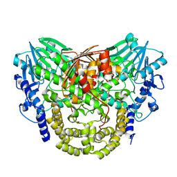 | | Sdsa sulfatase tetragonal | | 分子名称: | ALKYL SULFATASE, ZINC ION | | 著者 | De la Mora, E, Flores-Hernandez, E, Jakoncic, J, Stojanoff, V, Sanchez-Puig, N, Moreno, A. | | 登録日 | 2015-02-25 | | 公開日 | 2015-10-07 | | 最終更新日 | 2024-01-10 | | 実験手法 | X-RAY DIFFRACTION (3.45 Å) | | 主引用文献 | Sdsa Polymorph Isolation and Improvement of Their Crystal Quality Using Nonconventional Crystallization Techniques
J.Appl.Crystallogr., 48, 2015
|
|
5A23
 
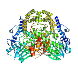 | | SdsA sulfatase triclinic form | | 分子名称: | SDS HYDROLASE SDSA1, ZINC ION | | 著者 | De la Mora, E, Flores-Hernandez, E, Jakoncik, J, Stojanoff, V, Sanchez-Puig, N, Moreno, A. | | 登録日 | 2015-05-11 | | 公開日 | 2015-10-07 | | 最終更新日 | 2024-01-10 | | 実験手法 | X-RAY DIFFRACTION (2.41 Å) | | 主引用文献 | Sdsa Polymorph Isolation and Improvement of Their Crystal Quality Using Nonconventional Crystallization Techniques
J.Appl.Crystallogr., 48, 2015
|
|
5AIJ
 
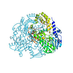 | | P. aeruginosa SdsA hexagonal polymorph | | 分子名称: | ALKYL SULFATASE, GLYCEROL, ZINC ION | | 著者 | De la Mora, E, Flores-Hernandez, E, Jakoncic, J, Stojanoff, V, Sanchez-Puig, N, Moreno, A. | | 登録日 | 2015-02-13 | | 公開日 | 2015-10-07 | | 最終更新日 | 2024-01-10 | | 実験手法 | X-RAY DIFFRACTION (1.95 Å) | | 主引用文献 | Sdsa Polymorph Isolation and Improvement of Their Crystal Quality Using Nonconventional Crystallization Techniques
J.Appl.Crystallogr., 48, 2015
|
|
2B4Z
 
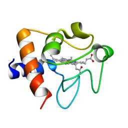 | | Crystal structure of cytochrome C from bovine heart at 1.5 A resolution. | | 分子名称: | Cytochrome c, PROTOPORPHYRIN IX CONTAINING FE | | 著者 | Mirkin, N, Jakoncic, J, Stojanoff, V, Moreno, A. | | 登録日 | 2005-09-27 | | 公開日 | 2005-10-11 | | 最終更新日 | 2023-08-23 | | 実験手法 | X-RAY DIFFRACTION (1.5 Å) | | 主引用文献 | High resolution X-ray crystallographic structure of bovine heart cytochrome c and its application to the design of an electron transfer biosensor.
Proteins, 70, 2008
|
|
4AFL
 
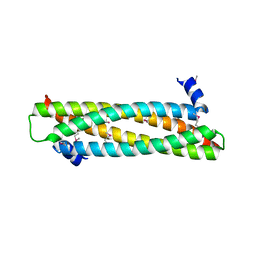 | | The crystal structure of the ING4 dimerization domain reveals the functional organization of the ING family of chromatin binding proteins. | | 分子名称: | INHIBITOR OF GROWTH PROTEIN 4 | | 著者 | Culurgioni, S, Munoz, I.G, Moreno, A, Palacios, A, Villate, M, Palmero, I, Montoya, G, Blanco, F.J. | | 登録日 | 2012-01-19 | | 公開日 | 2012-02-22 | | 最終更新日 | 2024-10-23 | | 実験手法 | X-RAY DIFFRACTION (2.275 Å) | | 主引用文献 | Crystal Structure of Inhibitor of Growth 4 (Ing4) Dimerization Domain Reveals Functional Organization of Ing Family of Chromatin-Binding Proteins.
J.Biol.Chem., 287, 2012
|
|
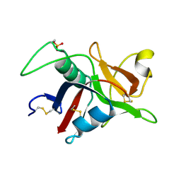
1GZ2
 
 | |
2P3X
 
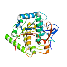 | | Crystal structure of Grenache (Vitis vinifera) Polyphenol Oxidase | | 分子名称: | CU-O-CU LINKAGE, Polyphenol oxidase, chloroplast | | 著者 | Reyes Grajeda, J.P, Virador, V.M, Blanco-Labra, A, Mendiola-Olaya, E, Smith, G.M, Moreno, A, Whitaker, J.R. | | 登録日 | 2007-03-09 | | 公開日 | 2008-03-11 | | 最終更新日 | 2024-10-30 | | 実験手法 | X-RAY DIFFRACTION (2.2 Å) | | 主引用文献 | Cloning, sequencing, purification, and crystal structure of Grenache (Vitis vinifera) polyphenol oxidase.
J.Agric.Food Chem., 58, 2010
|
|
6PAJ
 
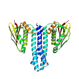 | |
