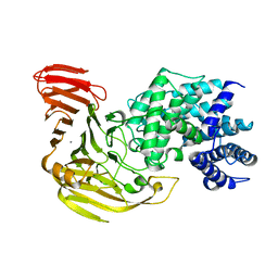
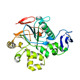
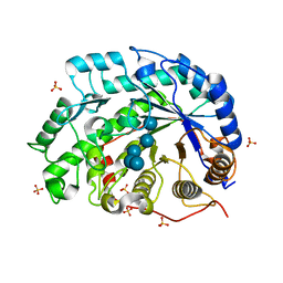
6A4U
 
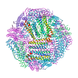 | | The first crystal structure of crustacean ferritin that is a hybrid type of H and L ferritin | | 分子名称: | 1,2-ETHANEDIOL, CHLORIDE ION, Ferritin, ... | | 著者 | Masuda, T, Mikami, B, Zang, J, Zhao, G. | | 登録日 | 2018-06-21 | | 公開日 | 2018-08-22 | | 最終更新日 | 2023-11-22 | | 実験手法 | X-RAY DIFFRACTION (1.16 Å) | | 主引用文献 | The first crystal structure of crustacean ferritin that is a hybrid type of H and L ferritin
Protein Sci., 27, 2018
|
|
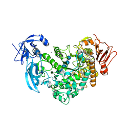
1VAV
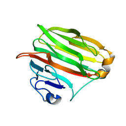 | | Crystal structure of alginate lyase PA1167 from Pseudomonas aeruginosa at 2.0 A resolution | | 分子名称: | Alginate lyase PA1167 | | 著者 | Yamasaki, M, Moriwaki, S, Miyake, O, Hashimoto, W, Murata, K, Mikami, B. | | 登録日 | 2004-02-19 | | 公開日 | 2004-05-25 | | 最終更新日 | 2023-12-27 | | 実験手法 | X-RAY DIFFRACTION (2 Å) | | 主引用文献 | Structure and function of a hypothetical Pseudomonas aeruginosa protein PA1167 classified into family PL-7: a novel alginate lyase with a beta-sandwich fold.
J.Biol.Chem., 279, 2004
|
|
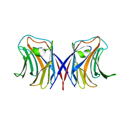
1WW6
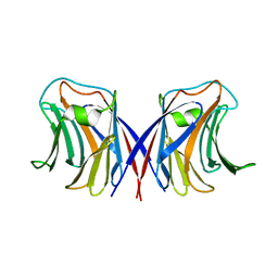 | | Agrocybe cylindracea galectin complexed with lactose | | 分子名称: | beta-D-galactopyranose-(1-4)-beta-D-glucopyranose, galectin | | 著者 | Ban, M, Yoon, H.J, Demirkan, E, Utsumi, S, Mikami, B, Yagi, F. | | 登録日 | 2005-01-03 | | 公開日 | 2005-08-23 | | 最終更新日 | 2024-03-13 | | 実験手法 | X-RAY DIFFRACTION (2.2 Å) | | 主引用文献 | Structural Basis of a Fungal Galectin from Agrocybe cylindracea for Recognizing Sialoconjugate
J.Mol.Biol., 351, 2005
|
|
1WW4
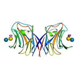 | | Agrocybe cylindracea galectin complexed with NeuAca2-3lactose | | 分子名称: | N-acetyl-alpha-neuraminic acid-(2-3)-alpha-D-galactopyranose-(1-4)-alpha-D-glucopyranose, N-acetyl-alpha-neuraminic acid-(2-3)-beta-D-galactopyranose-(1-4)-beta-D-glucopyranose, galectin | | 著者 | Ban, M, Yoon, H.J, Demirkan, E, Utsumi, S, Mikami, B, Yagi, F. | | 登録日 | 2004-12-31 | | 公開日 | 2005-08-23 | | 最終更新日 | 2024-03-13 | | 実験手法 | X-RAY DIFFRACTION (2.3 Å) | | 主引用文献 | Structural Basis of a Fungal Galectin from Agrocybe cylindracea for Recognizing Sialoconjugate
J.Mol.Biol., 351, 2005
|
|
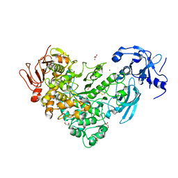
1J0M
 | | Crystal Structure of Bacillus sp. GL1 Xanthan Lyase that Acts on Side Chains of Xanthan | | 分子名称: | CALCIUM ION, XANTHAN LYASE | | 著者 | Hashimoto, W, Nankai, H, Mikami, B, Murata, K. | | 登録日 | 2002-11-19 | | 公開日 | 2003-04-01 | | 最終更新日 | 2023-12-27 | | 実験手法 | X-RAY DIFFRACTION (2.3 Å) | | 主引用文献 | Crystal Structure of Bacillus sp. GL1 Xanthan Lyase, Which Acts on the Side Chains of Xanthan.
J.Biol.Chem., 278, 2003
|
|
6J4H
 | | Crystal Structure of maltotriose-complex of PulA-G680L mutant from Klebsiella pneumoniae | | 分子名称: | CALCIUM ION, GLYCEROL, MAGNESIUM ION, ... | | 著者 | Saka, N, Iwamoto, H, Takahashi, N, Mizutani, K, Mikami, B. | | 登録日 | 2019-01-09 | | 公開日 | 2019-09-18 | | 最終更新日 | 2020-07-29 | | 実験手法 | X-RAY DIFFRACTION (1.641 Å) | | 主引用文献 | Relationship between the induced-fit loop and the activity of Klebsiella pneumoniae pullulanase.
Acta Crystallogr D Struct Biol, 75, 2019
|
|
1WW5
 | | Agrocybe cylindracea galectin complexed with 3'-sulfonyl lactose | | 分子名称: | 3-O-sulfo-beta-D-galactopyranose-(1-4)-beta-D-glucopyranose, galectin | | 著者 | Ban, M, Yoon, H.J, Demirkan, E, Utsumi, S, Mikami, B, Yagi, F. | | 登録日 | 2005-01-03 | | 公開日 | 2005-08-23 | | 最終更新日 | 2024-03-13 | | 実験手法 | X-RAY DIFFRACTION (2.2 Å) | | 主引用文献 | Structural Basis of a Fungal Galectin from Agrocybe cylindracea for Recognizing Sialoconjugate
J.Mol.Biol., 351, 2005
|
|
1KWH
 | | Structure Analysis AlgQ2, a Macromolecule(alginate)-Binding Periplasmic Protein of Sphingomonas sp. A1. | | 分子名称: | CALCIUM ION, Macromolecule-Binding Periplasmic Protein | | 著者 | Momma, K, Mikami, B, Mishima, Y, Hashimoto, W, Murata, K. | | 登録日 | 2002-01-29 | | 公開日 | 2002-02-13 | | 最終更新日 | 2024-03-13 | | 実験手法 | X-RAY DIFFRACTION (2 Å) | | 主引用文献 | Crystal structure of AlgQ2, a macromolecule (alginate)-binding protein of Sphingomonas sp. A1 at 2.0A resolution.
J.Mol.Biol., 316, 2002
|
|
6J34
 | | Crystal Structure of maltotriose-complex of PulA from Klebsiella pneumoniae | | 分子名称: | ACETATE ION, GLYCEROL, MAGNESIUM ION, ... | | 著者 | Saka, N, Iwamoto, H, Takahashi, N, Mizutani, K, Mikami, B. | | 登録日 | 2019-01-09 | | 公開日 | 2019-09-18 | | 最終更新日 | 2023-11-22 | | 実験手法 | X-RAY DIFFRACTION (1.498 Å) | | 主引用文献 | Relationship between the induced-fit loop and the activity of Klebsiella pneumoniae pullulanase.
Acta Crystallogr D Struct Biol, 75, 2019
|
|
1V73
 | | Crystal Structure of Cold-Active Protein-Tyrosine Phosphatase of a Psychrophile Shewanella SP. | | 分子名称: | ACETIC ACID, CALCIUM ION, psychrophilic phosphatase I | | 著者 | Tsuruta, H, Mikami, B, Aizono, Y. | | 登録日 | 2003-12-09 | | 公開日 | 2005-03-01 | | 最終更新日 | 2023-12-27 | | 実験手法 | X-RAY DIFFRACTION (1.82 Å) | | 主引用文献 | Crystal Structure of Cold-Active Protein-Tyrosine Phosphatase from a Psychrophile, Shewanella sp
J.Biochem.(Tokyo), 137, 2005
|
|
1IEJ
 | | OVOTRANSFERRIN, N-TERMINAL LOBE, HOLO FORM, AT 1.65 A RESOLUTION | | 分子名称: | CARBONATE ION, FE (III) ION, OVOTRANSFERRIN | | 著者 | Mizutani, K, Mikami, B, Hirose, M. | | 登録日 | 2001-04-10 | | 公開日 | 2001-06-20 | | 最終更新日 | 2017-10-04 | | 実験手法 | X-RAY DIFFRACTION (1.65 Å) | | 主引用文献 | Domain closure mechanism in transferrins: new viewpoints about the hinge structure and motion as deduced from high resolution crystal structures of ovotransferrin N-lobe.
J.Mol.Biol., 309, 2001
|
|
1J0N
 | | Crystal Structure of Bacillus sp. GL1 Xanthan Lyase that Acts on Side Chains of Xanthan | | 分子名称: | 4,6-O-[(1S)-1-carboxyethylidene]-beta-D-glucopyranose, CALCIUM ION, XANTHAN LYASE | | 著者 | Hashimoto, W, Nankai, H, Mikami, B, Murata, K. | | 登録日 | 2002-11-19 | | 公開日 | 2003-04-01 | | 最終更新日 | 2023-12-27 | | 実験手法 | X-RAY DIFFRACTION (2.4 Å) | | 主引用文献 | Crystal Structure of Bacillus sp. GL1 Xanthan Lyase, Which Acts on the Side Chains of Xanthan.
J.Biol.Chem., 278, 2003
|
|
1TFA
 | | OVOTRANSFERRIN, N-TERMINAL LOBE, APO FORM | | 分子名称: | PROTEIN (OVOTRANSFERRIN), SULFATE ION | | 著者 | Mizutani, K, Yamashita, H, Mikami, B, Hirose, M. | | 登録日 | 1999-01-07 | | 公開日 | 1999-01-13 | | 最終更新日 | 2018-04-04 | | 実験手法 | X-RAY DIFFRACTION (1.9 Å) | | 主引用文献 | Alternative structural state of transferrin. The crystallographic analysis of iron-loaded but domain-opened ovotransferrin N-lobe.
J.Biol.Chem., 274, 1999
|
|
1VD5
 | | Crystal Structure of Unsaturated Glucuronyl Hydrolase, Responsible for the Degradation of Glycosaminoglycan, from Bacillus sp. GL1 at 1.8 A Resolution | | 分子名称: | (4S)-2-METHYL-2,4-PENTANEDIOL, 2,3-DIHYDROXY-1,4-DITHIOBUTANE, GLYCINE, ... | | 著者 | Itoh, T, Akao, S, Hashimoto, W, Mikami, B, Murata, K. | | 登録日 | 2004-03-18 | | 公開日 | 2004-07-13 | | 最終更新日 | 2023-12-27 | | 実験手法 | X-RAY DIFFRACTION (1.8 Å) | | 主引用文献 | Crystal Structure of Unsaturated Glucuronyl Hydrolase, Responsible for the Degradation of Glycosaminoglycan, from Bacillus sp. GL1 at 1.8 A Resolution
J.Biol.Chem., 279, 2004
|
|
1VEN
 | | Crystal Structure Analysis of Y164E/maltose of Bacilus cereus Beta-amylase at pH 4.6 | | 分子名称: | Beta-amylase, CALCIUM ION, alpha-D-glucopyranose | | 著者 | Hirata, A, Adachi, M, Utsumi, S, Mikami, B. | | 登録日 | 2004-04-03 | | 公開日 | 2005-05-24 | | 最終更新日 | 2023-10-25 | | 実験手法 | X-RAY DIFFRACTION (2.02 Å) | | 主引用文献 | Engineering of the pH optimum of Bacillus cereus beta-amylase: conversion of the pH optimum from a bacterial type to a higher-plant type
Biochemistry, 43, 2004
|
|
5XCA
 | | Crystal structure of GH45 endoglucanase EG27II D137A mutant in complex with cellobiose | | 分子名称: | Endo-beta-1,4-glucanase, beta-D-glucopyranose-(1-4)-beta-D-glucopyranose | | 著者 | Nomura, T, Mizutani, K, Iwase, H, Takahashi, N, Mikami, B. | | 登録日 | 2017-03-22 | | 公開日 | 2018-03-28 | | 最終更新日 | 2023-11-22 | | 実験手法 | X-RAY DIFFRACTION (1.35 Å) | | 主引用文献 | High-resolution crystal structures of the glycoside hydrolase family 45 endoglucanase EG27II from the snail Ampullaria crossean.
Acta Crystallogr D Struct Biol, 75, 2019
|
|
5XC8
 | | Crystal structure of GH45 endoglucanase EG27II at pH5.5, in complex with cellobiose | | 分子名称: | Endo-beta-1,4-glucanase, beta-D-glucopyranose-(1-4)-beta-D-glucopyranose | | 著者 | Nomura, T, Mizutani, K, Iwase, H, Takahashi, N, Mikami, B. | | 登録日 | 2017-03-22 | | 公開日 | 2018-03-28 | | 最終更新日 | 2023-11-22 | | 実験手法 | X-RAY DIFFRACTION (1.45 Å) | | 主引用文献 | High-resolution crystal structures of the glycoside hydrolase family 45 endoglucanase EG27II from the snail Ampullaria crossean.
Acta Crystallogr D Struct Biol, 75, 2019
|
|
5XBX
 | | Crystal structure of GH45 endoglucanase EG27II in complex with cellobiose | | 分子名称: | Endo-beta-1,4-glucanase, beta-D-glucopyranose-(1-4)-beta-D-glucopyranose | | 著者 | Nomura, T, Mizutani, K, Iwase, H, Takahashi, N, Mikami, B. | | 登録日 | 2017-03-21 | | 公開日 | 2018-03-28 | | 最終更新日 | 2023-11-22 | | 実験手法 | X-RAY DIFFRACTION (1.04 Å) | | 主引用文献 | High-resolution crystal structures of the glycoside hydrolase family 45 endoglucanase EG27II from the snail Ampullaria crossean.
Acta Crystallogr D Struct Biol, 75, 2019
|
|
5XC4
 | | Crystal structure of GH45 endoglucanase EG27II at pH4.0, in complex with cellobiose | | 分子名称: | Endo-beta-1,4-glucanase, beta-D-glucopyranose-(1-4)-beta-D-glucopyranose | | 著者 | Nomura, T, Mizutani, K, Iwase, H, Takahashi, N, Mikami, B. | | 登録日 | 2017-03-22 | | 公開日 | 2018-03-28 | | 最終更新日 | 2023-11-22 | | 実験手法 | X-RAY DIFFRACTION (1.42 Å) | | 主引用文献 | High-resolution crystal structures of the glycoside hydrolase family 45 endoglucanase EG27II from the snail Ampullaria crossean.
Acta Crystallogr D Struct Biol, 75, 2019
|
|
5XC9
 | | Crystal structure of GH45 endoglucanase EG27II at pH8.0, in complex with cellobiose | | 分子名称: | Endo-beta-1,4-glucanase, beta-D-glucopyranose-(1-4)-beta-D-glucopyranose | | 著者 | Nomura, T, Mizutani, K, Iwase, H, Takahashi, N, Mikami, B. | | 登録日 | 2017-03-22 | | 公開日 | 2018-03-28 | | 最終更新日 | 2023-11-22 | | 実験手法 | X-RAY DIFFRACTION (1.28 Å) | | 主引用文献 | High-resolution crystal structures of the glycoside hydrolase family 45 endoglucanase EG27II from the snail Ampullaria crossean.
Acta Crystallogr D Struct Biol, 75, 2019
|
|
1V3H
 | | The roles of Glu186 and Glu380 in the catalytic reaction of soybean beta-amylase | | 分子名称: | Beta-amylase, SULFATE ION, alpha-D-glucopyranose-(1-4)-alpha-D-glucopyranose-(1-4)-alpha-D-glucopyranose-(1-4)-alpha-D-glucopyranose-(1-4)-alpha-D-glucopyranose | | 著者 | Kang, Y.N, Adachi, M, Utsumi, S, Mikami, B. | | 登録日 | 2003-11-02 | | 公開日 | 2004-06-22 | | 最終更新日 | 2023-10-25 | | 実験手法 | X-RAY DIFFRACTION (1.6 Å) | | 主引用文献 | The Roles of Glu186 and Glu380 in the Catalytic Reaction of Soybean beta-Amylase.
J.Mol.Biol., 339, 2004
|
|
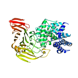
1Y3I
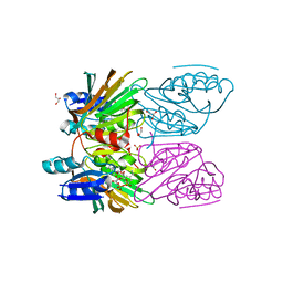 | | Crystal Structure of Mycobacterium tuberculosis NAD kinase-NAD complex | | 分子名称: | GLYCEROL, Inorganic polyphosphate/ATP-NAD kinase, NICOTINAMIDE-ADENINE-DINUCLEOTIDE, ... | | 著者 | Mori, S, Yamasaki, M, Maruyama, Y, Momma, K, Kawai, S, Hashimoto, W, Mikami, B, Murata, K. | | 登録日 | 2004-11-25 | | 公開日 | 2005-01-18 | | 最終更新日 | 2024-03-13 | | 実験手法 | X-RAY DIFFRACTION (2.6 Å) | | 主引用文献 | NAD-binding mode and the significance of intersubunit contact revealed by the crystal structure of Mycobacterium tuberculosis NAD kinase-NAD complex
Biochem.Biophys.Res.Commun., 327, 2005
|
|
5XS8
 | | Crystal structure of solute-binding protein complexed with unsaturated chondroitin disaccharide with two sulfate groups at C-4 and C-6 positions of GalNAc | | 分子名称: | 4-deoxy-alpha-L-threo-hex-4-enopyranuronic acid-(1-3)-2-acetamido-2-deoxy-4,6-di-O-sulfo-beta-D-galactopyranose, CALCIUM ION, Extracellular solute-binding protein family 1 | | 著者 | Oiki, S, Kamochi, R, Mikami, B, Murata, K, Hashimoto, W. | | 登録日 | 2017-06-12 | | 公開日 | 2018-01-17 | | 最終更新日 | 2023-11-22 | | 実験手法 | X-RAY DIFFRACTION (1.952 Å) | | 主引用文献 | Alternative substrate-bound conformation of bacterial solute-binding protein involved in the import of mammalian host glycosaminoglycans.
Sci Rep, 7, 2017
|
|
1VEM
 | | Crystal Structure Analysis of Bacillus Cereus Beta-Amylase at the optimum pH (6.5) | | 分子名称: | Beta-amylase, CALCIUM ION, alpha-D-glucopyranose-(1-4)-alpha-D-glucopyranose | | 著者 | Hirata, A, Adachi, M, Utsumi, S, Mikami, B. | | 登録日 | 2004-04-03 | | 公開日 | 2005-05-24 | | 最終更新日 | 2023-10-25 | | 実験手法 | X-RAY DIFFRACTION (1.85 Å) | | 主引用文献 | Engineering of the pH optimum of Bacillus cereus beta-amylase: conversion of the pH optimum from a bacterial type to a higher-plant type
Biochemistry, 43, 2004
|
|
1WDS
 | | The role of an inner loop in the catalytic mechanism of soybean beta-amylase | | 分子名称: | Beta-amylase, SULFATE ION, alpha-D-glucopyranose, ... | | 著者 | Kang, Y.N, Adachi, M, Utsumi, S, Mikami, B. | | 登録日 | 2004-05-17 | | 公開日 | 2005-04-05 | | 最終更新日 | 2024-05-29 | | 実験手法 | X-RAY DIFFRACTION (1.64 Å) | | 主引用文献 | Structural analysis of threonine 342 mutants of soybean beta-amylase: role of a conformational change of the inner loop in the catalytic mechanism.
Biochemistry, 44, 2005
|
|


