Loading
PDBj
MenuPDBj@FacebookPDBj@TwitterPDBj@YouTubewwPDB FoundationwwPDB
RCSB PDBPDBeBMRBAdv. SearchSearch help
PDB: 54 results

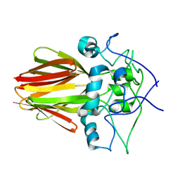
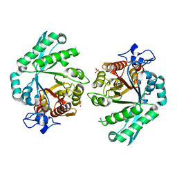
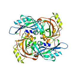
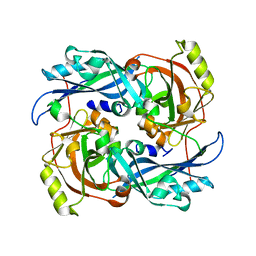
4MGU
DownloadVisualize
BU of 4mgu by Molmil
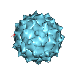
Crystal structure of Acheta domesticus Densovirus
Descriptor: DNA (5'-D(P*TP*TP*T)-3'), Structural protein VP2
Authors:Meng, G, Rossmann, M.G.
Deposit date:2013-08-28
Release date:2013-10-30
Last modified:2024-02-28
Method:X-RAY DIFFRACTION (3.5 Å)
Cite:The structure and host entry of an invertebrate parvovirus.
J.Virol., 87, 2013
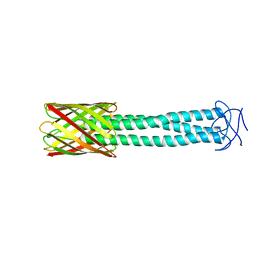
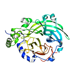
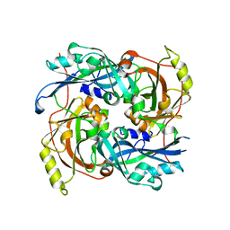
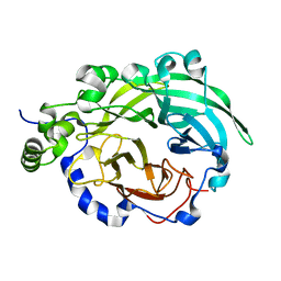
3SYJ
DownloadVisualize
BU of 3syj by Molmil
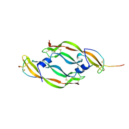
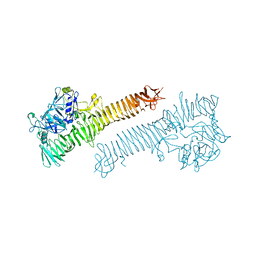
Crystal structure of the Haemophilus influenzae Hap adhesin
Descriptor: Adhesion and penetration protein autotransporter
Authors:Meng, G.
Deposit date:2011-07-18
Release date:2011-08-24
Last modified:2024-10-16
Method:X-RAY DIFFRACTION (2.2 Å)
Cite:Crystal structure of the Haemophilus influenzae Hap adhesin reveals an intercellular oligomerization mechanism for bacterial aggregation
Embo J., 30, 2011
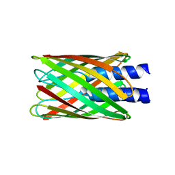
2GR7
DownloadVisualize
BU of 2gr7 by Molmil
Hia 992-1098
Descriptor: (HYDROXYETHYLOXY)TRI(ETHYLOXY)OCTANE, Adhesin
Authors:Meng, G, Waksman, G.
Deposit date:2006-04-23
Release date:2006-05-23
Last modified:2024-02-14
Method:X-RAY DIFFRACTION (2.3 Å)
Cite:Structure of the outer membrane translocator domain of the Haemophilus influenzae Hia trimeric autotransporter.
Embo J., 25, 2006
2GR8
DownloadVisualize
BU of 2gr8 by Molmil
Hia 1022-1098
Descriptor: Adhesin
Authors:Meng, G, Waksman, G.
Deposit date:2006-04-23
Release date:2006-05-23
Last modified:2024-02-14
Method:X-RAY DIFFRACTION (2 Å)
Cite:Structure of the outer membrane translocator domain of the Haemophilus influenzae Hia trimeric autotransporter.
Embo J., 25, 2006
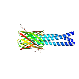
3EMO
DownloadVisualize
BU of 3emo by Molmil
Crystal structure of transmembrane Hia 973-1098
Descriptor: Hia (Adhesin)
Authors:Meng, G, Waksman, G.
Deposit date:2008-09-24
Release date:2008-11-04
Last modified:2023-11-01
Method:X-RAY DIFFRACTION (3 Å)
Cite:Repetitive Architecture of the Haemophilus influenzae Hia Trimeric Autotransporter
J.Mol.Biol., 384, 2008
3EMF
DownloadVisualize
BU of 3emf by Molmil
Crystal structure of Haemophilus influenzae HiaBD2
Descriptor: Hia (Adhesin)
Authors:Meng, G, Waksman, G.
Deposit date:2008-09-24
Release date:2008-11-04
Last modified:2023-11-01
Method:X-RAY DIFFRACTION (2 Å)
Cite:Repetitive Architecture of the Haemophilus influenzae Hia Trimeric Autotransporter
J.Mol.Biol., 384, 2008
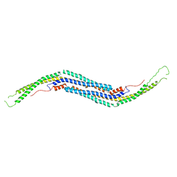
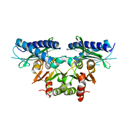
3QE6
DownloadVisualize
BU of 3qe6 by Molmil
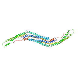
Mouse PACSIN 3 F-BAR domain structure
Descriptor: MAGNESIUM ION, Protein kinase C and casein kinase II substrate protein 3
Authors:Meng, G, Bai, X, Zheng, X.
Deposit date:2011-01-19
Release date:2012-01-25
Last modified:2023-11-01
Method:X-RAY DIFFRACTION (2.6 Å)
Cite:Rigidity of wedge loop in PACSIN 3 protein is a key factor in dictating diameters of tubules
J.Biol.Chem., 287, 2012
3QNI
DownloadVisualize
BU of 3qni by Molmil
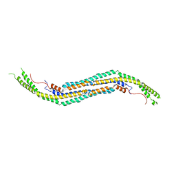
Crystal structure of human PACSIN 1 F-BAR domain
Descriptor: CALCIUM ION, Protein kinase C and casein kinase substrate in neurons protein 1
Authors:Meng, G, Bai, X, Zheng, X.
Deposit date:2011-02-08
Release date:2011-08-10
Last modified:2024-03-20
Method:X-RAY DIFFRACTION (2.8 Å)
Cite:Crystal structure of human PACSIN 1 F-BAR domain
to be published
3EMI
DownloadVisualize
BU of 3emi by Molmil
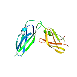
Crystal structure of Hia 307-422 non-adhesive domain
Descriptor: Hia (Adhesin)
Authors:Meng, G, Waksman, G.
Deposit date:2008-09-24
Release date:2008-11-04
Last modified:2024-05-29
Method:X-RAY DIFFRACTION (1.8 Å)
Cite:Repetitive Architecture of the Haemophilus influenzae Hia Trimeric Autotransporter
J.Mol.Biol., 384, 2008
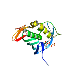
4EQK
DownloadVisualize
BU of 4eqk by Molmil
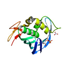
Crystal structure of the cap-binding domain of polymerase basic protein 2 from influenza virus A/Hong Kong/1/68 (h3n2) with bound m7GTP
Descriptor: 7N-METHYL-8-HYDROGUANOSINE-5'-TRIPHOSPHATE, NITRATE ION, Polymerase basic protein 2
Authors:Meng, G, Liu, Y, Zheng, X.
Deposit date:2012-04-19
Release date:2013-03-06
Last modified:2023-11-08
Method:X-RAY DIFFRACTION (1.95 Å)
Cite:Structural and functional characterization of K339T substitution identified in the PB2 subunit cap-binding pocket of influenza A virus
J.Biol.Chem., 288, 2013
4ENF
DownloadVisualize
BU of 4enf by Molmil
Crystal structure of the cap-binding domain of polymerase basic protein 2 from influenza virus A/Puerto Rico/8/34(h1n1)
Descriptor: 1,4-BUTANEDIOL, NITRATE ION, Polymerase basic protein 2
Authors:Meng, G, Liu, Y, Zheng, X.
Deposit date:2012-04-13
Release date:2013-03-06
Last modified:2024-03-20
Method:X-RAY DIFFRACTION (1.32 Å)
Cite:Structural and functional characterization of K339T substitution identified in the PB2 subunit cap-binding pocket of influenza A virus
J.Biol.Chem., 288, 2013
4ES5
DownloadVisualize
BU of 4es5 by Molmil
Crystal structure of the cap-binding domain of polymerase basic protein 2 from influenza virus A/Bar-headed Gs/Qinghai/15c/2005 (h5n1) with bound m7GTP
Descriptor: 7N-METHYL-8-HYDROGUANOSINE-5'-TRIPHOSPHATE, Polymerase basic subunit 2, SULFATE ION
Authors:Meng, G, Liu, Y, Zheng, X.
Deposit date:2012-04-22
Release date:2013-03-06
Last modified:2023-11-08
Method:X-RAY DIFFRACTION (1.8 Å)
Cite:Structural and functional characterization of K339T substitution identified in the PB2 subunit cap-binding pocket of influenza A virus
J.Biol.Chem., 288, 2013
3M3W
DownloadVisualize
BU of 3m3w by Molmil
Crystal structure of mouse PACSIN3 BAR domain mutant
Descriptor: Protein kinase C and casein kinase II substrate protein 3
Authors:Meng, G, Bai, X.Y.
Deposit date:2010-03-10
Release date:2011-03-16
Last modified:2024-03-20
Method:X-RAY DIFFRACTION (2.6 Å)
Cite:Rigidity of wedge loop in PACSIN 3 protein is a key factor in dictating diameters of tubules
J.Biol.Chem., 287, 2012
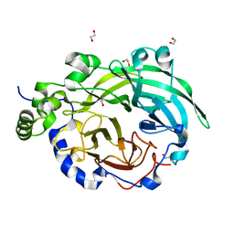
1OYG
DownloadVisualize
BU of 1oyg by Molmil
Crystal structure of Bacillus subtilis levansucrase
Descriptor: 1,2-ETHANEDIOL, CALCIUM ION, levansucrase
Authors:Meng, G, Futterer, K.
Deposit date:2003-04-04
Release date:2003-10-14
Last modified:2024-02-14
Method:X-RAY DIFFRACTION (1.5 Å)
Cite:Structural framework of fructosyl transfer in Bacillus subtilis levansucrase
Nat.Struct.Biol., 10, 2003
1PT2
DownloadVisualize
BU of 1pt2 by Molmil
Crystal structure of levansucrase (E342A) complexed with sucrose
Descriptor: CALCIUM ION, Levansucrase, beta-D-fructofuranose-(2-1)-alpha-D-glucopyranose
Authors:Meng, G, Futterer, K.
Deposit date:2003-06-22
Release date:2003-10-14
Last modified:2023-08-16
Method:X-RAY DIFFRACTION (2.1 Å)
Cite:Structural framework of fructosyl transfer in Bacillus subtilis levansucrase
Nat.Struct.Biol., 10, 2003
3BXW
DownloadVisualize
BU of 3bxw by Molmil
Crystal Structure of Stabilin-1 Interacting Chitinase-Like Protein, SI-CLP
Descriptor: Chitinase domain-containing protein 1, SULFATE ION
Authors:Meng, G, Green, T.J.
Deposit date:2008-01-15
Release date:2009-01-20
Last modified:2024-03-13
Method:X-RAY DIFFRACTION (2.7 Å)
Cite:Structure of human stabilin-1 interacting chitinase-like protein (SI-CLP) reveals a saccharide-binding cleft with lower sugar-binding selectivity.
J.Biol.Chem., 285, 2010
4G1L
DownloadVisualize
BU of 4g1l by Molmil
Crystal structure of Newcastle disease virus matrix protein
Descriptor: Matrix protein
Authors:Meng, G, Rossmann, M.G.
Deposit date:2012-07-10
Release date:2012-08-15
Last modified:2024-02-28
Method:X-RAY DIFFRACTION (2.207 Å)
Cite:Structure and assembly of a paramyxovirus matrix protein.
Proc.Natl.Acad.Sci.USA, 109, 2012
4G1G
DownloadVisualize
BU of 4g1g by Molmil
Crystal structure of Newcastle disease virus matrix protein
Descriptor: Matrix protein
Authors:Meng, G, Rossmann, M.G.
Deposit date:2012-07-10
Release date:2012-08-15
Last modified:2024-02-28
Method:X-RAY DIFFRACTION (2.2 Å)
Cite:Structure and assembly of a paramyxovirus matrix protein.
Proc.Natl.Acad.Sci.USA, 109, 2012
4G1O
DownloadVisualize
BU of 4g1o by Molmil
Crystal structure of Newcastle disease virus matrix protein
Descriptor: Matrix protein
Authors:Meng, G, Rossmann, M.G.
Deposit date:2012-07-11
Release date:2012-08-22
Last modified:2024-02-28
Method:X-RAY DIFFRACTION (2.2 Å)
Cite:Structure and assembly of a paramyxovirus matrix protein.
Proc.Natl.Acad.Sci.USA, 109, 2012
3BYL
DownloadVisualize
BU of 3byl by Molmil
Crystal structure of B. subtilis levansucrase mutant E342A
Descriptor: Levansucrase
Authors:Futterer, K, Meng, G.
Deposit date:2008-01-16
Release date:2008-02-12
Last modified:2023-11-01
Method:X-RAY DIFFRACTION (2.1 Å)
Cite:Donor substrate recognition in the raffinose-bound E342A mutant of fructosyltransferase Bacillus subtilis levansucrase
To be Published
2Q74
DownloadVisualize
BU of 2q74 by Molmil
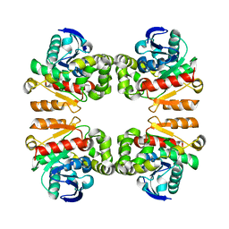
Mycobacterium tuberculosis SuhB
Descriptor: Inositol-1-monophosphatase
Authors:Brown, A.K, Meng, G, Ghadbane, H, Besra, G.S, Futterer, K.
Deposit date:2007-06-06
Release date:2007-10-23
Last modified:2023-08-30
Method:X-RAY DIFFRACTION (2.6 Å)
Cite:Dimerization of inositol monophosphatase Mycobacterium tuberculosis SuhB is not constitutive, but induced by binding of the activator Mg2+
Bmc Struct.Biol., 7, 2007
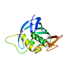
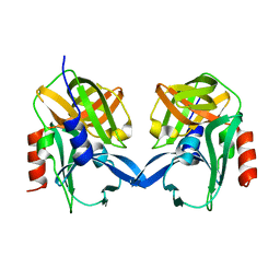
6L0T
DownloadVisualize
BU of 6l0t by Molmil
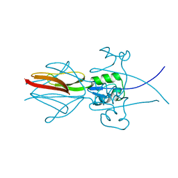
Crystal structure of senecavirus A 3C protease
Descriptor: 3C PROTEASE
Authors:Meng, K.W, Zhang, L.J, Meng, G.
Deposit date:2019-09-27
Release date:2020-09-30
Last modified:2023-11-22
Method:X-RAY DIFFRACTION (1.9 Å)
Cite:Structure of Senecavirus A 3C Protease Revealed the Cleavage Pattern of 3C Protease in Picornaviruses
J.Virol., 2022
6L7M
DownloadVisualize
BU of 6l7m by Molmil
Characterization and structural analysis of a thermostable zearalenone-degrading enzyme
Descriptor: AB hydrolase-1 domain-containing protein
Authors:Wei, X.H, Meng, G.
Deposit date:2019-11-01
Release date:2020-11-04
Last modified:2023-11-22
Method:X-RAY DIFFRACTION (2.4 Å)
Cite:Characterization and structural analysis of a thermostable zearalenone-degrading enzyme
To Be published
3Q48
DownloadVisualize
BU of 3q48 by Molmil
Crystal structure of Pseudomonas aeruginosa CupB2 chaperone
Descriptor: Chaperone CupB2
Authors:Cai, X, Wang, R, Filloux, A, Waksman, G, Meng, G.
Deposit date:2010-12-23
Release date:2011-02-09
Last modified:2023-11-01
Method:X-RAY DIFFRACTION (2.5 Å)
Cite:Structural and functional characterization of Pseudomonas aeruginosa CupB chaperones
Plos One, 6, 2011
1VPP
DownloadVisualize
BU of 1vpp by Molmil
COMPLEX BETWEEN VEGF AND A RECEPTOR BLOCKING PEPTIDE
Descriptor: PROTEIN (PEPTIDE V108), PROTEIN (VASCULAR ENDOTHELIAL GROWTH FACTOR)
Authors:Wiesmann, C, Christinger, H.W, Cochran, A.G, Cunningham, B.C, Fairbrother, W.J, Keenan, C.J, Meng, G, de Vos, A.M.
Deposit date:1998-10-09
Release date:1999-02-23
Last modified:2023-08-23
Method:X-RAY DIFFRACTION (1.9 Å)
Cite:Crystal structure of the complex between VEGF and a receptor-blocking peptide.
Biochemistry, 37, 1998


123>

226707

數據於2024-10-30公開中

PDB statisticsPDBj update infoContact PDBjnumon