Loading
PDBj
MenuPDBj@FacebookPDBj@TwitterPDBj@YouTubewwPDB FoundationwwPDB
RCSB PDBPDBeBMRBAdv. SearchSearch help
PDB: 60 results

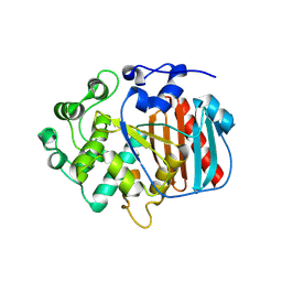
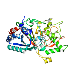
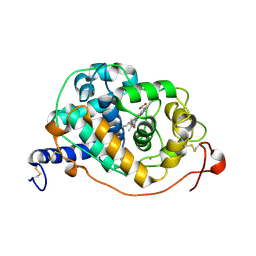
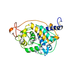
4W7O
DownloadVisualize
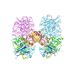
BU of 4w7o by Molmil
CRYSTAL STRUCTURE OF A DECOLORIZING PEROXIDASE (DYP) FROM AURICULARIA AURICULA-JUDAE. G169L, Y147S AND W377S TRIPLE MUTANT
Descriptor: Dye-decolorizing peroxidase, PROTOPORPHYRIN IX CONTAINING FE
Authors:Medrano, F.J, Romero, A.
Deposit date:2014-08-22
Release date:2015-03-11
Last modified:2024-01-10
Method:X-RAY DIFFRACTION (1.2 Å)
Cite:Catalytic surface radical in dye-decolorizing peroxidase: a computational, spectroscopic and site-directed mutagenesis study.
Biochem.J., 466, 2015
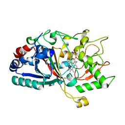
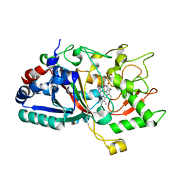
8OY1
DownloadVisualize
BU of 8oy1 by Molmil
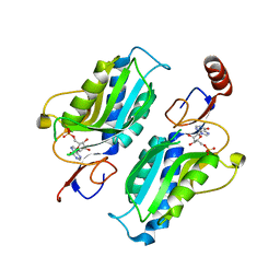
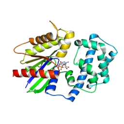
Structure of the human Guanine Nucleotide-Binding Protein G(K) Subunit Alpha
Descriptor: GUANOSINE-5'-DIPHOSPHATE, Guanine nucleotide-binding protein G(i) subunit alpha-3
Authors:Medrano, F.J, Blanco, F.J, Ferreras-Gutierrez, M.O.
Deposit date:2023-05-03
Release date:2024-05-15
Method:X-RAY DIFFRACTION (3.34 Å)
Cite:Structure of the human Guanine Nucleotide-Binding Protein G(K) Subunit Alpha
To Be Published
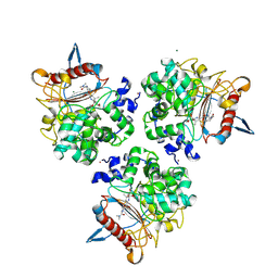
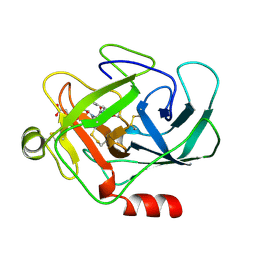
2YMW
DownloadVisualize
BU of 2ymw by Molmil
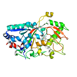
Structure of the epsilon-lysine oxidase from Marinomonas mediterranea
Descriptor: ETHANOL, GLYCEROL, L-LYSINE 6-OXIDASE, ...
Authors:Medrano, F.J, Romero, A.
Deposit date:2012-10-10
Release date:2013-10-23
Last modified:2017-11-08
Method:X-RAY DIFFRACTION (2.41 Å)
Cite:Structure of the Epsilon-Lysine Oxidase from Marinomonas Mediterranea
To be Published
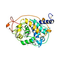
3UOR
DownloadVisualize
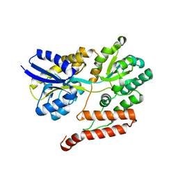
BU of 3uor by Molmil
The structure of the sugar-binding protein MalE from the phytopathogen Xanthomonas citri
Descriptor: ABC transporter sugar binding protein
Authors:Medrano, F.J, Souza, C.S, Balan, A.
Deposit date:2011-11-17
Release date:2011-12-14
Last modified:2024-02-28
Method:X-RAY DIFFRACTION (2.202 Å)
Cite:Structure determination of a sugar-binding protein from the phytopathogenic bacterium Xanthomonas citri.
Acta Crystallogr F Struct Biol Commun, 70, 2014
4W7L
DownloadVisualize
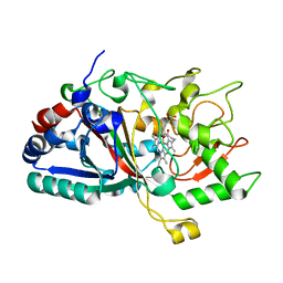
BU of 4w7l by Molmil
CRYSTAL STRUCTURE OF A DECOLORIZING PEROXIDASE (DYP) FROM AURICULARIA AURICULA-JUDAE. D168N MUTANT
Descriptor: Dye-decolorizing peroxidase, PROTOPORPHYRIN IX CONTAINING FE
Authors:Medrano, F.J, Romero, A.
Deposit date:2014-08-22
Release date:2015-03-11
Last modified:2024-01-10
Method:X-RAY DIFFRACTION (1.05 Å)
Cite:Catalytic surface radical in dye-decolorizing peroxidase: a computational, spectroscopic and site-directed mutagenesis study.
Biochem.J., 466, 2015
4W7N
DownloadVisualize
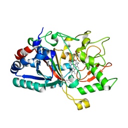
BU of 4w7n by Molmil
CRYSTAL STRUCTURE OF A DECOLORIZING PEROXIDASE (DYP) FROM AURICULARIA AURICULA-JUDAE. Y147S AND W377S DOUBLE MUTANT
Descriptor: Dye-decolorizing peroxidase, PROTOPORPHYRIN IX CONTAINING FE
Authors:Medrano, F.J, Romero, A.
Deposit date:2014-08-22
Release date:2015-03-11
Last modified:2024-01-10
Method:X-RAY DIFFRACTION (1.401 Å)
Cite:Catalytic surface radical in dye-decolorizing peroxidase: a computational, spectroscopic and site-directed mutagenesis study.
Biochem.J., 466, 2015
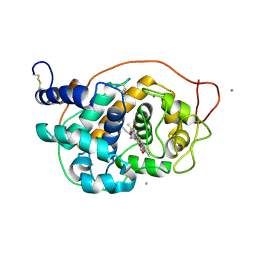
8PAQ
DownloadVisualize
BU of 8paq by Molmil
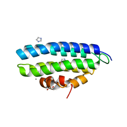
Structure of the small subunit of the laccase-like Nlac protein from Pleurotus eryngii
Descriptor: 1,2-ETHANEDIOL, CALCIUM ION, IMIDAZOLE, ...
Authors:Medrano, F.J, Camarero, S.
Deposit date:2023-06-08
Release date:2024-02-07
Method:X-RAY DIFFRACTION (1.6 Å)
Cite:Role and structure of the small subunit forming heterodimers with laccase-like enzymes.
Protein Sci., 32, 2023
1AZW
DownloadVisualize
BU of 1azw by Molmil
PROLINE IMINOPEPTIDASE FROM XANTHOMONAS CAMPESTRIS PV. CITRI
Descriptor: PROLINE IMINOPEPTIDASE
Authors:Medrano, F.J, Alonso, J, Garcia, J.L, Romero, A, Bode, W, Gomis-Ruth, F.X.
Deposit date:1997-11-22
Release date:1999-01-13
Last modified:2024-02-07
Method:X-RAY DIFFRACTION (2.7 Å)
Cite:Structure of proline iminopeptidase from Xanthomonas campestris pv. citri: a prototype for the prolyl oligopeptidase family.
EMBO J., 17, 1998
1BQB
DownloadVisualize
BU of 1bqb by Molmil
AUREOLYSIN, STAPHYLOCOCCUS AUREUS METALLOPROTEINASE
Descriptor: CALCIUM ION, PROTEIN (AUREOLYSIN), ZINC ION
Authors:Medrano, F.J, Banbula, A, Potempa, J, Travis, J, Bode, W.
Deposit date:1998-07-14
Release date:1999-01-13
Last modified:2023-08-09
Method:X-RAY DIFFRACTION (1.72 Å)
Cite:Amino-acid sequence and three-dimensional structure of the Staphylococcus aureus metalloproteinase at 1.72 A resolution.
Structure, 6, 1998
1P19
DownloadVisualize
BU of 1p19 by Molmil
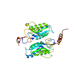
Hypoxanthine Phosphoribosyltransferase from Trypanosoma cruzi, in complex with the product IMP
Descriptor: INOSINIC ACID, hypoxanthine phosphoribosyltransferase
Authors:Medrano, F.J, Eakin, A.E, Craig III, S.P.
Deposit date:2003-04-11
Release date:2004-05-18
Last modified:2024-02-14
Method:X-RAY DIFFRACTION (2.3 Å)
Cite:Interactions at the dimer interface influence the relative efficiencies for purine nucleotide synthesis and pyrophosphorolysis in a phosphoribosyltransferase.
J.Mol.Biol., 335, 2004
1P17
DownloadVisualize
BU of 1p17 by Molmil
Hypoxanthine Phosphoribosyltransferase from Trypanosoma cruzi, K68R mutant, complexed with the product IMP
Descriptor: INOSINIC ACID, hypoxanthine phosphoribosyltransferase
Authors:Medrano, F.J, Eakin, A.E, Craig III, S.P.
Deposit date:2003-04-11
Release date:2004-05-18
Last modified:2024-02-14
Method:X-RAY DIFFRACTION (2.7 Å)
Cite:Interactions at the dimer interface influence the relative efficiencies for purine nucleotide synthesis and pyrophosphorolysis in a phosphoribosyltransferase.
J.Mol.Biol., 335, 2004
8RLJ
DownloadVisualize
BU of 8rlj by Molmil
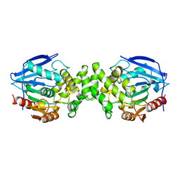
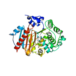
Structure of the apo form of PIB-1 in an Orthorombic space group
Descriptor: Class C beta-lactamase-related serine hydrolase
Authors:Medrano, F.J, Romero, A.
Deposit date:2024-01-03
Release date:2024-08-14
Last modified:2024-09-18
Method:X-RAY DIFFRACTION (2.45 Å)
Cite:A new type of Class C beta-lactamases defined by PIB-1. A metal-dependent carbapenem-hydrolyzing beta-lactamase, from Pseudomonas aeruginosa: Structural and functional analysis.
Int.J.Biol.Macromol., 277, 2024
8RLL
DownloadVisualize
BU of 8rll by Molmil
Structure of the apo form of PIB-1 in an Orthorombic space group
Descriptor: Class C beta-lactamase-related serine hydrolase
Authors:Medrano, F.J, Romero, A.
Deposit date:2024-01-03
Release date:2024-08-14
Last modified:2024-09-18
Method:X-RAY DIFFRACTION (2.72 Å)
Cite:A new type of Class C beta-lactamases defined by PIB-1. A metal-dependent carbapenem-hydrolyzing beta-lactamase, from Pseudomonas aeruginosa: Structural and functional analysis.
Int.J.Biol.Macromol., 277, 2024
8RLK
DownloadVisualize
BU of 8rlk by Molmil
Structure of the apo form of PIB-1 in an Orthorombic space group
Descriptor: (4R,5S)-3-{[(3S,5S)-5-(dimethylcarbamoyl)pyrrolidin-3-yl]sulfanyl}-5-[(2S,3R)-3-hydroxy-1-oxobutan-2-yl]-4-methyl-4,5-d ihydro-1H-pyrrole-2-carboxylic acid, Class C beta-lactamase-related serine hydrolase, MAGNESIUM ION, ...
Authors:Medrano, F.J, Romero, A.
Deposit date:2024-01-03
Release date:2024-08-14
Last modified:2024-09-18
Method:X-RAY DIFFRACTION (2 Å)
Cite:A new type of Class C beta-lactamases defined by PIB-1. A metal-dependent carbapenem-hydrolyzing beta-lactamase, from Pseudomonas aeruginosa: Structural and functional analysis.
Int.J.Biol.Macromol., 277, 2024
4W7J
DownloadVisualize
BU of 4w7j by Molmil
CRYSTAL STRUCTURE OF A DECOLORIZING PEROXIDASE (DYP) FROM AURICULARIA AURICULA-JUDAE
Descriptor: Dye-decolorizing peroxidase, PROTOPORPHYRIN IX CONTAINING FE
Authors:Medrano, F.J, Romero, A.
Deposit date:2014-08-22
Release date:2015-03-11
Last modified:2024-01-10
Method:X-RAY DIFFRACTION (1.79 Å)
Cite:Catalytic surface radical in dye-decolorizing peroxidase: a computational, spectroscopic and site-directed mutagenesis study.
Biochem.J., 466, 2015
4W7K
DownloadVisualize
BU of 4w7k by Molmil
CRYSTAL STRUCTURE OF A DECOLORIZING PEROXIDASE (DYP) FROM AURICULARIA AURICULA-JUDAE. Y147S MUTANT
Descriptor: Dye-decolorizing peroxidase, PROTOPORPHYRIN IX CONTAINING FE
Authors:Medrano, F.J, Romero, A.
Deposit date:2014-08-22
Release date:2015-03-11
Last modified:2024-01-10
Method:X-RAY DIFFRACTION (1.05 Å)
Cite:Catalytic surface radical in dye-decolorizing peroxidase: a computational, spectroscopic and site-directed mutagenesis study.
Biochem.J., 466, 2015
4W7M
DownloadVisualize
BU of 4w7m by Molmil
CRYSTAL STRUCTURE OF A DECOLORIZING PEROXIDASE (DYP) FROM AURICULARIA AURICULA-JUDAE. W377S MUTANT
Descriptor: Dye-decolorizing peroxidase, PROTOPORPHYRIN IX CONTAINING FE
Authors:Medrano, F.J, Romero, A.
Deposit date:2014-08-22
Release date:2015-03-11
Last modified:2024-01-10
Method:X-RAY DIFFRACTION (1.15 Å)
Cite:Catalytic surface radical in dye-decolorizing peroxidase: a computational, spectroscopic and site-directed mutagenesis study.
Biochem.J., 466, 2015
1AU8
DownloadVisualize
BU of 1au8 by Molmil
HUMAN CATHEPSIN G
Descriptor: CATHEPSIN G, N-(3-carboxypropanoyl)-L-valyl-N-[(1R)-5-amino-1-phosphonopentyl]-L-prolinamide
Authors:Medrano, F.J, Bode, W, Banbula, A, Potempa, J.
Deposit date:1997-09-12
Release date:1998-10-14
Last modified:2012-12-12
Method:X-RAY DIFFRACTION (1.9 Å)
Cite:HUMAN CATHEPSIN G
to be published
5ABN
DownloadVisualize
BU of 5abn by Molmil
CRYSTAL STRUCTURE ANALYSIS OF FUNGAL VERSATILE PEROXIDASE FROM PLEUROTUS ERYNGII. MUTANT VPi. MUTATED RESIDUES D69S, T70D, S86E, D146T, Q202L, H232E, Q239R AND S301K.
Descriptor: CALCIUM ION, MAGNESIUM ION, PROTOPORPHYRIN IX CONTAINING FE, ...
Authors:Medrano, F.J, Romero, A.
Deposit date:2015-08-07
Release date:2015-11-04
Last modified:2024-01-10
Method:X-RAY DIFFRACTION (2.194 Å)
Cite:Improving the Ph-Stability of Versatile Peroxidase by Comparative Structural Analysis with a Naturally-Stable Manganese Peroxidase.
Plos One, 10, 2015
5ABO
DownloadVisualize
BU of 5abo by Molmil
CRYSTAL STRUCTURE ANALYSIS OF FUNGAL VERSATILE PEROXIDASE FROM PLEUROTUS ERYNGII. MUTANT VPi-br. MUTATED RESIDUES T2K, D69S, T70D, S86E, A131K, D146T, Q202L, Q219K, H232E, Q239R, L288R, S301K, A308R, A309K AND A314R.
Descriptor: CALCIUM ION, PROTOPORPHYRIN IX CONTAINING FE, VERSATILE PEROXIDASE VPL2
Authors:Medrano, F.J, Romero, A.
Deposit date:2015-08-07
Release date:2015-11-04
Last modified:2024-10-23
Method:X-RAY DIFFRACTION (1.095 Å)
Cite:Improving the Ph-Stability of Versatile Peroxidase by Comparative Structural Analysis with a Naturally-Stable Manganese Peroxidase.
Plos One, 10, 2015
5ABQ
DownloadVisualize
BU of 5abq by Molmil
CRYSTAL STRUCTURE ANALYSIS OF FUNGAL VERSATILE PEROXIDASE FROM PLEUROTUS ERYNGII. MUTANT VPi-SS. MUTATED RESIDUES T2K, A49C, A61C, D69S, T70D, S86E, A131K, D146T, Q202L, Q219K, H232E, Q239R, L288R, S301K, A308R,A309K AND A314R.
Descriptor: CALCIUM ION, PROTOPORPHYRIN IX CONTAINING FE, VERSATILE PEROXIDASE
Authors:Medrano, F.J, Romero, A.
Deposit date:2015-08-07
Release date:2015-11-04
Last modified:2024-10-23
Method:X-RAY DIFFRACTION (2.293 Å)
Cite:Improving the Ph-Stability of Versatile Peroxidase by Comparative Structural Analysis with a Naturally-Stable Manganese Peroxidase.
Plos One, 10, 2015
5ANH
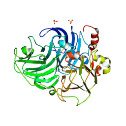
DownloadVisualize
BU of 5anh by Molmil
CRYSTAL STRUCTURE OF LACCASE FROM BASIDIOMYCETE PM1 (CECT 2971)
Descriptor: COPPER (II) ION, LACCASE, SULFATE ION
Authors:Medrano, F.J, Romero, A.
Deposit date:2015-09-07
Release date:2016-08-24
Last modified:2024-01-10
Method:X-RAY DIFFRACTION (2.492 Å)
Cite:Re-Designing the Substrate Binding Pocket of Laccase for Enhanced Oxidation of Sinapic Acid
Catal.Sci..Technol, 6, 2016
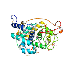
5FNE
DownloadVisualize
BU of 5fne by Molmil
CRYSTAL STRUCTURE OF FUNGAL VERSATILE PEROXIDASE FROM PLEUROTUS ERYNGII TRIPLE MUTANT E37K, H39R & G330R
Descriptor: CALCIUM ION, PROTOPORPHYRIN IX CONTAINING FE, SULFATE ION, ...
Authors:Medrano, F.J, Romero, A.
Deposit date:2015-11-13
Release date:2016-07-13
Last modified:2024-01-10
Method:X-RAY DIFFRACTION (1.499 Å)
Cite:Unveiling the Basis of Alkaline Stability of an Evolved Versatile Peroxidase.
Biochem.J., 473, 2016
5FNB
DownloadVisualize
BU of 5fnb by Molmil
CRYSTAL STRUCTURE OF FUNGAL VERSATILE PEROXIDASE FROM PLEUROTUS ERYNGII SEPTUPLE MUTANT E37K, H39R, V160A, T184M, Q202L, D213A & G330R
Descriptor: CALCIUM ION, PROTOPORPHYRIN IX CONTAINING FE, SULFATE ION, ...
Authors:Medrano, F.J, Romero, A.
Deposit date:2015-11-13
Release date:2016-07-13
Last modified:2024-01-10
Method:X-RAY DIFFRACTION (1.792 Å)
Cite:Unveiling the Basis of Alkaline Stability of an Evolved Versatile Peroxidase.
Biochem.J., 473, 2016
8BFO
DownloadVisualize
BU of 8bfo by Molmil
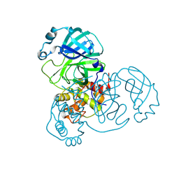
Structure of the apo form of Mpro from SARS-CoV-2
Descriptor: 3C-like proteinase nsp5
Authors:Medrano, F.J, Romero, A.
Deposit date:2022-10-26
Release date:2023-11-08
Method:X-RAY DIFFRACTION (1.99 Å)
Cite:Peptidyl Nitroalkenes Inhibit SARS-CoV-2 Main Protease and Block Virus Replication
To Be Published


123>

226707

건을2024-10-30부터공개중

PDB statisticsPDBj update infoContact PDBjnumon