Loading
PDBj
MenuPDBj@FacebookPDBj@TwitterPDBj@YouTubewwPDB FoundationwwPDB
RCSB PDBPDBeBMRBAdv. SearchSearch help
PDB: 62 results

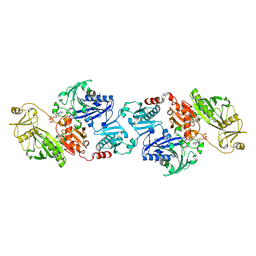
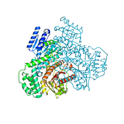
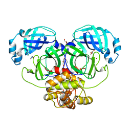
2YNB
DownloadVisualize
BU of 2ynb by Molmil
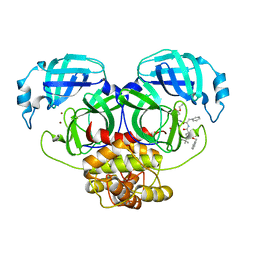
Crystal structure of the main protease of coronavirus HKU4 in complex with a Michael acceptor SG85
Descriptor: 3C-LIKE PROTEINASE, N-[(benzyloxy)carbonyl]-O-tert-butyl-L-seryl-N-{(2R)-5-ethoxy-5-oxo-1-[(3S)-2-oxopyrrolidin-3-yl]pentan-2-yl}-L-phenylalaninamide, NICKEL (II) ION
Authors:Ma, Q, Xiao, Y, Hilgenfeld, R.
Deposit date:2012-10-13
Release date:2013-10-23
Last modified:2024-10-23
Method:X-RAY DIFFRACTION (1.96 Å)
Cite:Inhibitor for the Main Protease of Coronavirus Hku4
To be Published
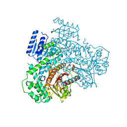
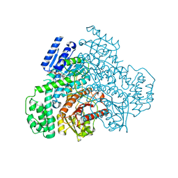
2YNA
DownloadVisualize
BU of 2yna by Molmil
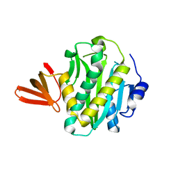
Crystal structure of the main protease of coronavirus HKU4
Descriptor: 3C-LIKE PROTEINASE, GLYCEROL, IMIDAZOLE, ...
Authors:Ma, Q, Xiao, Y, Hilgenfeld, R.
Deposit date:2012-10-13
Release date:2013-10-23
Last modified:2023-12-20
Method:X-RAY DIFFRACTION (1.5 Å)
Cite:Inhibitor for the Main Protease of Coronavirus Hku4
To be Published
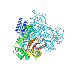
2CGQ
DownloadVisualize
BU of 2cgq by Molmil
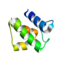
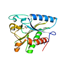
a putative acyl carrier protein(Rv0033) from Mycobacterium tuberculosis
Descriptor: ACYL CARRIER PROTEIN ACPA
Authors:Ma, Q, Wilmanns, M.
Deposit date:2006-03-09
Release date:2007-05-01
Last modified:2023-12-13
Method:X-RAY DIFFRACTION (1.83 Å)
Cite:The Structure of a Putative Acyl Carrier Protein (Rv0033) from Mycobacterium Tuberculosis
To be Published
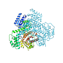
2CGH
DownloadVisualize
BU of 2cgh by Molmil
crystal structure of biotin ligase from Mycobacterium tuberculosis
Descriptor: BIOTIN LIGASE
Authors:Ma, Q, Wilmanns, M.
Deposit date:2006-03-06
Release date:2007-05-01
Last modified:2023-12-13
Method:X-RAY DIFFRACTION (1.8 Å)
Cite:Active Site Conformational Changes Upon Reaction Intermediate Biotinyl-5'-AMP Binding in Biotin Protein Ligase from Mycobacterium Tuberculosis.
Protein Sci., 23, 2014
2X3X
DownloadVisualize
BU of 2x3x by Molmil
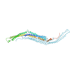
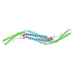
structure of mouse syndapin I (crystal form 1)
Descriptor: PROTEIN KINASE C AND CASEIN KINASE SUBSTRATE IN NEURONS PROTEIN 1
Authors:Ma, Q, Rao, Y, Vahedi-Faridi, A, Saenger, W, Haucke, V.
Deposit date:2010-01-28
Release date:2010-04-07
Last modified:2024-05-08
Method:X-RAY DIFFRACTION (3.35 Å)
Cite:Molecular Basis for SH3 Domain Regulation of F-Bar-Mediated Membrane Deformation.
Proc.Natl.Acad.Sci.USA, 107, 2010
2CHC
DownloadVisualize
BU of 2chc by Molmil
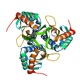
Structure of Rv3472(D26N), a function unknown protein from Mycobacterium tuberculosis
Descriptor: PROTEIN RV3472
Authors:Ma, Q, Wilmanns, M.
Deposit date:2006-03-13
Release date:2007-05-01
Last modified:2024-05-08
Method:X-RAY DIFFRACTION (1.69 Å)
Cite:Structure of Rv3472(D26N), a Function Unknown Protein from Mycobacterium Tuberculosis
To be Published
2FGH
DownloadVisualize
BU of 2fgh by Molmil
ATP bound gelsolin
Descriptor: ADENOSINE-5'-TRIPHOSPHATE, gelsolin
Authors:Ma, Q, Robinson, R.C, Burtnick, L.D, Urosev, D.
Deposit date:2005-12-22
Release date:2006-04-18
Last modified:2017-12-20
Method:X-RAY DIFFRACTION (2.8 Å)
Cite:The structure of gelsolin bound to ATP
J.Mol.Biol., 357, 2006
8J29
DownloadVisualize
BU of 8j29 by Molmil
Structures of the C-terminal subenzyme of the malonyl-CoA reductase from Chloroflexus aurantiacus
Descriptor: GLYCEROL, L(+)-TARTARIC ACID, Short-chain dehydrogenase/reductase SDR
Authors:Ma, Q, Liu, C.
Deposit date:2023-04-14
Release date:2024-04-17
Method:X-RAY DIFFRACTION (1.88 Å)
Cite:Structures of the C-terminal subenzyme of the malonyl-CoA reductase from Chloroflexus aurantiacus
To Be Published
8J2A
DownloadVisualize
BU of 8j2a by Molmil
Structure of the C-terminal subenzyme of the malonyl-CoA reductase from Chloroflexus aurantiacus, in complex with NADP+
Descriptor: GLYCEROL, MAGNESIUM ION, NADP NICOTINAMIDE-ADENINE-DINUCLEOTIDE PHOSPHATE, ...
Authors:Ma, Q, Liu, C.
Deposit date:2023-04-14
Release date:2024-04-17
Method:X-RAY DIFFRACTION (1.7 Å)
Cite:Structures of the C-terminal subenzyme of the malonyl-CoA reductase from Chloroflexus aurantiacus
To Be Published
8J2D
DownloadVisualize
BU of 8j2d by Molmil
Structure of the C-terminal subenzyme of the malonyl-CoA reductase from Chloroflexus aurantiacus, mutant N940V/K1106W/S1114R in complex with NADP+
Descriptor: GLYCEROL, NADP NICOTINAMIDE-ADENINE-DINUCLEOTIDE PHOSPHATE, Short-chain dehydrogenase/reductase SDR
Authors:Ma, Q, Liu, C.
Deposit date:2023-04-14
Release date:2024-04-17
Method:X-RAY DIFFRACTION (2.21 Å)
Cite:Structures of the C-terminal subenzyme of the malonyl-CoA reductase from Chloroflexus aurantiacus
To Be Published
8J2C
DownloadVisualize
BU of 8j2c by Molmil
Structure of the C-terminal subenzyme of the malonyl-CoA reductase from Chloroflexus aurantiacus, mutant N940V/K1106W/S1114R
Descriptor: GLYCEROL, L(+)-TARTARIC ACID, Short-chain dehydrogenase/reductase SDR
Authors:Ma, Q, Liu, C.
Deposit date:2023-04-14
Release date:2024-04-17
Method:X-RAY DIFFRACTION (1.7 Å)
Cite:Structures of the C-terminal subenzyme of the malonyl-CoA reductase from Chloroflexus aurantiacus
To Be Published
8J2E
DownloadVisualize
BU of 8j2e by Molmil
Structure of the C-terminal subenzyme of the malonyl-CoA reductase from Chloroflexus aurantiacus, mutant N940V/K1106W/S1114R in complex with NADP+ and malonate
Descriptor: MALONIC ACID, NADP NICOTINAMIDE-ADENINE-DINUCLEOTIDE PHOSPHATE, Short-chain dehydrogenase/reductase SDR
Authors:Ma, Q, Liu, C.
Deposit date:2023-04-14
Release date:2024-04-17
Method:X-RAY DIFFRACTION (2.12 Å)
Cite:Structures of the C-terminal subenzyme of the malonyl-CoA reductase from Chloroflexus aurantiacus
To Be Published
8J2B
DownloadVisualize
BU of 8j2b by Molmil
Structure of the C-terminal subenzyme of the malonyl-CoA reductase from Chloroflexus aurantiacus, in complex with NADP+ and malonate
Descriptor: MALONIC ACID, NADP NICOTINAMIDE-ADENINE-DINUCLEOTIDE PHOSPHATE, Short-chain dehydrogenase/reductase SDR
Authors:Ma, Q, Liu, C.
Deposit date:2023-04-14
Release date:2024-04-17
Method:X-RAY DIFFRACTION (2.13 Å)
Cite:Structures of the C-terminal subenzyme of the malonyl-CoA reductase from Chloroflexus aurantiacus
To Be Published
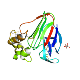
1RQW
DownloadVisualize
BU of 1rqw by Molmil
Thaumatin Structure at 1.05 A Resolution
Descriptor: L(+)-TARTARIC ACID, thaumatin I
Authors:Ma, Q, Sheldrick, G.M.
Deposit date:2003-12-07
Release date:2003-12-23
Last modified:2024-10-16
Method:X-RAY DIFFRACTION (1.05 Å)
Cite:Thaumatin Structure at 1.05 A Resolution
To be Published
2X3V
DownloadVisualize
BU of 2x3v by Molmil
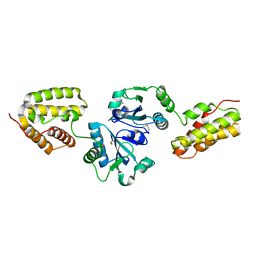
Structure of The F-BAR Domain of Mouse Syndapin I
Descriptor: PROTEIN KINASE C AND CASEIN KINASE SUBSTRATE IN NEURONS PROTEIN 1
Authors:Ma, Q, Rao, Y, Vahedi-Faridi, A, Saenger, W, Haucke, V.
Deposit date:2010-01-27
Release date:2010-04-07
Last modified:2024-05-08
Method:X-RAY DIFFRACTION (2.45 Å)
Cite:Molecular Basis for SH3 Domain Regulation of F-Bar-Mediated Membrane Deformation.
Proc.Natl.Acad.Sci.USA, 107, 2010
1OVN
DownloadVisualize
BU of 1ovn by Molmil
Crystal Structure and Functional Analysis of Drosophila Wind-- a PDI-Related Protein
Descriptor: CESIUM ION, Windbeutel
Authors:Ma, Q, Guo, C, Barnewitz, K, Sheldrick, G.M, Soling, H.D, Uson, I, Ferrari, D.M.
Deposit date:2003-03-27
Release date:2004-02-24
Last modified:2017-10-11
Method:X-RAY DIFFRACTION (1.9 Å)
Cite:Crystal structure and functional analysis of Drosophila Wind, a protein-disulfide isomerase-related protein.
J.Biol.Chem., 278, 2003
2X3W
DownloadVisualize
BU of 2x3w by Molmil
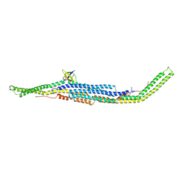
structure of mouse syndapin I (crystal form 2)
Descriptor: PROTEIN KINASE C AND CASEIN KINASE SUBSTRATE IN NEURONS PROTEIN 1
Authors:Ma, Q, Rao, Y, Saenger, W, Haucke, V.
Deposit date:2010-01-28
Release date:2010-04-07
Last modified:2024-05-08
Method:X-RAY DIFFRACTION (2.64 Å)
Cite:Molecular Basis for SH3 Domain Regulation of F-Bar-Mediated Membrane Deformation.
Proc.Natl.Acad.Sci.USA, 107, 2010
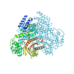
5NFS
DownloadVisualize
BU of 5nfs by Molmil
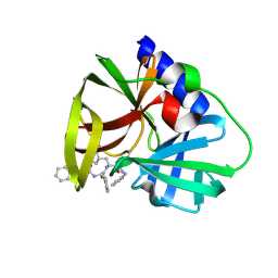
Structure of coxsackievirus B3 3C protease in complex with the alpha-ketoamide (S)-N-benzyl-3-((S)-2-cinnamamido-3-phenylpropanamido)-2-oxo-4-((S)-2-oxopyrrolidin-3-yl)butanamide (cinnamoyl-phenylalanine-GlnLactam-CO-CO-NH-benzyl)
Descriptor: (S)-N-benzyl-3-((S)-2-cinnamamido-3-phenylpropanamido)-2-oxo-4-((S)-2-oxopyrrolidin-3-yl)butanamide, Genome polyprotein
Authors:Ma, Q, Zhang, L, Hilgenfeld, R.
Deposit date:2017-03-15
Release date:2018-05-16
Last modified:2024-10-23
Method:X-RAY DIFFRACTION (1.8 Å)
Cite:Alpha-ketoamides as broad-spectrum inhibitors of coronavirus and enterovirus replication
To Be Published
1W66
DownloadVisualize
BU of 1w66 by Molmil
Structure of a lipoate-protein ligase b from Mycobacterium tuberculosis
Descriptor: DECANOIC ACID, LIPOYLTRANSFERASE
Authors:Ma, Q, Wilmanns, M.
Deposit date:2004-08-13
Release date:2005-12-08
Last modified:2019-07-24
Method:X-RAY DIFFRACTION (1.08 Å)
Cite:The Mycobacterium Tuberculosis Lipb Enzyme Functions as a Cysteine/Lysine Dyad Acyltransferase.
Proc.Natl.Acad.Sci.USA, 103, 2006
7XJM
DownloadVisualize
BU of 7xjm by Molmil
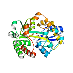
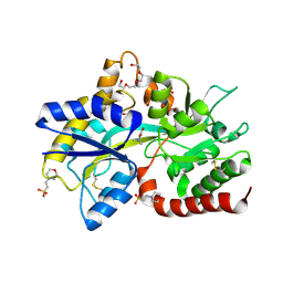
Structure of VcPotD1 in complex with spermidine
Descriptor: HEXAETHYLENE GLYCOL, Putrescine-binding periplasmic protein, SPERMIDINE, ...
Authors:Ma, Q, Liu, C.
Deposit date:2022-04-18
Release date:2023-04-26
Last modified:2023-11-29
Method:X-RAY DIFFRACTION (1.755 Å)
Cite:structure of VcPotD1 in complex with spermidine
To Be Published
7XJN
DownloadVisualize
BU of 7xjn by Molmil
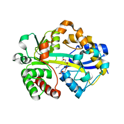
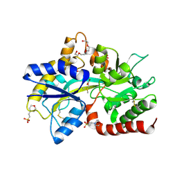
Structure of VcPotD1 in complex with norspermidine
Descriptor: HEXAETHYLENE GLYCOL, N-(3-aminopropyl)propane-1,3-diamine, Putrescine-binding periplasmic protein, ...
Authors:Ma, Q, Liu, C.
Deposit date:2022-04-18
Release date:2023-04-26
Last modified:2023-11-29
Method:X-RAY DIFFRACTION (1.79 Å)
Cite:Structure of VcPotD1 in complex with norspermidine
To Be Published
7XLZ
DownloadVisualize
BU of 7xlz by Molmil
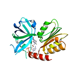
Structure of siderophore-interacting protein from Vibrio anguillarum
Descriptor: FLAVIN-ADENINE DINUCLEOTIDE, Siderophore-interacting protein
Authors:Ma, Q, Liu, C, Han, Y.
Deposit date:2022-04-23
Release date:2023-05-31
Last modified:2023-11-29
Method:X-RAY DIFFRACTION (1.19 Å)
Cite:Structure of siderophore-interacting protein from Vibrio anguillarum
To Be Published
8JV3
DownloadVisualize
BU of 8jv3 by Molmil
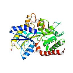
Structure of the SAR11 PotD in complex with glycine betaine
Descriptor: 2-(N-MORPHOLINO)-ETHANESULFONIC ACID, GLYCEROL, Spermidine/putrescine-binding periplasmic protein, ...
Authors:Ma, Q, Liu, C.
Deposit date:2023-06-27
Release date:2024-07-03
Method:X-RAY DIFFRACTION (1.41 Å)
Cite:Structure of the SAR11 PotD in complex with glycine betaine
To Be Published
8JV4
DownloadVisualize
BU of 8jv4 by Molmil
Structure of the SAR11 PotD in complex with DMSP
Descriptor: 2-(N-MORPHOLINO)-ETHANESULFONIC ACID, 3-(dimethyl-lambda~4~-sulfanyl)propanoic acid, GLYCEROL, ...
Authors:Ma, Q, Liu, C.
Deposit date:2023-06-27
Release date:2024-07-03
Last modified:2024-10-23
Method:X-RAY DIFFRACTION (1.445 Å)
Cite:Structure of the SAR11 PotD in complex with DMSP
To Be Published
8JV1
DownloadVisualize
BU of 8jv1 by Molmil
Structure of the SAR11 PotD in complex with GABA
Descriptor: 2-(N-MORPHOLINO)-ETHANESULFONIC ACID, CHLORIDE ION, GAMMA-AMINO-BUTANOIC ACID, ...
Authors:Ma, Q, Liu, C.
Deposit date:2023-06-27
Release date:2024-07-03
Method:X-RAY DIFFRACTION (1.309 Å)
Cite:Structure of the SAR11 PotD in complex with GABA
To Be Published


123>

226707

数据于2024-10-30公开中

PDB statisticsPDBj update infoContact PDBjnumon