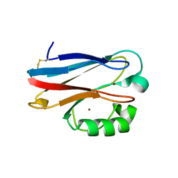
6QP2
 
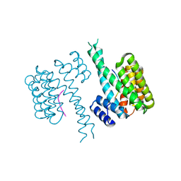
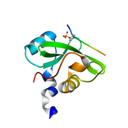
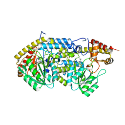 | | Crystal structure of the PLP-bound C-S lyase from Staphylococcus hominis | | 分子名称: | Aminotransferase, PYRIDOXAL-5'-PHOSPHATE, Polyhistidine tag | | 著者 | Herman, R, Rudden, M, Wilkinson, A.J, Hanai, S, Thomas, G.H. | | 登録日 | 2019-02-13 | | 公開日 | 2020-03-04 | | 最終更新日 | 2024-01-24 | | 実験手法 | X-RAY DIFFRACTION (1.6 Å) | | 主引用文献 | The molecular basis of thioalcohol production in human body odour.
Sci Rep, 10, 2020
|
|
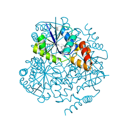
3MTC
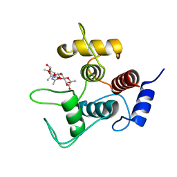
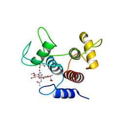
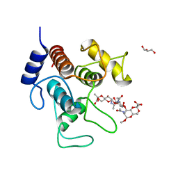
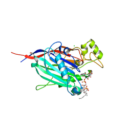 | | Crystal Structure of INPP5B in complex with phosphatidylinositol 4-phosphate | | 分子名称: | (2R)-3-{[(S)-hydroxy{[(1R,2R,3R,4R,5S,6R)-2,3,5,6-tetrahydroxy-4-(phosphonooxy)cyclohexyl]oxy}phosphoryl]oxy}propane-1,2-diyl dioctanoate, CHLORIDE ION, GLYCEROL, ... | | 著者 | Tresaugues, L, Welin, M, Arrowsmith, C.H, Berglund, H, Bountra, C, Collins, R, Edwards, A.M, Flodin, S, Flores, A, Graslund, S, Hammarstrom, M, Johansson, I, Karlberg, T, Kol, S, Kotenyova, T, Moche, M, Nyman, T, Persson, C, Schuler, H, Schutz, P, Siponen, M.I, Thorsell, A.G, van der Berg, S, Wahlberg, E, Weigelt, J, Wisniewska, M, Nordlund, P, Structural Genomics Consortium (SGC) | | 登録日 | 2010-04-30 | | 公開日 | 2010-06-30 | | 最終更新日 | 2023-09-06 | | 実験手法 | X-RAY DIFFRACTION (2.4 Å) | | 主引用文献 | Structural basis for phosphoinositide substrate recognition, catalysis, and membrane interactions in human inositol polyphosphate 5-phosphatases
Structure, 22, 2014
|
|
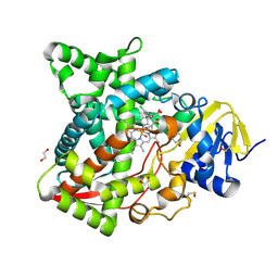
6AY6
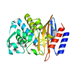
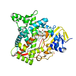 | | Naegleria fowleri CYP51-voriconazole complex | | 分子名称: | CYP51, sterol 14alpha-demethylase, PROTOPORPHYRIN IX CONTAINING FE, ... | | 著者 | Debnath, A, Calvet, C.M, Jennings, G, Zhou, W, Aksenov, A, Luth, M, Abagyan, R, Nes, W.D, McKerrow, J.H, Podust, L.M. | | 登録日 | 2017-09-07 | | 公開日 | 2017-11-22 | | 最終更新日 | 2023-11-15 | | 実験手法 | X-RAY DIFFRACTION (2.4 Å) | | 主引用文献 | CYP51 is an essential drug target for the treatment of primary amoebic meningoencephalitis (PAM).
PLoS Negl Trop Dis, 11, 2017
|
|
6QRO
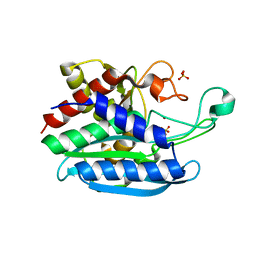 | | Crystal structure of Tannerella forsythia glutaminyl cyclase | | 分子名称: | Glutamine cyclotransferase, SULFATE ION, ZINC ION | | 著者 | Linnert, M, Piechotta, A, Parthier, C, Taudte, N, Kolenko, P, Rahfeld, J, Potempa, J, Stubbs, M.T. | | 登録日 | 2019-02-19 | | 公開日 | 2019-03-06 | | 最終更新日 | 2024-01-24 | | 実験手法 | X-RAY DIFFRACTION (2.1 Å) | | 主引用文献 | Mammalian-like type II glutaminyl cyclases in Porphyromonas gingivalis and other oral pathogenic bacteria as targets for treatment of periodontitis.
J.Biol.Chem., 296, 2021
|
|
3N2J
 | | Azurin H117G, oxidized form | | 分子名称: | Azurin, COPPER (II) ION | | 著者 | Hoffmann, M, Alagaratnam, S, Canters, G.W, Einsle, O. | | 登録日 | 2010-05-18 | | 公開日 | 2011-04-06 | | 最終更新日 | 2023-09-06 | | 実験手法 | X-RAY DIFFRACTION (1.35 Å) | | 主引用文献 | Probing the reactivity of different forms of azurin by flavin photoreduction.
Febs J., 278, 2011
|
|
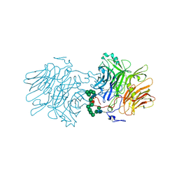
4PPH
 | | Crystal structure of conglutin gamma, a unique basic 7S globulin from lupine seeds | | 分子名称: | 1,2-ETHANEDIOL, 2-acetamido-2-deoxy-beta-D-glucopyranose, 2-acetamido-2-deoxy-beta-D-glucopyranose-(1-4)-2-acetamido-2-deoxy-beta-D-glucopyranose, ... | | 著者 | Czubinski, J, Barciszewski, J, Gilski, M, Lampart-Szczapa, E, Jaskolski, M. | | 登録日 | 2014-02-27 | | 公開日 | 2015-02-11 | | 最終更新日 | 2023-09-20 | | 実験手法 | X-RAY DIFFRACTION (2.009 Å) | | 主引用文献 | Structure of gamma-conglutin: insight into the quaternary structure of 7S basic globulins from legumes.
Acta Crystallogr.,Sect.D, 71, 2015
|
|
6QMG
 | |
5FKB
 | |
6AYB
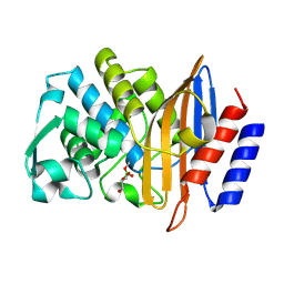
 | | Naegleria fowleri CYP51-ketoconazole complex | | 分子名称: | 1,2-ETHANEDIOL, 1-acetyl-4-(4-{[(2R,4S)-2-(2,4-dichlorophenyl)-2-(1H-imidazol-1-ylmethyl)-1,3-dioxolan-4-yl]methoxy}phenyl)piperazine, CALCIUM ION, ... | | 著者 | Debnath, A, Calvet, C.M, Jennings, G, Zhou, W, Aksenov, A, Luth, M, Abagyan, R, Nes, W.D, McKerrow, J.H, Podust, L.M. | | 登録日 | 2017-09-08 | | 公開日 | 2017-11-22 | | 最終更新日 | 2023-11-15 | | 実験手法 | X-RAY DIFFRACTION (1.87 Å) | | 主引用文献 | CYP51 is an essential drug target for the treatment of primary amoebic meningoencephalitis (PAM).
PLoS Negl Trop Dis, 11, 2017
|
|
6QZS
 | | 14-3-3 sigma in complex with FOXO1 pS256 peptide | | 分子名称: | 14-3-3 protein sigma, 2-[3-(2-HYDROXY-1,1-DIHYDROXYMETHYL-ETHYLAMINO)-PROPYLAMINO]-2-HYDROXYMETHYL-PROPANE-1,3-DIOL, FOXO1 pS256 site, ... | | 著者 | Ottmann, C, Wolter, M, Lau, R.A. | | 登録日 | 2019-03-12 | | 公開日 | 2019-07-31 | | 最終更新日 | 2024-01-24 | | 実験手法 | X-RAY DIFFRACTION (1.9 Å) | | 主引用文献 | AMPK and AKT protein kinases hierarchically phosphorylate the N-terminus of the FOXO1 transcription factor, modulating interactions with 14-3-3 proteins.
J.Biol.Chem., 294, 2019
|
|
6DWU
 | |
7SV6
 | |
7SV5
 | |
7SV3
 | |
7CIO
 | | Molecular interactions of cytoplasmic region of CTLA-4 with SH2 domains of PI3-kinase | | 分子名称: | Cytotoxic T-lymphocyte protein 4, Phosphatidylinositol 3-kinase regulatory subunit alpha | | 著者 | Iiyama, M, Numoto, N, Ogawa, S, Kuroda, M, Morii, H, Abe, R, Ito, N, Oda, M. | | 登録日 | 2020-07-08 | | 公開日 | 2020-12-09 | | 最終更新日 | 2023-11-29 | | 実験手法 | X-RAY DIFFRACTION (1.1 Å) | | 主引用文献 | Molecular interactions of the CTLA-4 cytoplasmic region with the phosphoinositide 3-kinase SH2 domains.
Mol.Immunol., 131, 2021
|
|
7SV4
 | | Crystal structure of SpaA-SLH in complex with 4,6-Pyr-beta-D-ManNAc-(1->4)-beta-D-GlcNAc-(1->3)-4,6-Pyr-beta-D-ManNAcOMe | | 分子名称: | GLYCEROL, Surface (S-) layer glycoprotein, methyl 2-acetamido-4,6-O-[(1S)-1-carboxyethylidene]-2-deoxy-beta-D-mannopyranosyl-(1->4)-2-acetamido-2-deoxy-beta-D-glucopyranosyl-(1->3)-2-acetamido-4,6-O-[(1S)-1-carboxyethylidene]-2-deoxy-beta-D-mannopyranoside | | 著者 | Legg, M.S.G, Evans, S.V. | | 登録日 | 2021-11-18 | | 公開日 | 2022-03-09 | | 最終更新日 | 2023-10-18 | | 実験手法 | X-RAY DIFFRACTION (2.06 Å) | | 主引用文献 | The S-layer homology domains of Paenibacillus alvei surface protein SpaA bind to cell wall polysaccharide through the terminal monosaccharide residue.
J.Biol.Chem., 298, 2022
|
|
8PCD
 | | Structure of serine-beta-lactamase CTX-M-14 following the time-resolved active site binding of boric acid, 150 ms | | 分子名称: | BORATE ION, Beta-lactamase, SULFATE ION | | 著者 | Prester, A, Perbandt, M, Galchenkova, M, Oberthuer, D, Yefanov, O, Hinrichs, W, Rohde, H, Betzel, C. | | 登録日 | 2023-06-11 | | 公開日 | 2024-06-26 | | 最終更新日 | 2024-07-17 | | 実験手法 | X-RAY DIFFRACTION (1.97 Å) | | 主引用文献 | Time-resolved crystallography of boric acid binding to the active site serine of the beta-lactamase CTX-M-14 and subsequent 1,2-diol esterification.
Commun Chem, 7, 2024
|
|
8PCM
 | | Structure of serine-beta-lactamase CTX-M-14 following the time-resolved active site binding of boric acid and subsequent glycerol-boric acid-ester formation, 80 ms | | 分子名称: | BORATE ION, Beta-lactamase, SULFATE ION, ... | | 著者 | Prester, A, Perbandt, M, Galchenkova, M, Oberthuer, D, Yefanov, O, Hinrichs, W, Rohde, H, Betzel, C. | | 登録日 | 2023-06-11 | | 公開日 | 2024-06-26 | | 最終更新日 | 2024-07-17 | | 実験手法 | X-RAY DIFFRACTION (1.84 Å) | | 主引用文献 | Time-resolved crystallography of boric acid binding to the active site serine of the beta-lactamase CTX-M-14 and subsequent 1,2-diol esterification.
Commun Chem, 7, 2024
|
|
8PCR
 | | Structure of serine-beta-lactamase CTX-M-14 following the time-resolved active site binding of boric acid and subsequent glycerol-boric acid-ester formation, 750 ms | | 分子名称: | BORATE ION, Beta-lactamase, SULFATE ION, ... | | 著者 | Prester, A, Perbandt, M, Galchenkova, M, Oberthuer, D, Yefanov, O, Hinrichs, W, Rohde, H, Betzel, C. | | 登録日 | 2023-06-11 | | 公開日 | 2024-06-26 | | 最終更新日 | 2024-07-17 | | 実験手法 | X-RAY DIFFRACTION (1.7 Å) | | 主引用文献 | Time-resolved crystallography of boric acid binding to the active site serine of the beta-lactamase CTX-M-14 and subsequent 1,2-diol esterification.
Commun Chem, 7, 2024
|
|
5M4R
 | | Structural tuning of CD81LEL (space group C2) | | 分子名称: | 1,2-ETHANEDIOL, CD81 antigen, SULFATE ION | | 著者 | Cunha, E.S, Sfriso, P, Rojas, A.L, Roversi, P, Hospital, A, Orozco, M, Abrescia, N.G. | | 登録日 | 2016-10-19 | | 公開日 | 2016-12-14 | | 最終更新日 | 2024-01-17 | | 実験手法 | X-RAY DIFFRACTION (3.1 Å) | | 主引用文献 | Mechanism of Structural Tuning of the Hepatitis C Virus Human Cellular Receptor CD81 Large Extracellular Loop.
Structure, 25, 2017
|
|
8PCA
 | | Structure of serine-beta-lactamase CTX-M-14 following the time-resolved active site binding of boric acid, 50 ms | | 分子名称: | Beta-lactamase, SULFATE ION | | 著者 | Prester, A, Perbandt, M, Galchenkova, M, Oberthuer, D, Yefanov, O, Hinrichs, W, Rohde, H, Betzel, C. | | 登録日 | 2023-06-11 | | 公開日 | 2024-06-26 | | 最終更新日 | 2024-07-17 | | 実験手法 | X-RAY DIFFRACTION (1.58 Å) | | 主引用文献 | Time-resolved crystallography of boric acid binding to the active site serine of the beta-lactamase CTX-M-14 and subsequent 1,2-diol esterification.
Commun Chem, 7, 2024
|
|
8PCE
 | | Structure of serine-beta-lactamase CTX-M-14 following the time-resolved active site binding of boric acid, 250 ms | | 分子名称: | BORATE ION, Beta-lactamase, SULFATE ION | | 著者 | Prester, A, Perbandt, M, Galchenkova, M, Oberthuer, D, Yefanov, O, Hinrichs, W, Rohde, H, Betzel, C. | | 登録日 | 2023-06-11 | | 公開日 | 2024-06-26 | | 最終更新日 | 2024-07-17 | | 実験手法 | X-RAY DIFFRACTION (1.65 Å) | | 主引用文献 | Time-resolved crystallography of boric acid binding to the active site serine of the beta-lactamase CTX-M-14 and subsequent 1,2-diol esterification.
Commun Chem, 7, 2024
|
|
8PCT
 | | Structure of serine-beta-lactamase CTX-M-14 following the time-resolved active site binding of boric acid and subsequent glycerol-boric acid-ester formation, 2500 ms | | 分子名称: | BORATE ION, Beta-lactamase, SULFATE ION, ... | | 著者 | Prester, A, Perbandt, M, Galchenkova, M, Oberthuer, D, Yefanov, O, Hinrichs, W, Rohde, H, Betzel, C. | | 登録日 | 2023-06-11 | | 公開日 | 2024-06-26 | | 最終更新日 | 2024-07-17 | | 実験手法 | X-RAY DIFFRACTION (1.55 Å) | | 主引用文献 | Time-resolved crystallography of boric acid binding to the active site serine of the beta-lactamase CTX-M-14 and subsequent 1,2-diol esterification.
Commun Chem, 7, 2024
|
|
8PCP
 | | Structure of serine-beta-lactamase CTX-M-14 following the time-resolved active site binding of boric acid and subsequent glycerol-boric acid-ester formation, 250 ms | | 分子名称: | BORATE ION, Beta-lactamase, SULFATE ION, ... | | 著者 | Prester, A, Perbandt, M, Galchenkova, M, Oberthuer, D, Yefanov, O, Hinrichs, W, Rohde, H, Betzel, C. | | 登録日 | 2023-06-11 | | 公開日 | 2024-06-26 | | 最終更新日 | 2024-07-17 | | 実験手法 | X-RAY DIFFRACTION (1.76 Å) | | 主引用文献 | Time-resolved crystallography of boric acid binding to the active site serine of the beta-lactamase CTX-M-14 and subsequent 1,2-diol esterification.
Commun Chem, 7, 2024
|
|
8PCI
 | | Structure of serine-beta-lactamase CTX-M-14 following the time-resolved active site binding of boric acid, 5000 ms | | 分子名称: | BORATE ION, Beta-lactamase, SULFATE ION | | 著者 | Prester, A, Perbandt, M, Galchenkova, M, Oberthuer, D, Yefanov, O, Hinrichs, W, Rohde, H, Betzel, C. | | 登録日 | 2023-06-11 | | 公開日 | 2024-06-26 | | 最終更新日 | 2024-07-17 | | 実験手法 | X-RAY DIFFRACTION (1.5 Å) | | 主引用文献 | Time-resolved crystallography of boric acid binding to the active site serine of the beta-lactamase CTX-M-14 and subsequent 1,2-diol esterification.
Commun Chem, 7, 2024
|
|


