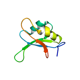
2RVC
 
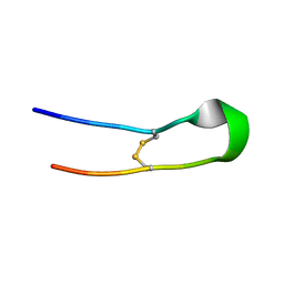
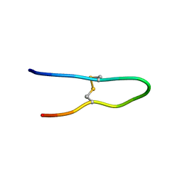
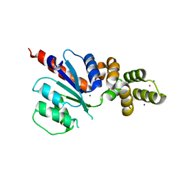
 | | Solution structure of Zalpha domain of goldfish ZBP-containing protein kinase | | 分子名称: | Interferon-inducible and double-stranded-dependent eIF-2kinase | | 著者 | Lee, A, Park, C, Park, J, Kwon, M, Choi, Y, Kim, K, Choi, B, Lee, J. | | 登録日 | 2015-07-08 | | 公開日 | 2016-02-03 | | 最終更新日 | 2024-05-01 | | 実験手法 | SOLUTION NMR | | 主引用文献 | Solution structure of the Z-DNA binding domain of PKR-like protein kinase from Carassius auratus and quantitative analyses of the intermediate complex during B-Z transition.
Nucleic Acids Res., 44, 2016
|
|
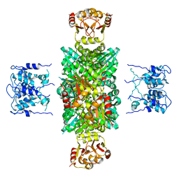
1LYW
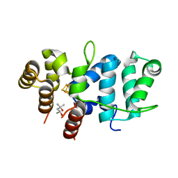
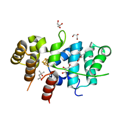
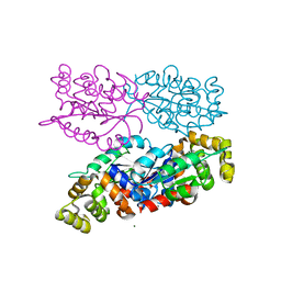
 | | CATHEPSIN D AT PH 7.5 | | 分子名称: | 4-(2-HYDROXYETHYL)-1-PIPERAZINE ETHANESULFONIC ACID, CATHEPSIN D | | 著者 | Lee, A.Y, Gulnik, S.V, Erickson, J.W. | | 登録日 | 1998-06-30 | | 公開日 | 1999-07-22 | | 最終更新日 | 2023-08-09 | | 実験手法 | X-RAY DIFFRACTION (2.5 Å) | | 主引用文献 | Conformational switching in an aspartic proteinase.
Nat.Struct.Biol., 5, 1998
|
|
1BJ2
 | |
1TPS
 | |
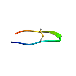
1TYN
 | | ATOMIC STRUCTURE OF THE TRYPSIN-CYCLOTHEONAMIDE A COMPLEX: LESSONS FOR THE DESIGN OF SERINE PROTEASE INHIBITORS | | 分子名称: | BETA-TRYPSIN, CYCLOTHEONAMIDE A | | 著者 | Lee, A.Y, Hagihara, M, Karmacharya, R, Albers, M.W, Schreiber, S.L, Clardy, J. | | 登録日 | 1994-09-19 | | 公開日 | 1995-01-26 | | 最終更新日 | 2011-07-13 | | 実験手法 | X-RAY DIFFRACTION (2 Å) | | 主引用文献 | Atomic Structure of the Trypsin-Cyclotheonamide a Complex: Lessons for the Design of Serine Protease Inhibitors
J.Am.Chem.Soc., 115, 1993
|
|
1SXL
 | |
2BJ2
 | |
8XTH
 | |
7TL3
 | | Crystal Structure of Yeast p58C Multi-Tyrosine Mutant 5YF431 | | 分子名称: | (4S)-2-METHYL-2,4-PENTANEDIOL, DNA primase large subunit, IRON/SULFUR CLUSTER | | 著者 | Blee, A.M, Salay, L.E, Chazin, W.J. | | 登録日 | 2022-01-18 | | 公開日 | 2022-06-29 | | 最終更新日 | 2023-10-18 | | 実験手法 | X-RAY DIFFRACTION (2.066 Å) | | 主引用文献 | Modification of the 4Fe-4S Cluster Charge Transport Pathway Alters RNA Synthesis by Yeast DNA Primase.
Biochemistry, 61, 2022
|
|
7TL4
 | | Crystal Structure of Yeast p58C Multi-Tyrosine Mutant 6YF | | 分子名称: | (4S)-2-METHYL-2,4-PENTANEDIOL, DNA primase large subunit, IRON/SULFUR CLUSTER | | 著者 | Blee, A.M, Salay, L.E, Chazin, W.J. | | 登録日 | 2022-01-18 | | 公開日 | 2022-06-29 | | 最終更新日 | 2023-10-18 | | 実験手法 | X-RAY DIFFRACTION (1.805 Å) | | 主引用文献 | Modification of the 4Fe-4S Cluster Charge Transport Pathway Alters RNA Synthesis by Yeast DNA Primase.
Biochemistry, 61, 2022
|
|
7TL2
 | | Crystal Structure of Yeast p58C Multi-Tyrosine Mutant 5YF412 | | 分子名称: | (4S)-2-METHYL-2,4-PENTANEDIOL, DNA primase large subunit, GLYCEROL, ... | | 著者 | Blee, A.M, Salay, L.E, Chazin, W.J. | | 登録日 | 2022-01-18 | | 公開日 | 2022-06-29 | | 最終更新日 | 2023-10-18 | | 実験手法 | X-RAY DIFFRACTION (1.529 Å) | | 主引用文献 | Modification of the 4Fe-4S Cluster Charge Transport Pathway Alters RNA Synthesis by Yeast DNA Primase.
Biochemistry, 61, 2022
|
|
8IL2
 | |
8IL6
 | |
8IL1
 | |
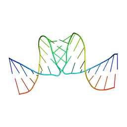
1LNQ
 | | CRYSTAL STRUCTURE OF MTHK AT 3.3 A | | 分子名称: | CALCIUM ION, POTASSIUM CHANNEL RELATED PROTEIN | | 著者 | Jiang, Y, Lee, A, Chen, J, Cadene, M, Chait, B.T, Mackinnon, R. | | 登録日 | 2002-05-03 | | 公開日 | 2002-06-19 | | 最終更新日 | 2024-02-14 | | 実験手法 | X-RAY DIFFRACTION (3.3 Å) | | 主引用文献 | CRYSTAL STRUCTURE AND MECHANISM OF A CALCIUM-GATED POTASSIUM CHANNEL
Nature, 417, 2002
|
|
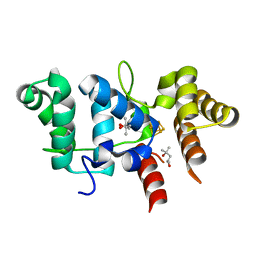
5CFL
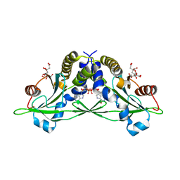 | | Crystal structure of anemone STING (Nematostella vectensis) in complex with 3', 3' c-di-GMP, c[G(3', 5')pG(3', 5')p] | | 分子名称: | 9,9'-[(2R,3R,3aS,5S,7aR,9R,10R,10aS,12S,14aR)-3,5,10,12-tetrahydroxy-5,12-dioxidooctahydro-2H,7H-difuro[3,2-d:3',2'-j][1,3,7,9,2,8]tetraoxadiphosphacyclododecine-2,9-diyl]bis(2-amino-1,9-dihydro-6H-purin-6-one), CITRATE ANION, Stimulator of Interferon Genes | | 著者 | Kranzusch, P.J, Wilson, S.C, Lee, A.S.Y, Berger, J.M, Doudna, J.A, Vance, R.E. | | 登録日 | 2015-07-08 | | 公開日 | 2015-08-26 | | 最終更新日 | 2024-03-06 | | 実験手法 | X-RAY DIFFRACTION (1.836 Å) | | 主引用文献 | Ancient Origin of cGAS-STING Reveals Mechanism of Universal 2',3' cGAMP Signaling.
Mol.Cell, 59, 2015
|
|
5CFO
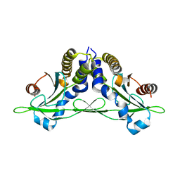 | | Crystal structure of anemone STING (Nematostella vectensis) in apo 'rotated' open conformation | | 分子名称: | Stimulator of Interferon Genes | | 著者 | Kranzusch, P.J, Wilson, S.C, Lee, A.S.Y, Berger, J.M, Doudna, J.A, Vance, R.E. | | 登録日 | 2015-07-08 | | 公開日 | 2015-08-26 | | 最終更新日 | 2024-03-06 | | 実験手法 | X-RAY DIFFRACTION (2.102 Å) | | 主引用文献 | Ancient Origin of cGAS-STING Reveals Mechanism of Universal 2',3' cGAMP Signaling.
Mol.Cell, 59, 2015
|
|
5CFR
 | | Crystal structure of anemone STING (Nematostella vectensis) in apo 'unrotated' closed conformation | | 分子名称: | CALCIUM ION, Stimulator of Interferon Genes | | 著者 | Kranzusch, P.J, Wilson, S.C, Lee, A.S.Y, Berger, J.M, Doudna, J.A, Vance, R.E. | | 登録日 | 2015-07-08 | | 公開日 | 2015-08-26 | | 最終更新日 | 2024-03-06 | | 実験手法 | X-RAY DIFFRACTION (2.85 Å) | | 主引用文献 | Ancient Origin of cGAS-STING Reveals Mechanism of Universal 2',3' cGAMP Signaling.
Mol.Cell, 59, 2015
|
|
5CFQ
 | | Crystal structure of anemone STING (Nematostella vectensis) in complex with 2',3' cGAMP, c[G(2',5')pA(3',5')p] | | 分子名称: | SULFATE ION, Stimulator of Interferon Genes, cGAMP | | 著者 | Kranzusch, P.J, Wilson, S.C, Lee, A.S.Y, Berger, J.M, Doudna, J.A, Vance, R.E. | | 登録日 | 2015-07-08 | | 公開日 | 2015-08-26 | | 最終更新日 | 2024-03-06 | | 実験手法 | X-RAY DIFFRACTION (2.099 Å) | | 主引用文献 | Ancient Origin of cGAS-STING Reveals Mechanism of Universal 2',3' cGAMP Signaling.
Mol.Cell, 59, 2015
|
|
5CFM
 | | Crystal structure of anemone STING (Nematostella vectensis) in complex with 3', 3' cGAMP, c[G(3', 5')pA(3', 5')p] | | 分子名称: | 2-amino-9-[(2R,3R,3aS,5R,7aR,9R,10R,10aS,12R,14aR)-9-(6-amino-9H-purin-9-yl)-3,5,10,12-tetrahydroxy-5,12-dioxidooctahydro-2H,7H-difuro[3,2-d:3',2'-j][1,3,7,9,2,8]tetraoxadiphosphacyclododecin-2-yl]-1,9-dihydro-6H-purin-6-one, CITRATE ANION, Stimulator of Interferon Genes | | 著者 | Kranzusch, P.J, Wilson, S.C, Lee, A.S.Y, Berger, J.M, Doudna, J.A, Vance, R.E. | | 登録日 | 2015-07-08 | | 公開日 | 2015-08-26 | | 最終更新日 | 2024-03-06 | | 実験手法 | X-RAY DIFFRACTION (1.994 Å) | | 主引用文献 | Ancient Origin of cGAS-STING Reveals Mechanism of Universal 2',3' cGAMP Signaling.
Mol.Cell, 59, 2015
|
|
6NJX
 | | C-terminal region of the Xanthomonas campestris pv. campestris OLD protein phased with mercury | | 分子名称: | IODIDE ION, MERCURY (II) ION, Xcc_ctr_Hg | | 著者 | Schiltz, C.J, Lee, A, Partlow, E.A, Hosford, C.J, Chappie, J.S. | | 登録日 | 2019-01-04 | | 公開日 | 2019-08-07 | | 最終更新日 | 2024-03-13 | | 実験手法 | X-RAY DIFFRACTION (1.95 Å) | | 主引用文献 | Structural characterization of Class 2 OLD family nucleases supports a two-metal catalysis mechanism for cleavage.
Nucleic Acids Res., 47, 2019
|
|
6NK8
 | | C-terminal region of the Burkholderia pseudomallei OLD protein | | 分子名称: | Class 2 OLD family nuclease, MAGNESIUM ION | | 著者 | Schiltz, C.J, Lee, A, Partlow, E.A, Hosford, C.J, Chappie, J.S. | | 登録日 | 2019-01-05 | | 公開日 | 2019-08-07 | | 最終更新日 | 2024-03-13 | | 実験手法 | X-RAY DIFFRACTION (2.24 Å) | | 主引用文献 | Structural characterization of Class 2 OLD family nucleases supports a two-metal catalysis mechanism for cleavage.
Nucleic Acids Res., 47, 2019
|
|
6NJW
 | | C-terminal region of the Xanthomonas campestris pv. campestris OLD protein phased with platinum | | 分子名称: | IODIDE ION, MAGNESIUM ION, PLATINUM (II) ION, ... | | 著者 | Schiltz, C.J, Lee, A, Partlow, E.A, Hosford, C.J, Chappie, J.S. | | 登録日 | 2019-01-04 | | 公開日 | 2019-08-07 | | 最終更新日 | 2024-03-13 | | 実験手法 | X-RAY DIFFRACTION (1.86 Å) | | 主引用文献 | Structural characterization of Class 2 OLD family nucleases supports a two-metal catalysis mechanism for cleavage.
Nucleic Acids Res., 47, 2019
|
|
6NJV
 | | C-terminal region of the Xanthomonas campestris pv. campestris OLD protein phased with iodine | | 分子名称: | IODIDE ION, MAGNESIUM ION, Xcc_CTR_I | | 著者 | Schiltz, C.J, Lee, A, Partlow, E.A, Hosford, C.J, Chappie, J.S. | | 登録日 | 2019-01-04 | | 公開日 | 2019-08-07 | | 最終更新日 | 2024-03-13 | | 実験手法 | X-RAY DIFFRACTION (2.3 Å) | | 主引用文献 | Structural characterization of Class 2 OLD family nucleases supports a two-metal catalysis mechanism for cleavage.
Nucleic Acids Res., 47, 2019
|
|
3PSH
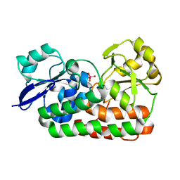 | | Classification of a Haemophilus influenzae ABC transporter HI1470/71 through its cognate molybdate periplasmic binding protein MolA (MolA bound to Molybdate) | | 分子名称: | MOLYBDATE ION, protein HI_1472 | | 著者 | Tirado-Lee, L, Lee, A, Rees, D.C, Pinkett, H.W. | | 登録日 | 2010-12-01 | | 公開日 | 2011-11-30 | | 最終更新日 | 2024-02-21 | | 実験手法 | X-RAY DIFFRACTION (1.5 Å) | | 主引用文献 | Classification of a Haemophilus influenzae ABC Transporter HI1470/71 through Its Cognate Molybdate Periplasmic Binding Protein, MolA.
Structure, 19, 2011
|
|


