Loading
PDBj
MenuPDBj@FacebookPDBj@TwitterPDBj@YouTubewwPDB FoundationwwPDB
RCSB PDBPDBeBMRBAdv. SearchSearch help
PDB: 318 results

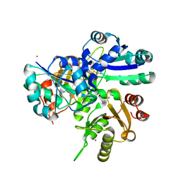
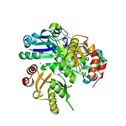
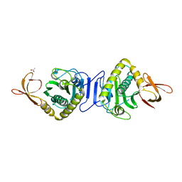
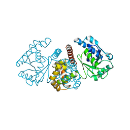
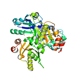
2HUV
DownloadVisualize
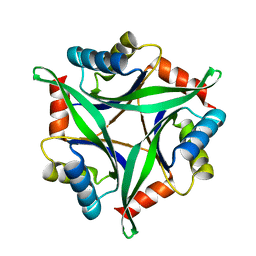
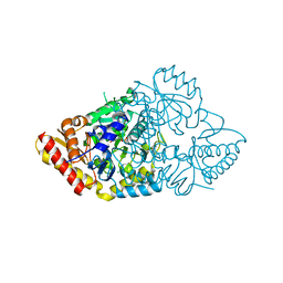
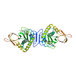
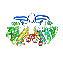
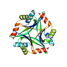
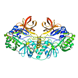
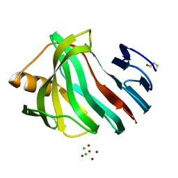
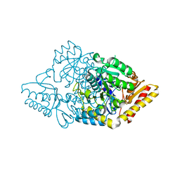
BU of 2huv by Molmil
Crystal structure of PH0725 from Pyrococcus horikoshii OT3
Descriptor: PLATINUM (II) ION, Probable diphthine synthase, S-ADENOSYL-L-HOMOCYSTEINE, ...
Authors:Sugahara, M, Saraboji, K, Malathy sony, S.M, Ponnuswamy, M.N, Kumarevel, T.S, Kunishima, N, RIKEN Structural Genomics/Proteomics Initiative (RSGI)
Deposit date:2006-07-27
Release date:2007-01-27
Last modified:2023-10-25
Method:X-RAY DIFFRACTION (2.1 Å)
Cite:Crystal structure of PH0725 from Pyrococcus horikoshii OT3
To be Published
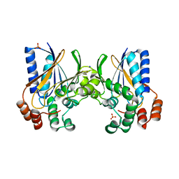
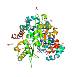
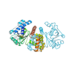
2HR8
DownloadVisualize
BU of 2hr8 by Molmil
Crystal structure of PH0725 from Pyrococcus horikoshii OT3
Descriptor: PLATINUM (II) ION, S-ADENOSYL-L-HOMOCYSTEINE, diphthine synthase
Authors:Yamamoto, H, Taketa, M, Kunishima, N, RIKEN Structural Genomics/Proteomics Initiative (RSGI)
Deposit date:2006-07-20
Release date:2007-01-20
Last modified:2023-10-25
Method:X-RAY DIFFRACTION (2.8 Å)
Cite:Crystal structure of PH0725 from Pyrococcus horikoshii OT3
To be Published
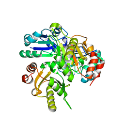
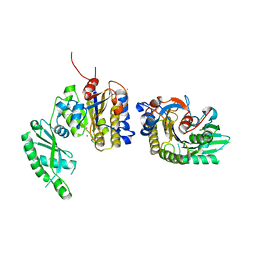
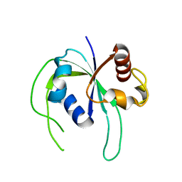
2GJU
DownloadVisualize
BU of 2gju by Molmil
Crystal structure of hypothetical protein PH1004 from Pyrococcus horikoshii OT3
Descriptor: 252aa long hypothetical protein, PHOSPHATE ION, SODIUM ION
Authors:Yamamoto, H, Kunishima, N, RIKEN Structural Genomics/Proteomics Initiative (RSGI)
Deposit date:2006-03-31
Release date:2006-10-10
Last modified:2023-10-25
Method:X-RAY DIFFRACTION (2 Å)
Cite:Crystal structure of hypothetical protein PH1004 from Pyrococcus horikoshii OT3
To be published
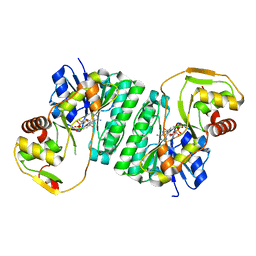
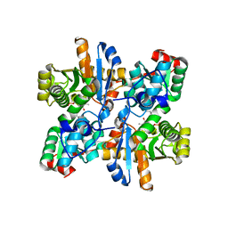
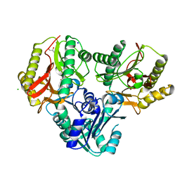
2HUQ
DownloadVisualize
BU of 2huq by Molmil
Crystal structure of PH0725 from Pyrococcus horikoshii OT3
Descriptor: PLATINUM (II) ION, Probable diphthine synthase, S-ADENOSYL-L-HOMOCYSTEINE
Authors:Sugahara, M, Karthe, P, Kumarevel, T.S, Kunishima, N, RIKEN Structural Genomics/Proteomics Initiative (RSGI)
Deposit date:2006-07-27
Release date:2007-01-27
Last modified:2023-10-25
Method:X-RAY DIFFRACTION (2.2 Å)
Cite:Crystal structure of PH0725 from Pyrococcus horikoshii OT3
To be Published
2HUN
DownloadVisualize
BU of 2hun by Molmil
Crystal structure of hypothetical protein PH0414 from Pyrococcus horikoshii OT3
Descriptor: 336aa long hypothetical dTDP-glucose 4,6-dehydratase, NICOTINAMIDE-ADENINE-DINUCLEOTIDE
Authors:Yamamoto, H, Kunishima, N, RIKEN Structural Genomics/Proteomics Initiative (RSGI)
Deposit date:2006-07-26
Release date:2007-01-26
Last modified:2023-10-25
Method:X-RAY DIFFRACTION (2.07 Å)
Cite:Crystal structure of hypothetical protein PH0414 from Pyrococcus horikoshii OT3
To be Published
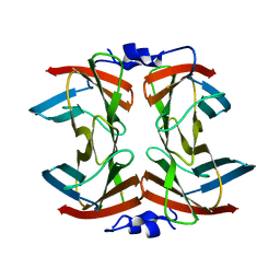
2HUX
DownloadVisualize
BU of 2hux by Molmil
Crystal structure of PH0725 from Pyrococcus horikoshii OT3
Descriptor: PLATINUM (II) ION, Probable diphthine synthase, S-ADENOSYL-L-HOMOCYSTEINE
Authors:Sugahara, M, Kunishima, N, RIKEN Structural Genomics/Proteomics Initiative (RSGI)
Deposit date:2006-07-27
Release date:2007-01-27
Last modified:2023-10-25
Method:X-RAY DIFFRACTION (2.4 Å)
Cite:Crystal structure of PH0725 from Pyrococcus horikoshii OT3
To be Published
2HVB
DownloadVisualize
BU of 2hvb by Molmil
Crystal structure of hypothetical protein PH1083 from Pyrococcus horikoshii OT3
Descriptor: FE (III) ION, Superoxide reductase
Authors:Yamamoto, H, Kunishima, N, RIKEN Structural Genomics/Proteomics Initiative (RSGI)
Deposit date:2006-07-28
Release date:2007-01-28
Last modified:2023-10-25
Method:X-RAY DIFFRACTION (2.5 Å)
Cite:Crystal structure of hypothetical protein PH1083 from Pyrococcus horikoshii OT3
To be Published
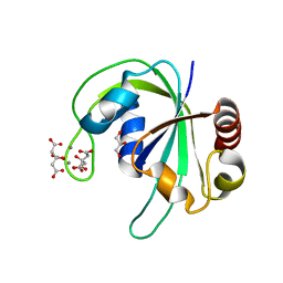
2HD9
DownloadVisualize
BU of 2hd9 by Molmil
Crystal structure of PH1033 from Pyrococcus horikoshii OT3
Descriptor: CALCIUM ION, CITRIC ACID, GLYCEROL, ...
Authors:Sugahara, M, Kunishima, N, RIKEN Structural Genomics/Proteomics Initiative (RSGI)
Deposit date:2006-06-20
Release date:2006-12-20
Last modified:2023-10-25
Method:X-RAY DIFFRACTION (1.35 Å)
Cite:Nucleant-mediated protein crystallization with the application of microporous synthetic zeolites.
Acta Crystallogr.,Sect.D, 64, 2008
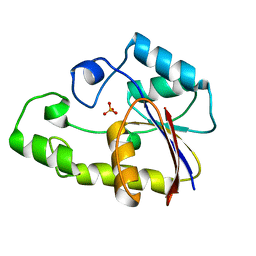
2HIA
DownloadVisualize
BU of 2hia by Molmil
Crystal structure of TTHB049 from Thermus thermophilus HB8
Descriptor: Alpha-ribazole-5'-phosphate phosphatase, PHOSPHATE ION
Authors:Sugahara, M, Kunishima, N, RIKEN Structural Genomics/Proteomics Initiative (RSGI)
Deposit date:2006-06-29
Release date:2006-12-29
Last modified:2023-10-25
Method:X-RAY DIFFRACTION (1.6 Å)
Cite:Crystal structure of TTHB049 from Thermus thermophilus HB8
To be Published
2HNI
DownloadVisualize
BU of 2hni by Molmil
Crystal Structure Of Biotin Protein Ligase From Pyrococcus Horikoshii OT3, K111A mutation
Descriptor: 235aa long hypothetical biotin-[acetyl-CoA-carboxylase] ligase, ACETIC ACID
Authors:Bagautdinov, B, Kunishima, N, RIKEN Structural Genomics/Proteomics Initiative (RSGI)
Deposit date:2006-07-13
Release date:2007-01-13
Last modified:2023-10-25
Method:X-RAY DIFFRACTION (1.9 Å)
Cite:Ligand Structures Of Biotin Protein Ligase From Pyrococcus Horikoshii OT3
To be Published
2Z6R
DownloadVisualize
BU of 2z6r by Molmil
Crystal structure of Lys49 to Arg mutant of Diphthine synthase
Descriptor: 2-(N-MORPHOLINO)-ETHANESULFONIC ACID, GLYCEROL, S-ADENOSYL-L-HOMOCYSTEINE, ...
Authors:Mizutani, H, Matsuura, Y, Krishna Swamy, B.S, Simanshu, D.K, Murthy, M.R.N, Kunishima, N.
Deposit date:2007-08-08
Release date:2007-08-28
Last modified:2023-11-01
Method:X-RAY DIFFRACTION (1.5 Å)
Cite:Crystal structure of diphthine synthase from Pyrococcus horikoshii OT3
To be Published
2ZKT
DownloadVisualize
BU of 2zkt by Molmil
Structure of PH0037 protein from Pyrococcus horikoshii
Descriptor: 2,3-bisphosphoglycerate-independent phosphoglycerate mutase, CALCIUM ION, ZINC ION
Authors:Lokanath, N.K, Pampa, K.J, Kunishima, N, RIKEN Structural Genomics/Proteomics Initiative (RSGI)
Deposit date:2008-03-29
Release date:2009-04-21
Last modified:2024-03-13
Method:X-RAY DIFFRACTION (2.4 Å)
Cite:Structure of PH0037 protein from Pyrococcus horikoshii
To be Published
2ZSJ
DownloadVisualize
BU of 2zsj by Molmil
Crystal structure of threonine synthase from Aquifex aeolicus VF5
Descriptor: PYRIDOXAL-5'-PHOSPHATE, Threonine synthase
Authors:Mizutani, H, Kunishima, N.
Deposit date:2008-09-11
Release date:2008-10-21
Last modified:2023-11-01
Method:X-RAY DIFFRACTION (1.8 Å)
Cite:Crystal structure of threonine synthase from Aquifex aeolicus VF5
To be Published
3ADO
DownloadVisualize
BU of 3ado by Molmil
Crystal Structure of the Rabbit L-Gulonate 3-Dehydrogenase
Descriptor: Lambda-crystallin
Authors:Asada, Y, Hara, A, Kunishima, N, RIKEN Structural Genomics/Proteomics Initiative (RSGI)
Deposit date:2010-01-26
Release date:2010-02-09
Last modified:2023-11-01
Method:X-RAY DIFFRACTION (1.7 Å)
Cite:Crystal Structure of the Rabbit L-Gulonate 3-Dehydrogenase
To be Published
3ADP
DownloadVisualize
BU of 3adp by Molmil
Crystal Structure of the Rabbit L-Gulonate 3-Dehydrogenase (NADH Form)
Descriptor: 1,4-DIHYDRONICOTINAMIDE ADENINE DINUCLEOTIDE, Lambda-crystallin
Authors:Asada, Y, Hara, A, Kunishima, N, RIKEN Structural Genomics/Proteomics Initiative (RSGI)
Deposit date:2010-01-26
Release date:2010-02-09
Last modified:2023-11-01
Method:X-RAY DIFFRACTION (1.85 Å)
Cite:Crystal Structure of the Rabbit L-Gulonate 3-Dehydrogenase
To be Published
2ZBN
DownloadVisualize
BU of 2zbn by Molmil
Crystal structure of PH1033 from Pyrococcus horikoshii OT3
Descriptor: UPF0310 protein PH1033
Authors:Sugahara, M, Kunishima, N, RIKEN Structural Genomics/Proteomics Initiative (RSGI)
Deposit date:2007-10-26
Release date:2008-04-29
Last modified:2023-11-01
Method:X-RAY DIFFRACTION (2 Å)
Cite:Nucleant-mediated protein crystallization with the application of microporous synthetic zeolites.
Acta Crystallogr.,Sect.D, 64, 2008
2ZSG
DownloadVisualize
BU of 2zsg by Molmil
Crystal structure of X-Pro aminopeptidase from Thermotoga maritima MSB8
Descriptor: Aminopeptidase P, putative, CHLORIDE ION, ...
Authors:Mizutani, H, Kunishima, N.
Deposit date:2008-09-10
Release date:2008-10-14
Last modified:2023-11-01
Method:X-RAY DIFFRACTION (1.65 Å)
Cite:Crystal structure of X-Pro aminopeptidase from Thermotoga maritima MSB8
To be Published
3AA9
DownloadVisualize
BU of 3aa9 by Molmil
Crystal Structure Analysis of the Mutant CutA1 (E61V) from E. coli
Descriptor: Divalent-cation tolerance protein cutA
Authors:Matsuura, Y, Tanaka, T, Bagautdinov, B, Kunishima, N, Yutani, K.
Deposit date:2009-11-12
Release date:2010-08-11
Last modified:2023-11-01
Method:X-RAY DIFFRACTION (2.3 Å)
Cite:Remarkable improvement in the heat stability of CutA1 from Escherichia coli by rational protein design
J.Biochem., 148, 2010
2ZSM
DownloadVisualize
BU of 2zsm by Molmil
Crystal structure of glutamate-1-semialdehyde 2,1-aminomutase from Aeropyrum pernix, hexagonal form
Descriptor: 4'-DEOXY-4'-AMINOPYRIDOXAL-5'-PHOSPHATE, CHLORIDE ION, Glutamate-1-semialdehyde 2,1-aminomutase
Authors:Mizutani, H, Kunishima, N.
Deposit date:2008-09-17
Release date:2008-10-21
Last modified:2023-11-01
Method:X-RAY DIFFRACTION (2.3 Å)
Cite:Crystal structure of glutamate-1-semialdehyde 2,1-aminomutase from Aeropyrum pernix
To be Published
2ZGW
DownloadVisualize
BU of 2zgw by Molmil
Crystal Structure Of Biotin Protein Ligase From Pyrococcus Horikoshii Complexed with Adenosine and Biotin, Mutations R48A and K111A
Descriptor: ADENOSINE, BIOTIN, biotin--[acetyl-CoA-carboxylase] ligase
Authors:Bagautdinov, B, Matsuura, Y, Bagautdinova, S, Kunishima, N, RIKEN Structural Genomics/Proteomics Initiative (RSGI)
Deposit date:2008-01-28
Release date:2008-02-05
Last modified:2023-11-01
Method:X-RAY DIFFRACTION (1.5 Å)
Cite:Protein biotinylation visualized by a complex structure of biotin protein ligase with a substrate
J.Biol.Chem., 283, 2008
2ZSU
DownloadVisualize
BU of 2zsu by Molmil
Crystal structure of spermidine synthase from Pyrococcus horikoshii OT3, P1 form
Descriptor: 1-{4-[(3-aminopropyl)amino]butyl}guanidine, spermidine synthase
Authors:Mizutani, H, Kunishima, N.
Deposit date:2008-09-17
Release date:2008-10-21
Last modified:2023-11-01
Method:X-RAY DIFFRACTION (2.2 Å)
Cite:Crystal structure of spermidine synthase from Pyrococcus horikoshii OT3
To be Published
3AH6
DownloadVisualize
BU of 3ah6 by Molmil
Remarkable improvement of the heat stability of CutA1 from E.coli by rational protein designing
Descriptor: Divalent-cation tolerance protein cutA
Authors:Matsuura, Y, Tanaka, T, Bagautdinov, B, Kunishima, N, Yutani, K.
Deposit date:2010-04-15
Release date:2010-08-11
Last modified:2023-11-01
Method:X-RAY DIFFRACTION (2.4 Å)
Cite:Remarkable improvement in the heat stability of CutA1 from Escherichia coli by rational protein design
J.Biochem., 148, 2010
1V5V
DownloadVisualize
BU of 1v5v by Molmil
Crystal Structure of a Component of Glycine Cleavage System: T-protein from Pyrococcus horikoshii OT3 at 1.5 A Resolution
Descriptor: DI(HYDROXYETHYL)ETHER, GLYCEROL, aminomethyltransferase
Authors:Lokanath, N.K, Kunishima, N, RIKEN Structural Genomics/Proteomics Initiative (RSGI)
Deposit date:2003-11-26
Release date:2004-10-26
Last modified:2023-12-27
Method:X-RAY DIFFRACTION (1.5 Å)
Cite:Crystal structure of a component of glycine cleavage system: T-protein from Pyrococcus horikoshii OT3 at 1.5 A resolution
Proteins, 58, 2004
3AKQ
DownloadVisualize
BU of 3akq by Molmil
Crystal structure of xylanase from Trichoderma longibrachiatum
Descriptor: CHLORIDE ION, ZINC ION, xylanase
Authors:Sugahara, M, Kunishima, N.
Deposit date:2010-07-15
Release date:2011-06-01
Last modified:2023-11-01
Method:X-RAY DIFFRACTION (0.97 Å)
Cite:Packing Space Expansion of Protein Crystallization Screening with Synthetic Zeolite as a Heteroepitaxic Nucleant
Cryst.Growth Des., 11, 2011
2ZSL
DownloadVisualize
BU of 2zsl by Molmil
Crystal structure of glutamate-1-semialdehyde 2,1-aminomutase from Aeropyrum pernix
Descriptor: 4'-DEOXY-4'-AMINOPYRIDOXAL-5'-PHOSPHATE, Glutamate-1-semialdehyde 2,1-aminomutase
Authors:Mizutani, H, Kunishima, N.
Deposit date:2008-09-17
Release date:2008-10-21
Last modified:2023-11-01
Method:X-RAY DIFFRACTION (1.7 Å)
Cite:Crystal structure of glutamate-1-semialdehyde 2,1-aminomutase from Aeropyrum pernix
To be Published

222624

PDB entries from 2024-07-17

PDB statisticsPDBj update infoContact PDBjnumon