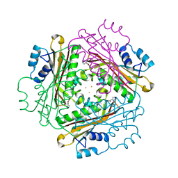
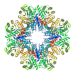
1WPT
 
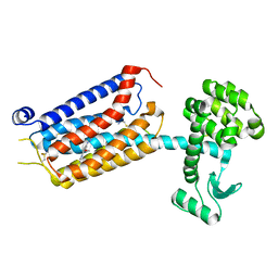
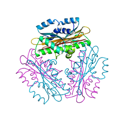 | | Crystal Structure of HutP, an RNA binding anti-termination protein | | 分子名称: | Hut operon positive regulatory protein | | 著者 | Kumarevel, T, Mizuno, H, Kumar, P.K.R. | | 登録日 | 2004-09-13 | | 公開日 | 2005-08-30 | | 最終更新日 | 2023-10-25 | | 実験手法 | X-RAY DIFFRACTION (2.7 Å) | | 主引用文献 | Characterization of the metal ion binding site in the anti-terminator protein, HutP, of Bacillus subtilis
Nucleic Acids Res., 33, 2005
|
|
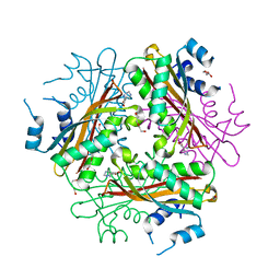
1WMQ
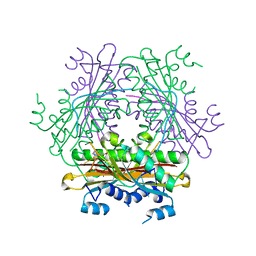 | | Structure of the HutP antitermination complex bound to a single stranded region of hut mRNA | | 分子名称: | 5'-R(P*UP*UP*UP*AP*GP*UP*U)-3', HISTIDINE, Hut operon positive regulatory protein, ... | | 著者 | Kumarevel, T.S, Mizuno, H, Kumar, P.K.R. | | 登録日 | 2004-07-14 | | 公開日 | 2005-03-15 | | 最終更新日 | 2023-10-25 | | 実験手法 | X-RAY DIFFRACTION (1.6 Å) | | 主引用文献 | Structural basis of HutP-mediated anti-termination and roles of the Mg2+ ion and L-histidine ligand.
Nature, 434, 2005
|
|
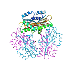
1WPV
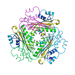 | | Crystal Structure of Activated Binary complex of HutP, an RNA binding anti-termination protein | | 分子名称: | HISTIDINE, Hut operon positive regulatory protein, MAGNESIUM ION | | 著者 | Kumarevel, T.S, Mizuno, H, Kumar, P.K.R. | | 登録日 | 2004-09-14 | | 公開日 | 2005-03-15 | | 最終更新日 | 2023-10-25 | | 実験手法 | X-RAY DIFFRACTION (1.7 Å) | | 主引用文献 | Structural basis of HutP-mediated anti-termination and roles of the Mg2+ ion and L-histidine ligand.
Nature, 434, 2005
|
|
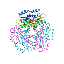
1WPU
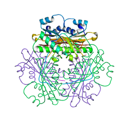 | |
1WRO
 | | Metal Ion dependency of the antiterminator protein, HutP, for binding to the terminator region of hut mRNA- A structural basis | | 分子名称: | BARIUM ION, HISTIDINE, Hut operon positive regulatory protein | | 著者 | Kumarevel, T, Mizuno, H, Kumar, P.K.R. | | 登録日 | 2004-10-25 | | 公開日 | 2005-08-30 | | 最終更新日 | 2023-10-25 | | 実験手法 | X-RAY DIFFRACTION (2.35 Å) | | 主引用文献 | Characterization of the metal ion binding site in the anti-terminator protein, HutP, of Bacillus subtilis
Nucleic Acids Res., 33, 2005
|
|
1WRN
 | | Metal Ion dependency of the antiterminator protein, HutP, for binding to the terminator region of hut mRNA- A structural basis | | 分子名称: | DI(HYDROXYETHYL)ETHER, HISTIDINE, Hut operon positive regulatory protein, ... | | 著者 | Kumarevel, T, Mizuno, H, Kumar, P.K.R. | | 登録日 | 2004-10-25 | | 公開日 | 2005-08-30 | | 最終更新日 | 2024-05-29 | | 実験手法 | X-RAY DIFFRACTION (2.3 Å) | | 主引用文献 | Characterization of the metal ion binding site in the anti-terminator protein, HutP, of Bacillus subtilis
Nucleic Acids Res., 33, 2005
|
|
1WPS
 | |
1VEA
 | | Crystal Structure of HutP, an RNA binding antitermination protein | | 分子名称: | Hut operon positive regulatory protein, N-(2-NAPHTHYL)HISTIDINAMIDE | | 著者 | Kumarevel, T.S, Fujimoto, Z, Karthe, P, Oda, M, Mizuno, H, Kumar, P.K.R. | | 登録日 | 2004-03-29 | | 公開日 | 2004-07-20 | | 最終更新日 | 2023-12-27 | | 実験手法 | X-RAY DIFFRACTION (2.8 Å) | | 主引用文献 | Crystal Structure of Activated HutP; An RNA Binding Protein that Regulates Transcription of the hut Operon in Bacillus subtilis
Structure, 12, 2004
|
|
1NBK
 | | The structure of RNA aptamer for HIV Tat complexed with two argininamide molecules | | 分子名称: | 2-AMINO-5-GUANIDINO-PENTANOIC ACID, RNA aptamer | | 著者 | Matsugami, A, Kobayashi, S, Ouhashi, K, Uesugi, S, Yamamoto, R, Taira, K, Nishikawa, S, Kumar, P.K.R, Katahira, M. | | 登録日 | 2002-12-03 | | 公開日 | 2003-12-03 | | 最終更新日 | 2024-05-29 | | 実験手法 | SOLUTION NMR | | 主引用文献 | Structural Basis of the Highly Efficient Trapping of the HIV Tat Protein by an RNA Aptamer
Structure, 11, 2003
|
|
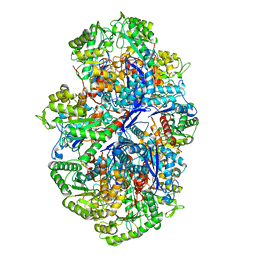
2ZFA
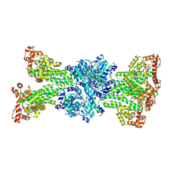
 | | Structure of Lactate Oxidase at pH4.5 from AEROCOCCUS VIRIDANS | | 分子名称: | 1,2-ETHANEDIOL, FLAVIN MONONUCLEOTIDE, Lactate oxidase | | 著者 | Furuichi, M, Balasundaresan, D, Suzuki, N, Yoshida, Y, Minagawa, H, Kaneko, H, Waga, I, Kumar, P.K.R, Mizuno, H. | | 登録日 | 2007-12-26 | | 公開日 | 2008-04-22 | | 最終更新日 | 2023-11-01 | | 実験手法 | X-RAY DIFFRACTION (1.81 Å) | | 主引用文献 | X-ray structures of Aerococcus viridans lactate oxidase and its complex with D-lactate at pH 4.5 show an alpha-hydroxyacid oxidation mechanism
J.Mol.Biol., 378, 2008
|
|
4H4L
 | | Crystal Structure of ternary complex of HutP(HutP-L-His-Zn) | | 分子名称: | HISTIDINE, Hut operon positive regulatory protein, ZINC ION | | 著者 | Dhakshnamoorthy, B, Misono, T.S, Mizuno, H, Kumar, P.K.R. | | 登録日 | 2012-09-17 | | 公開日 | 2013-09-04 | | 最終更新日 | 2023-11-08 | | 実験手法 | X-RAY DIFFRACTION (2.5 Å) | | 主引用文献 | Alternative binding modes of l-histidine guided by metal ions for the activation of the antiterminator protein HutP of Bacillus subtilis.
J.Struct.Biol., 183, 2013
|
|
2NLI
 | | Crystal Structure of the complex between L-lactate oxidase and a substrate analogue at 1.59 angstrom resolution | | 分子名称: | FLAVIN MONONUCLEOTIDE, HYDROGEN PEROXIDE, LACTIC ACID, ... | | 著者 | Furuichi, M, Suzuki, N, Balasundaresan, D, Yoshida, Y, Minagawa, H, Watanabe, Y, Kaneko, H, Waga, I, Kumar, P.K.R, Mizuno, H. | | 登録日 | 2006-10-20 | | 公開日 | 2007-10-23 | | 最終更新日 | 2023-11-15 | | 実験手法 | X-RAY DIFFRACTION (1.59 Å) | | 主引用文献 | X-ray structures of Aerococcus viridans lactate oxidase and its complex with D-lactate at pH 4.5 show an alpha-hydroxyacid oxidation mechanism
J.Mol.Biol., 378, 2008
|
|
2MJX
 | | Solution NMR structure of a mismatch DNA | | 分子名称: | DNA (5'-D(*CP*GP*CP*GP*TP*AP*CP*GP*AP*TP*GP*CP*GP*C)-3'), DNA (5'-D(*GP*CP*GP*CP*AP*TP*GP*CP*TP*AP*CP*GP*CP*G)-3') | | 著者 | Ghosh, A, Kumar, K.R, Bhunia, A, Chatterjee, S. | | 登録日 | 2014-01-21 | | 公開日 | 2014-03-05 | | 最終更新日 | 2024-05-15 | | 実験手法 | SOLUTION NMR | | 主引用文献 | Double GC:GC mismatch in dsDNA enhances local dynamics retaining the DNA footprint: a high-resolution NMR study
Chemmedchem, 9, 2014
|
|
7P8W
 | | Human erythrocyte catalase cryoEM | | 分子名称: | Catalase, NADPH DIHYDRO-NICOTINAMIDE-ADENINE-DINUCLEOTIDE PHOSPHATE, PROTOPORPHYRIN IX CONTAINING FE | | 著者 | Chen, S, Li, J, Vinothkumar, K.R, Henderson, R. | | 登録日 | 2021-07-23 | | 公開日 | 2021-08-25 | | 最終更新日 | 2024-07-17 | | 実験手法 | ELECTRON MICROSCOPY (2.2 Å) | | 主引用文献 | Interaction of human erythrocyte catalase with air-water interface in cryoEM.
Microscopy (Oxf), 71, 2022
|
|
7P2L
 | | thermostabilised 7TM domain of human mGlu5 receptor bound to photoswitchable ligand alloswitch-1 | | 分子名称: | 2-chloranyl-~{N}-[2-methoxy-4-[(~{E})-pyridin-2-yldiazenyl]phenyl]benzamide, Metabotropic glutamate receptor 5,Endolysin,Metabotropic glutamate receptor 5 | | 著者 | Huang, C.Y, Vinothkumar, K.R, Lebon, G. | | 登録日 | 2021-07-06 | | 公開日 | 2021-09-08 | | 最終更新日 | 2024-01-31 | | 実験手法 | X-RAY DIFFRACTION (2.54 Å) | | 主引用文献 | Agonists and allosteric modulators promote signaling from different metabotropic glutamate receptor 5 conformations.
Cell Rep, 36, 2021
|
|
7Y0D
 | |
7V2Q
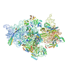 | | T.thermophilus 30S ribosome with KsgA, class K6 | | 分子名称: | 16s ribosomal RNA, 30S ribosomal protein S10, 30S ribosomal protein S11, ... | | 著者 | Raina, R, Singh, J, Anand, R, Vinothkumar, K.R. | | 登録日 | 2021-08-09 | | 公開日 | 2022-04-06 | | 最終更新日 | 2024-06-12 | | 実験手法 | ELECTRON MICROSCOPY (3.24 Å) | | 主引用文献 | Decoding the Mechanism of Specific RNA Targeting by Ribosomal Methyltransferases.
Acs Chem.Biol., 17, 2022
|
|
7V2N
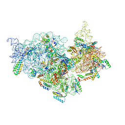 | | T.thermophilus 30S ribosome with KsgA, class K2 | | 分子名称: | 16s ribosomal RNA, 30S ribosomal protein S10, 30S ribosomal protein S11, ... | | 著者 | Raina, R, Singh, J, Anand, R, Vinothkumar, K.R. | | 登録日 | 2021-08-09 | | 公開日 | 2022-04-06 | | 最終更新日 | 2024-06-12 | | 実験手法 | ELECTRON MICROSCOPY (3.6 Å) | | 主引用文献 | Decoding the Mechanism of Specific RNA Targeting by Ribosomal Methyltransferases.
Acs Chem.Biol., 17, 2022
|
|
7V2O
 | | T.thermophilus 30S ribosome with KsgA, class K4 | | 分子名称: | 16s ribosomal RNA, 30S ribosomal protein S10, 30S ribosomal protein S11, ... | | 著者 | Raina, R, Singh, J, Anand, R, Vinothkumar, K.R. | | 登録日 | 2021-08-09 | | 公開日 | 2022-04-06 | | 最終更新日 | 2024-06-12 | | 実験手法 | ELECTRON MICROSCOPY (3.5 Å) | | 主引用文献 | Decoding the Mechanism of Specific RNA Targeting by Ribosomal Methyltransferases.
Acs Chem.Biol., 17, 2022
|
|
7V2L
 | | T.thermophilus 30S ribosome with KsgA, class K1k2 | | 分子名称: | 16s ribosomal RNA, 30S ribosomal protein S10, 30S ribosomal protein S11, ... | | 著者 | Raina, R, Singh, J, Anand, R, Vinothkumar, K.R. | | 登録日 | 2021-08-09 | | 公開日 | 2022-04-06 | | 最終更新日 | 2024-06-12 | | 実験手法 | ELECTRON MICROSCOPY (3.3 Å) | | 主引用文献 | Decoding the Mechanism of Specific RNA Targeting by Ribosomal Methyltransferases.
Acs Chem.Biol., 17, 2022
|
|
7V2M
 | | T.thermophilus 30S ribosome with KsgA, class K1k4 | | 分子名称: | 16s ribosomal RNA, 30S ribosomal protein S10, 30S ribosomal protein S11, ... | | 著者 | Raina, R, Singh, J, Anand, R, Vinothkumar, K.R. | | 登録日 | 2021-08-09 | | 公開日 | 2022-04-06 | | 最終更新日 | 2024-06-12 | | 実験手法 | ELECTRON MICROSCOPY (3.4 Å) | | 主引用文献 | Decoding the Mechanism of Specific RNA Targeting by Ribosomal Methyltransferases.
Acs Chem.Biol., 17, 2022
|
|
7V2P
 | | T.thermophilus 30S ribosome with KsgA, class K5 | | 分子名称: | 16s ribosomal RNA, 30S ribosomal protein S10, 30S ribosomal protein S11, ... | | 著者 | Raina, R, Singh, J, Anand, R, Vinothkumar, K.R. | | 登録日 | 2021-08-09 | | 公開日 | 2022-04-06 | | 最終更新日 | 2024-06-12 | | 実験手法 | ELECTRON MICROSCOPY (3.3 Å) | | 主引用文献 | Decoding the Mechanism of Specific RNA Targeting by Ribosomal Methyltransferases.
Acs Chem.Biol., 17, 2022
|
|
7E51
 | |
7E4X
 | |
5MWV
 | | Solid-state NMR Structure of outer membrane protein G in lipid bilayers | | 分子名称: | Outer membrane protein G | | 著者 | Retel, J.S, Nieuwkoop, A.J, Hiller, M, Higman, V.A, Barbet-Massin, E, Stanek, J, Andreas, L.B, Franks, W.T, van Rossum, B.-J, Vinothkumar, K.R, Handel, L, de Palma, G.G, Bardiaux, B, Pintacuda, G, Emsley, L, Kuelbrandt, W, Oschkinat, H. | | 登録日 | 2017-01-20 | | 公開日 | 2017-12-27 | | 最終更新日 | 2024-05-15 | | 実験手法 | SOLID-STATE NMR | | 主引用文献 | Structure of outer membrane protein G in lipid bilayers.
Nat Commun, 8, 2017
|
|


