Loading
PDBj
MenuPDBj@FacebookPDBj@X(formerly Twitter)PDBj@BlueSkyPDBj@YouTubewwPDB FoundationwwPDB
RCSB PDBPDBeBMRBAdv. SearchSearch help
PDB: 55 results

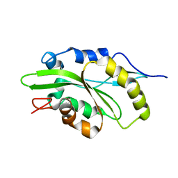
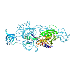
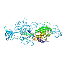
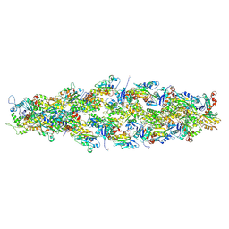
7U8K
DownloadVisualize
BU of 7u8k by Molmil
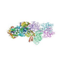
Magic Angle Spinning NMR Structure of Human Cofilin-2 Assembled on Actin Filaments
Descriptor: Actin, alpha skeletal muscle, Cofilin-2
Authors:Kraus, J, Russell, R, Kudryashova, E, Xu, C, Katyal, N, Kudryashov, D, Perilla, J.R, Polenova, T.
Deposit date:2022-03-08
Release date:2023-08-16
Last modified:2024-10-23
Method:SOLID-STATE NMR
Cite:Magic angle spinning NMR structure of human cofilin-2 assembled on actin filaments reveals isoform-specific conformation and binding mode.
Nat Commun, 13, 2022
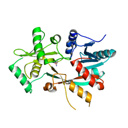
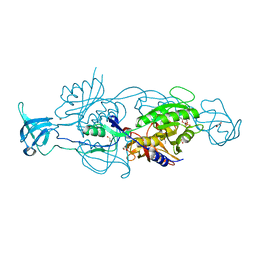
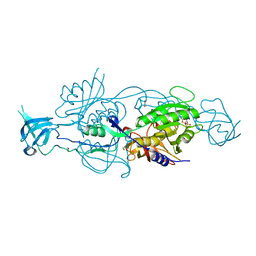
7M0G
DownloadVisualize
BU of 7m0g by Molmil
Magic Angle Spinning NMR Structure of Human Cofilin-2 Assembled on Actin Filaments
Descriptor: Cofilin-2
Authors:Kraus, J, Polenova, T, Perilla, J.P, Xu, C.
Deposit date:2021-03-10
Release date:2022-04-20
Last modified:2024-05-15
Method:SOLID-STATE NMR
Cite:Magic angle spinning NMR structure of human cofilin-2 assembled on actin filaments reveals isoform-specific conformation and binding mode.
Nat Commun, 13, 2022
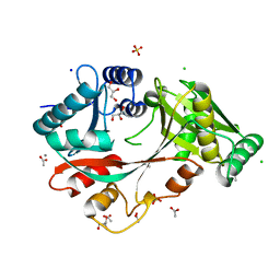
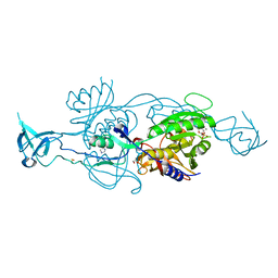
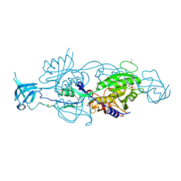
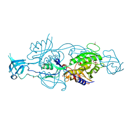
4V36
DownloadVisualize
BU of 4v36 by Molmil
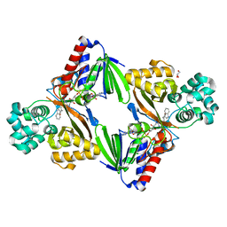
The structure of L-PGS from Bacillus licheniformis
Descriptor: 2,6-DIAMINO-HEXANOIC ACID AMIDE, 2-AMINO-2-HYDROXYMETHYL-PROPANE-1,3-DIOL, LYSYL-TRNA-DEPENDENT L-YSYL-PHOSPHATIDYLGYCEROL SYNTHASE
Authors:Krausze, J, Hebecker, S, Heinz, D.W, Moser, J.
Deposit date:2014-10-16
Release date:2015-08-19
Last modified:2024-05-08
Method:X-RAY DIFFRACTION (2.1 Å)
Cite:Structures of Two Bacterial Resistance Factors Mediating tRNA-Dependent Aminoacylation of Phosphatidylglycerol with Lysine or Alanine.
Proc.Natl.Acad.Sci.USA, 112, 2015
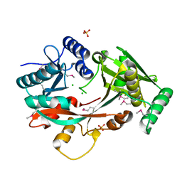
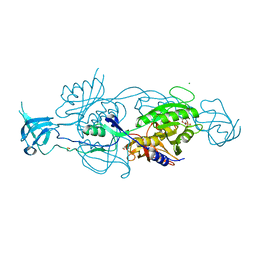
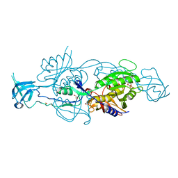
4V35
DownloadVisualize
BU of 4v35 by Molmil
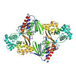
The Structure of A-PGS from Pseudomonas aeruginosa
Descriptor: ACETATE ION, ALANYL-TRNA-DEPENDENT L-ALANYL- PHOPHATIDYLGLYCEROL SYNTHASE, CALCIUM ION, ...
Authors:Krausze, J, Hebecker, S, Hasenkampf, T, Heinz, D.W, Moser, J.
Deposit date:2014-10-16
Release date:2015-08-19
Last modified:2024-05-08
Method:X-RAY DIFFRACTION (2.3 Å)
Cite:Structures of Two Bacterial Resistance Factors Mediating tRNA-Dependent Aminoacylation of Phosphatidylglycerol with Lysine or Alanine.
Proc.Natl.Acad.Sci.USA, 112, 2015
4V34
DownloadVisualize
BU of 4v34 by Molmil
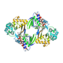
The Structure of A-PGS from Pseudomonas aeruginosa (SeMet derivative)
Descriptor: ALANYL-TRNA-DEPENDENT L-ALANYL- PHOPHATIDYLGLYCEROL SYNTHASE, CHLORIDE ION, SULFATE ION
Authors:Krausze, J, Hebecker, S, Hasenkampf, T, Heinz, D.W, Moser, J.
Deposit date:2014-10-16
Release date:2015-08-19
Last modified:2024-10-09
Method:X-RAY DIFFRACTION (3.1 Å)
Cite:Structures of Two Bacterial Resistance Factors Mediating tRNA-Dependent Aminoacylation of Phosphatidylglycerol with Lysine or Alanine.
Proc.Natl.Acad.Sci.USA, 112, 2015
6Y01
DownloadVisualize
BU of 6y01 by Molmil
The structure of the molybdenum cofactor binding protein from the phototrophic bacterium Rippkaea orientalis
Descriptor: CHLORIDE ION, IMIDAZOLE, p450 cytochrome, ...
Authors:Krausze, J.
Deposit date:2020-02-05
Release date:2020-09-16
Last modified:2024-01-24
Method:X-RAY DIFFRACTION (1.23 Å)
Cite:The structure of the Moco carrier protein from Rippkaea orientalis.
Acta Crystallogr.,Sect.F, 76, 2020
6Q32
DownloadVisualize
BU of 6q32 by Molmil
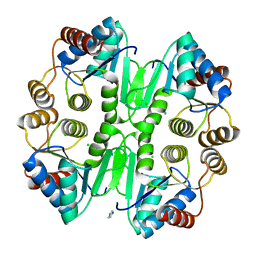
The structure of the Mo-insertase domain Cnx1E (variant S269DD274S) from Arabidopsis thaliana in complex with Moco-AMP
Descriptor: 2-(N-MORPHOLINO)-ETHANESULFONIC ACID, MAGNESIUM ION, Moco-AMP, ...
Authors:Krausze, J.
Deposit date:2018-12-03
Release date:2021-02-17
Last modified:2024-01-24
Method:X-RAY DIFFRACTION (1.39 Å)
Cite:Mechanism of molybdate insertion into pterin-based molybdenum cofactors.
Nat.Chem., 13, 2021
6ROZ
DownloadVisualize
BU of 6roz by Molmil
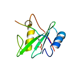
Structure of the N-SH2 domain of the human tyrosine-protein phosphatase non-receptor type 11 in complex with the phosphorylated immune receptor tyrosine-based switch motif
Descriptor: Tyrosine-protein phosphatase non-receptor type 11, immune receptor tyrosine-based switch motif (ITSM)
Authors:Krausze, J, Sikorska, J, Carlomagno, T.
Deposit date:2019-05-13
Release date:2020-02-19
Last modified:2024-10-23
Method:X-RAY DIFFRACTION (2.89 Å)
Cite:Molecular mechanism of SHP2 activation by PD-1 stimulation.
Sci Adv, 6, 2020
6ROY
DownloadVisualize
BU of 6roy by Molmil
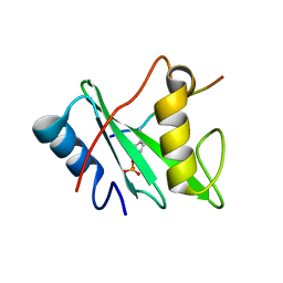
Structure of the N-SH2 domain of the human tyrosine-protein phosphatase non-receptor type 11 in complex with the phosphorylated immune receptor tyrosine-based inhibitory motif
Descriptor: Tyrosine-protein phosphatase non-receptor type 11, immune receptor tyrosine-based inhibitory motif (ITIM)
Authors:Krausze, J, Sikorska, J, Carlomagno, T.
Deposit date:2019-05-13
Release date:2020-02-19
Last modified:2024-10-16
Method:X-RAY DIFFRACTION (2.1 Å)
Cite:Molecular mechanism of SHP2 activation by PD-1 stimulation.
Sci Adv, 6, 2020
6RMS
DownloadVisualize
BU of 6rms by Molmil
The Structure of variant D274E of the Mo-insertase domain Cnx1E from Arabidopsis thaliana in complex with AMP
Descriptor: 1,2-ETHANEDIOL, ADENOSINE MONOPHOSPHATE, IMIDAZOLE, ...
Authors:Krausze, J.
Deposit date:2019-05-07
Release date:2020-02-19
Last modified:2024-01-24
Method:SOLUTION SCATTERING (1.74 Å), X-RAY DIFFRACTION
Cite:Insights into the Cnx1E catalyzed MPT-AMP hydrolysis.
Biosci.Rep., 40, 2020
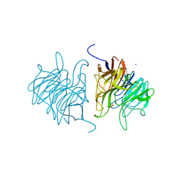
5A10
DownloadVisualize
BU of 5a10 by Molmil
The crystal structure of Ta-TFP, a thiocyanate-forming protein involved in glucosinolate breakdown (space group C2)
Descriptor: ACETATE ION, SODIUM ION, THIOCYANATE FORMING PROTEIN
Authors:Krausze, J, Gumz, F, Wittstock, U.
Deposit date:2015-04-27
Release date:2015-08-26
Last modified:2024-01-10
Method:X-RAY DIFFRACTION (1.423 Å)
Cite:The Crystal Structure of the Thiocyanate-Forming Protein from Thlaspi Arvense, a Kelch Protein Involved in Glucosinolate Breakdown.
Plant Mol.Biol., 89, 2015
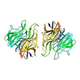
5A11
DownloadVisualize
BU of 5a11 by Molmil
The crystal structure of Ta-TFP, a thiocyanate-forming protein involved in glucosinolate breakdown (space group P21)
Descriptor: IODIDE ION, THIOCYANATE FORMING PROTEIN
Authors:Krausze, J, Gumz, F, Wittstock, U.
Deposit date:2015-04-27
Release date:2015-08-26
Last modified:2024-01-10
Method:X-RAY DIFFRACTION (2.47 Å)
Cite:The Crystal Structure of the Thiocyanate-Forming Protein from Thlaspi Arvense, a Kelch Protein Involved in Glucosinolate Breakdown.
Plant Mol.Biol., 89, 2015
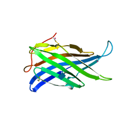
4AKM
DownloadVisualize
BU of 4akm by Molmil
Crystal structure of the human lysosome-associated membrane protein LAMP-3 (aka DC-LAMP)
Descriptor: 2-acetamido-2-deoxy-beta-D-glucopyranose, IRIDIUM (III) ION, LYSOSOME-ASSOCIATED MEMBRANE GLYCOPROTEIN 3
Authors:Krausze, J, Wilke, S, Buessow, K.
Deposit date:2012-02-24
Release date:2012-08-15
Last modified:2024-10-09
Method:X-RAY DIFFRACTION (2.687 Å)
Cite:Crystal Structure of the Conserved Domain of the Dc Lysosomal Associated Membrane Protein: Implications for the Lysosomal Glycocalyx.
Bmc Biol., 10, 2012
5G3U
DownloadVisualize
BU of 5g3u by Molmil
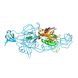
The structure of the L-tryptophan oxidase VioA from Chromobacterium violaceum in complex with its inhibitor 2-(1H-indol-3-ylmethyl)prop-2- enoic acid
Descriptor: 2-[(1H-indol-3-yl)methyl]prop-2-enoic acid, DIHYDROFLAVINE-ADENINE DINUCLEOTIDE, GLYCEROL, ...
Authors:Krausze, J, Rabe, J, Moser, J.
Deposit date:2016-05-01
Release date:2016-08-03
Last modified:2024-10-23
Method:X-RAY DIFFRACTION (2.377 Å)
Cite:Biosynthesis of Violacein, Structure and Function of l-Tryptophan Oxidase VioA from Chromobacterium violaceum.
J.Biol.Chem., 291, 2016
5G3S
DownloadVisualize
BU of 5g3s by Molmil
The structure of the L-tryptophan oxidase VioA from Chromobacterium violaceum - Samarium derivative
Descriptor: 2-AMINO-2-HYDROXYMETHYL-PROPANE-1,3-DIOL, CHLORIDE ION, DIHYDROFLAVINE-ADENINE DINUCLEOTIDE, ...
Authors:Krausze, J, Rabe, J, Moser, J.
Deposit date:2016-05-01
Release date:2016-08-03
Last modified:2019-03-06
Method:X-RAY DIFFRACTION (2.076 Å)
Cite:Biosynthesis of Violacein: Structure and Function of L-Tryptophan Oxidase Vioa Chromobacterium Violaceum
J.Biol.Chem., 291, 2016
5G3T
DownloadVisualize
BU of 5g3t by Molmil
The structure of the L-tryptophan oxidase VioA from Chromobacterium violaceum
Descriptor: 2-AMINO-2-HYDROXYMETHYL-PROPANE-1,3-DIOL, CHLORIDE ION, DIHYDROFLAVINE-ADENINE DINUCLEOTIDE, ...
Authors:Krausze, J, Rabe, J, Moser, J.
Deposit date:2016-05-01
Release date:2016-08-03
Last modified:2024-05-08
Method:X-RAY DIFFRACTION (1.8 Å)
Cite:Biosynthesis of Violacein: Structure and Function of L-Tryptophan Oxidase Vioa Chromobacterium Violaceum
J.Biol.Chem., 291, 2016
2YNM
DownloadVisualize
BU of 2ynm by Molmil
Structure of the ADPxAlF3-Stabilized Transition State of the Nitrogenase-like Dark-Operative Protochlorophyllide Oxidoreductase Complex from Prochlorococcus marinus with Its Substrate Protochlorophyllide a
Descriptor: 4-(2-HYDROXYETHYL)-1-PIPERAZINE ETHANESULFONIC ACID, ADENOSINE-5'-DIPHOSPHATE, ALUMINUM FLUORIDE, ...
Authors:Krausze, J, Lange, C, Heinz, D.W, Moser, J.
Deposit date:2012-10-16
Release date:2013-01-30
Last modified:2023-12-20
Method:X-RAY DIFFRACTION (2.1 Å)
Cite:Structure of Adp-Aluminium Fluoride-Stabilized Protochlorophyllide Oxidoreductase Complex.
Proc.Natl.Acad.Sci.USA, 110, 2013
6ETF
DownloadVisualize
BU of 6etf by Molmil
The Structure of the Mo-insertase domain Cnx1E from Arabidopsis thaliana in complex with AMP and molybdate
Descriptor: 1,2-ETHANEDIOL, ADENOSINE MONOPHOSPHATE, MAGNESIUM ION, ...
Authors:Krausze, J.
Deposit date:2017-10-26
Release date:2018-07-18
Last modified:2024-01-17
Method:X-RAY DIFFRACTION (1.781 Å)
Cite:The functional principle of eukaryotic molybdenum insertases.
Biochem. J., 475, 2018
6ETH
DownloadVisualize
BU of 6eth by Molmil
The Structure of the Mo-insertase domain Cnx1E from Arabidopsis thaliana in complex with AMP and tungstate
Descriptor: 1,2-ETHANEDIOL, ACETYL GROUP, ADENOSINE MONOPHOSPHATE, ...
Authors:Krausze, J.
Deposit date:2017-10-26
Release date:2018-07-18
Last modified:2024-01-17
Method:X-RAY DIFFRACTION (1.64 Å)
Cite:The functional principle of eukaryotic molybdenum insertases.
Biochem. J., 475, 2018
6ETD
DownloadVisualize
BU of 6etd by Molmil
The Structure of the Mo-insertase domain Cnx1E from Arabidopsis thaliana in complex with AMP
Descriptor: 1,2-ETHANEDIOL, ACETYL GROUP, ADENOSINE MONOPHOSPHATE, ...
Authors:Krausze, J.
Deposit date:2017-10-26
Release date:2018-07-18
Last modified:2024-01-17
Method:X-RAY DIFFRACTION (1.723 Å)
Cite:The functional principle of eukaryotic molybdenum insertases.
Biochem. J., 475, 2018
6GB0
DownloadVisualize
BU of 6gb0 by Molmil
The Structure of variant K294A of the Mo-insertase domain Cnx1E from Arabidopsis thaliana in complex with AMP
Descriptor: 1,2-ETHANEDIOL, ADENOSINE MONOPHOSPHATE, CHLORIDE ION, ...
Authors:Krausze, J.
Deposit date:2018-04-13
Release date:2018-07-18
Last modified:2024-01-17
Method:X-RAY DIFFRACTION (1.814 Å)
Cite:The functional principle of eukaryotic molybdenum insertases.
Biochem. J., 475, 2018
6GBC
DownloadVisualize
BU of 6gbc by Molmil
The Structure of variant R369A of the Mo-insertase domain Cnx1E from Arabidopsis thaliana in complex with AMP and molybdate
Descriptor: 1,2-ETHANEDIOL, ADENOSINE MONOPHOSPHATE, MAGNESIUM ION, ...
Authors:Krausze, J.
Deposit date:2018-04-13
Release date:2018-07-18
Last modified:2024-01-17
Method:X-RAY DIFFRACTION (1.59 Å)
Cite:The functional principle of eukaryotic molybdenum insertases.
Biochem. J., 475, 2018
6GAX
DownloadVisualize
BU of 6gax by Molmil
The Structure of variant K294A of the Mo-insertase domain Cnx1E from Arabidopsis thaliana in complex with AMP and molybdate
Descriptor: 1,2-ETHANEDIOL, ADENOSINE MONOPHOSPHATE, MAGNESIUM ION, ...
Authors:Krausze, J.
Deposit date:2018-04-13
Release date:2018-07-18
Last modified:2024-01-17
Method:X-RAY DIFFRACTION (1.77 Å)
Cite:The functional principle of eukaryotic molybdenum insertases.
Biochem. J., 475, 2018
6GBF
DownloadVisualize
BU of 6gbf by Molmil
The Structure of variant R369A of the Mo-insertase domain Cnx1E from Arabidopsis thaliana in complex with AMP
Descriptor: 1,2-ETHANEDIOL, ACETATE ION, ADENOSINE MONOPHOSPHATE, ...
Authors:Krausze, J.
Deposit date:2018-04-13
Release date:2018-07-18
Last modified:2024-01-17
Method:X-RAY DIFFRACTION (1.712 Å)
Cite:The functional principle of eukaryotic molybdenum insertases.
Biochem. J., 475, 2018
6GB4
DownloadVisualize
BU of 6gb4 by Molmil
The Structure of variant S328A of the Mo-insertase domain Cnx1E from Arabidopsis thaliana in complex with AMP and molybdate
Descriptor: 1,2-ETHANEDIOL, ADENOSINE MONOPHOSPHATE, MAGNESIUM ION, ...
Authors:Krausze, J.
Deposit date:2018-04-13
Release date:2018-07-18
Last modified:2024-01-17
Method:X-RAY DIFFRACTION (1.648 Å)
Cite:The functional principle of eukaryotic molybdenum insertases.
Biochem. J., 475, 2018


123>

227344

건을2024-11-13부터공개중

PDB statisticsPDBj update infoContact PDBjnumon