Loading
PDBj
MenuPDBj@FacebookPDBj@TwitterPDBj@YouTubewwPDB FoundationwwPDB
RCSB PDBPDBeBMRBAdv. SearchSearch help
PDB: 121 results

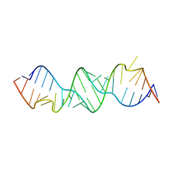
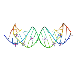
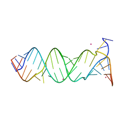
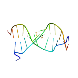
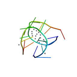
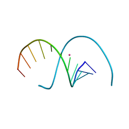
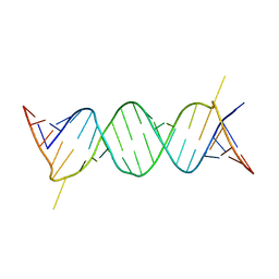
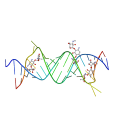
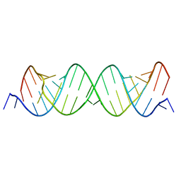
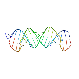
3BNP
DownloadVisualize
BU of 3bnp by Molmil
Crystal Structure of the Homo sapiens Mitochondrial Ribosomal Decoding Site (A1555G Mutant)
Descriptor: A site of human mitochondrial ribosome, A chain, B chain, ...
Authors:Kondo, J, Westhof, E.
Deposit date:2007-12-14
Release date:2008-06-24
Last modified:2024-02-21
Method:X-RAY DIFFRACTION (2.7 Å)
Cite:The bacterial and mitochondrial ribosomal A-site molecular switches possess different conformational substates
Nucleic Acids Res., 36, 2008
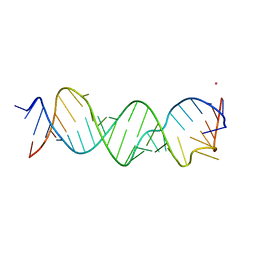
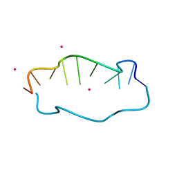
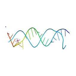
3BNT
DownloadVisualize
BU of 3bnt by Molmil
Crystal Structure of the Homo sapiens Mitochondrial Ribosomal Decoding Site in the Presence of [Co(NH3)6]Cl3 (A1555G mutant, Br-derivative)
Descriptor: A site of human mitochondrial ribosome, COBALT HEXAMMINE(III), SODIUM ION
Authors:Kondo, J, Westhof, E.
Deposit date:2007-12-14
Release date:2008-06-24
Last modified:2024-02-21
Method:X-RAY DIFFRACTION (2.3 Å)
Cite:The bacterial and mitochondrial ribosomal A-site molecular switches possess different conformational substates
Nucleic Acids Res., 36, 2008
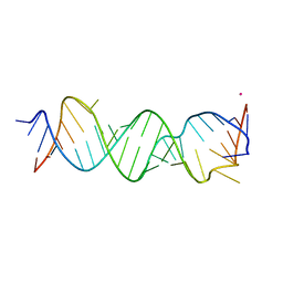
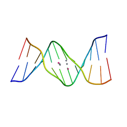
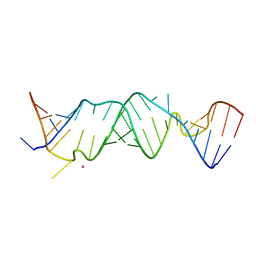
3BNN
DownloadVisualize
BU of 3bnn by Molmil
Crystal Structure of the Homo sapiens Mitochondrial Ribosomal Decoding Site
Descriptor: A site of human ribosome
Authors:Kondo, J, Westhof, E.
Deposit date:2007-12-14
Release date:2008-06-24
Last modified:2024-02-21
Method:X-RAY DIFFRACTION (2 Å)
Cite:The bacterial and mitochondrial ribosomal A-site molecular switches possess different conformational substates
Nucleic Acids Res., 36, 2008
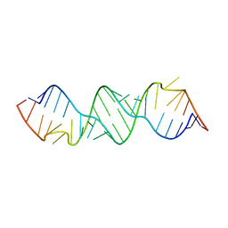
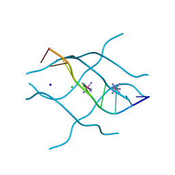
3BNS
DownloadVisualize
BU of 3bns by Molmil
Crystal Structure of the Homo sapiens Mitochondrial Ribosomal Decoding Site (A1555G mutant, Br-derivative)
Descriptor: A site of human mitochondrial ribosome, chain three, chain two, ...
Authors:Kondo, J, Westhof, E.
Deposit date:2007-12-14
Release date:2008-06-24
Last modified:2024-02-21
Method:X-RAY DIFFRACTION (1.9 Å)
Cite:The bacterial and mitochondrial ribosomal A-site molecular switches possess different conformational substates
Nucleic Acids Res., 36, 2008
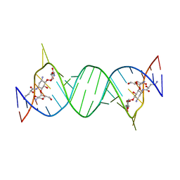
3BNR
DownloadVisualize
BU of 3bnr by Molmil
Crystal Structure of the Homo sapiens Mitochondrial Ribosomal Decoding Site in the presence of nonspecifically bound paromomycin (A1555G mutant, Br-derivative)
Descriptor: A site of human mitochondrial ribosome, chain one, chain three, ...
Authors:Kondo, J, Westhof, E.
Deposit date:2007-12-14
Release date:2008-06-24
Last modified:2024-02-21
Method:X-RAY DIFFRACTION (2.1 Å)
Cite:The bacterial and mitochondrial ribosomal A-site molecular switches possess different conformational substates
Nucleic Acids Res., 36, 2008
3BNO
DownloadVisualize
BU of 3bno by Molmil
Crystal Structure of the Homo sapiens Mitochondrial Ribosomal Decoding Site (Br-derivative)
Descriptor: A site of human mitochondrial ribosome
Authors:Kondo, J, Westhof, E.
Deposit date:2007-12-14
Release date:2008-06-24
Last modified:2024-02-21
Method:X-RAY DIFFRACTION (2.35 Å)
Cite:The bacterial and mitochondrial ribosomal A-site molecular switches possess different conformational substates
Nucleic Acids Res., 36, 2008
3BNL
DownloadVisualize
BU of 3bnl by Molmil
Crystal structure of the bacterial ribosomal decoding A site in the presence of [Co(NH3)6]Cl3
Descriptor: A site of bacterial ribosome, COBALT HEXAMMINE(III)
Authors:Kondo, J, Westhof, E.
Deposit date:2007-12-14
Release date:2008-06-24
Last modified:2024-02-21
Method:X-RAY DIFFRACTION (2.602 Å)
Cite:The bacterial and mitochondrial ribosomal A-site molecular switches possess different conformational substates
Nucleic Acids Res., 36, 2008
3BNQ
DownloadVisualize
BU of 3bnq by Molmil
Crystal Structure of the Homo sapiens Mitochondrial Ribosomal Decoding Site in the Presence of SrCl2 (A1555G mutant, Br-derivative)
Descriptor: A site of human mitochondrial ribosome, A chain, B chain, ...
Authors:Kondo, J, Westhof, E.
Deposit date:2007-12-14
Release date:2008-06-24
Last modified:2024-02-21
Method:X-RAY DIFFRACTION (2 Å)
Cite:The bacterial and mitochondrial ribosomal A-site molecular switches possess different conformational substates
Nucleic Acids Res., 36, 2008
6IYQ
DownloadVisualize
BU of 6iyq by Molmil
Crystal structure of a DNA duplex cross-linked by 6-thioguanine-6-thioguanine disulfides
Descriptor: DNA (5'-D(*CP*GP*CP*GP*AP*(S6G)P*(S6G)P*(BRU)P*CP*GP*CP*G)-3')
Authors:Kondo, J, Ono, A, Atsugi, T.
Deposit date:2018-12-17
Release date:2019-10-30
Last modified:2024-03-27
Method:X-RAY DIFFRACTION (2.01 Å)
Cite:Crystal structure of a DNA duplex cross-linked by 6-thioguanine-6-thioguanine disulfides: reversible formation and cleavage catalyzed by Cu(II) ion and glutathione
Rsc Adv, 2019
6JR4
DownloadVisualize
BU of 6jr4 by Molmil
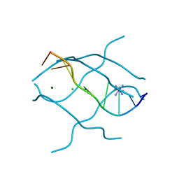
Crystal structure of a NIR-emitting DNA-stabilized Ag16 nanocluster
Descriptor: CALCIUM ION, DNA (5'-D(*CP*AP*CP*CP*TP*AP*GP*CP*GP*A)-3'), SILVER ION
Authors:Kondo, J, Cerretani, C, Kanazawa, H, Vosch, T.
Deposit date:2019-04-02
Release date:2019-08-28
Last modified:2024-03-27
Method:X-RAY DIFFRACTION (1.798 Å)
Cite:Crystal structure of a NIR-Emitting DNA-Stabilized Ag16Nanocluster.
Angew.Chem.Int.Ed.Engl., 58, 2019
7XKM
DownloadVisualize
BU of 7xkm by Molmil
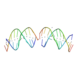
Crystal structure of DNA-Ag(I) rod comprising a one-dimensional array of 11 silver ions
Descriptor: DNA (5'-D(*CP*GP*CP*GP*CP*(BRU)P*CP*(BRU)P*CP*GP*CP*G)-3'), SILVER ION
Authors:Kondo, J, Ono, A, Atsugi, T.
Deposit date:2022-04-20
Release date:2022-07-06
Last modified:2024-05-29
Method:X-RAY DIFFRACTION (2.79 Å)
Cite:A Novel Ag I -DNA Rod Comprising a One-Dimensional Array of 11 Silver Ions within a Double Helical Structure.
Angew.Chem.Int.Ed.Engl., 61, 2022
7XLW
DownloadVisualize
BU of 7xlw by Molmil
Crystal structure of a NIR-emitting DNA-stabilized Ag16 nanocluster (G7I/G9I mutant)
Descriptor: DNA (5'-D(*CP*AP*CP*CP*TP*AP*IP*CP*IP*A)-3'), SILVER ION
Authors:Kondo, J, Cerretani, C, Vosch, T.
Deposit date:2022-04-23
Release date:2022-07-13
Last modified:2023-11-29
Method:X-RAY DIFFRACTION (1.89 Å)
Cite:The effect of inosine on the spectroscopic properties and crystal structure of a NIR-emitting DNA-stabilized silver nanocluster.
Nanoscale Adv, 4, 2022
7XLV
DownloadVisualize
BU of 7xlv by Molmil
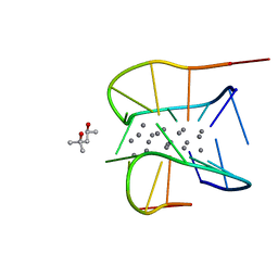
Crystal structure of a NIR-emitting DNA-stabilized Ag16 nanocluster (G7I mutant)
Descriptor: (4S)-2-METHYL-2,4-PENTANEDIOL, DNA (5'-D(*CP*AP*CP*CP*TP*AP*IP*CP*GP*A)-3'), SILVER ION
Authors:Kondo, J, Cerretani, C, Vosch, T.
Deposit date:2022-04-23
Release date:2022-07-13
Last modified:2023-11-29
Method:X-RAY DIFFRACTION (1.1 Å)
Cite:The effect of inosine on the spectroscopic properties and crystal structure of a NIR-emitting DNA-stabilized silver nanocluster.
Nanoscale Adv, 4, 2022
7Y8P
DownloadVisualize
BU of 7y8p by Molmil
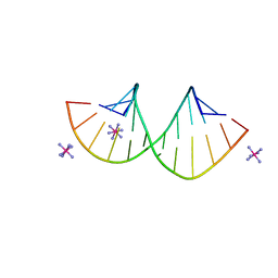
Crystal structure of 4'-selenoRNA duplex
Descriptor: COBALT HEXAMMINE(III), RNA (5'-R(*GP*GP*AP*(IKS)P*(ILK)P*(IKS)P*GP*AP*GP*UP*CP*C)-3')
Authors:Kondo, J, Minakawa, N, Ohta, M, Takahashi, H, Tarashima, N.
Deposit date:2022-06-24
Release date:2023-05-03
Last modified:2023-11-29
Method:X-RAY DIFFRACTION (1.5 Å)
Cite:Synthesis and properties of fully-modified 4'-selenoRNA, an endonuclease-resistant RNA analog.
Bioorg.Med.Chem., 76, 2022
7YGP
DownloadVisualize
BU of 7ygp by Molmil
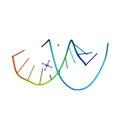
DNA duplex containing 5OHU-T base pairs
Descriptor: COBALT (II) ION, COBALT HEXAMMINE(III), DNA (5'-D(*GP*GP*AP*CP*CP*TP*(OHU)P*GP*GP*TP*CP*C)-3')
Authors:Kondo, J, Torigoe, H, Arakawa, F.
Deposit date:2022-07-12
Release date:2023-05-24
Last modified:2024-05-29
Method:X-RAY DIFFRACTION (1.997 Å)
Cite:Specific binding of Hg 2+ to mismatched base pairs involving 5-hydroxyuracil in duplex DNA.
J.Inorg.Biochem., 241, 2023
7YGO
DownloadVisualize
BU of 7ygo by Molmil
DNA duplex containing 5OHU-Hg(II)-T base pairs
Descriptor: DNA (5'-D(*GP*GP*AP*CP*CP*TP*(6HU)P*GP*GP*TP*CP*C)-3'), MERCURY (II) ION
Authors:Kondo, J, Torigoe, H, Arakawa, F.
Deposit date:2022-07-12
Release date:2023-05-24
Last modified:2024-05-29
Method:X-RAY DIFFRACTION (2.403 Å)
Cite:Specific binding of Hg 2+ to mismatched base pairs involving 5-hydroxyuracil in duplex DNA.
J.Inorg.Biochem., 241, 2023
7YVX
DownloadVisualize
BU of 7yvx by Molmil
Left-handed DNA duplex containing consecutive G-G base pairs
Descriptor: COBALT (II) ION, DNA (5'-D(*CP*GP*(CBR)P*GP*GP*GP*CP*GP*CP*G)-3')
Authors:Kondo, J.
Deposit date:2022-08-19
Release date:2022-10-12
Last modified:2024-05-29
Method:X-RAY DIFFRACTION (2.99 Å)
Cite:Left-handed DNA duplex containing consecutive G-G base pairs
To Be Published
5XUV
DownloadVisualize
BU of 5xuv by Molmil
Crystal structure of DNA duplex containing 4-thiothymine-2Ag(I)-4-thiothymine base pairs
Descriptor: DNA (5'-D(*CP*GP*CP*GP*AP*(8RO)P*(8RO)P*TP*CP*GP*CP*G)-3'), POTASSIUM ION, SILVER ION
Authors:Kondo, J, Sugawara, T, Saneyoshi, H, Ono, A.
Deposit date:2017-06-26
Release date:2017-09-20
Last modified:2023-11-22
Method:X-RAY DIFFRACTION (1.9 Å)
Cite:Crystal structure of a DNA duplex containing four Ag(i) ions in consecutive dinuclear Ag(i)-mediated base pairs: 4-thiothymine-2Ag(i)-4-thiothymine
Chem. Commun. (Camb.), 53, 2017
1UHY
DownloadVisualize
BU of 1uhy by Molmil
Crystal structure of d(GCGATAGC): the base-intercalated duplex
Descriptor: 5'-D(*GP*(CBR)P*GP*AP*TP*AP*GP*C)-3', CHLORIDE ION, COBALT HEXAMMINE(III), ...
Authors:Kondo, J, Umeda, S.I, Fujita, K, Sunami, T, Takenaka, A.
Deposit date:2003-07-13
Release date:2004-02-03
Last modified:2023-12-27
Method:X-RAY DIFFRACTION (1.7 Å)
Cite:X-ray analyses of d(GCGAXAGC) containing G and T at X: the base-intercalated duplex is still stable even in point mutants at the fifth residue.
J.Synchrotron Radiat., 11, 2004
1UHX
DownloadVisualize
BU of 1uhx by Molmil
Crystal structure of d(GCGAGAGC): the base-intercalated duplex
Descriptor: 5'-D(*GP*(CBR)P*GP*AP*GP*AP*GP*C)-3', CHLORIDE ION, COBALT HEXAMMINE(III), ...
Authors:Kondo, J, Umeda, S.I, Fujita, K, Sunami, T, Takenaka, A.
Deposit date:2003-07-13
Release date:2004-02-03
Last modified:2023-12-27
Method:X-RAY DIFFRACTION (2 Å)
Cite:X-ray analyses of d(GCGAXAGC) containing G and T at X: the base-intercalated duplex is still stable even in point mutants at the fifth residue.
J.Synchrotron Radiat., 11, 2004
4P3U
DownloadVisualize
BU of 4p3u by Molmil
Crystal structure of the bacterial A1408U-mutant ribosomal decoding site (C2 form 1)
Descriptor: 5'-R(*UP*UP*GP*CP*GP*UP*CP*UP*CP*GP*(5BU)P*CP*GP*AP*CP*GP*AP*AP*GP*UP*CP*GP*C)-3'
Authors:Kondo, J, Koganei, M.
Deposit date:2014-03-10
Release date:2016-05-04
Last modified:2023-12-27
Method:X-RAY DIFFRACTION (3 Å)
Cite:Crystal structure of the bacterial A1408U-mutant ribosomal decoding site (C2 form 1)
To Be Published
4P3S
DownloadVisualize
BU of 4p3s by Molmil
Crystal structure of the bacterial A1408C-mutant ribosomal decoding site in complex with geneticin
Descriptor: 5'-R(*UP*UP*GP*CP*GP*UP*CP*CP*CP*GP*(5BU)P*CP*GP*AP*CP*GP*AP*AP*GP*UP*CP*GP*C)-3', GENETICIN
Authors:Kondo, J, Koganei, M.
Deposit date:2014-03-10
Release date:2016-05-04
Last modified:2023-12-27
Method:X-RAY DIFFRACTION (2.3 Å)
Cite:Crystal structure of the bacterial A1408C-mutant ribosomal decoding site in complex with geneticin
To Be Published
4P3T
DownloadVisualize
BU of 4p3t by Molmil
Crystal structure of the bacterial A1408C-mutant ribosomal decoding site
Descriptor: 5'-D(*UP*UP*GP*CP*GP*UP*CP*CP*CP*GP*(5BU)P*CP*GP*AP*CP*GP*AP*AP*GP*UP*CP*GP*C)-3'
Authors:Kondo, J, Koganei, M.
Deposit date:2014-03-10
Release date:2016-05-04
Last modified:2023-12-27
Method:X-RAY DIFFRACTION (1.6 Å)
Cite:Crystal structure of the bacterial A1408C-mutant ribosomal decoding site
To Be Published
4P43
DownloadVisualize
BU of 4p43 by Molmil
Crystal structure of the bacterial A1408U-mutant ribosomal decoding site (C2 form 2)
Descriptor: 5'-R(*UP*UP*GP*CP*GP*UP*CP*UP*CP*GP*UP*CP*GP*AP*CP*GP*AP*AP*GP*UP*CP*GP*C)-3', CALCIUM ION, SODIUM ION
Authors:Kondo, J, Koganei, M.
Deposit date:2014-03-11
Release date:2016-05-04
Last modified:2023-12-27
Method:X-RAY DIFFRACTION (2 Å)
Cite:Crystal structure of the bacterial A1408U-mutant ribosomal decoding site (C2 form 2)
To Be Published
4P20
DownloadVisualize
BU of 4p20 by Molmil
Crystal structures of the bacterial ribosomal decoding site complexed with amikacin
Descriptor: (2S)-N-[(1R,2S,3S,4R,5S)-4-[(2R,3R,4S,5S,6R)-6-(aminomethyl)-3,4,5-tris(oxidanyl)oxan-2-yl]oxy-5-azanyl-2-[(2S,3R,4S,5S ,6R)-4-azanyl-6-(hydroxymethyl)-3,5-bis(oxidanyl)oxan-2-yl]oxy-3-oxidanyl-cyclohexyl]-4-azanyl-2-oxidanyl-butanamide, 5'-R(*UP*UP*GP*CP*GP*UP*CP*AP*CP*AP*CP*CP*GP*GP*UP*GP*AP*AP*GP*UP*CP*GP*C)-3'
Authors:Kondo, J, Francois, B, Russell, R.J.M, Murray, J.B, Westhof, E.
Deposit date:2014-02-28
Release date:2014-05-07
Last modified:2023-12-27
Method:X-RAY DIFFRACTION (2.7 Å)
Cite:Crystal structure of the bacterial ribosomal decoding site complexed with amikacin containing the gamma-amino-alpha-hydroxybutyryl (haba) group.
Biochimie, 88, 2006

222926

數據於2024-07-24公開中

PDB statisticsPDBj update infoContact PDBjnumon