Loading
PDBj
MenuPDBj@FacebookPDBj@TwitterPDBj@YouTubewwPDB FoundationwwPDB
RCSB PDBPDBeBMRBAdv. SearchSearch help
PDB: 141 results

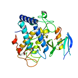
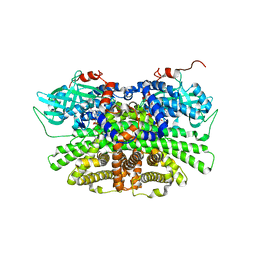
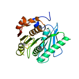
7DZT
DownloadVisualize
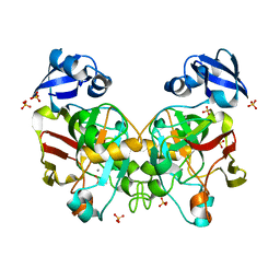
BU of 7dzt by Molmil
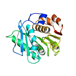
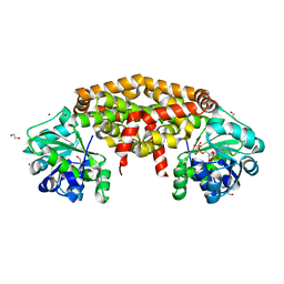
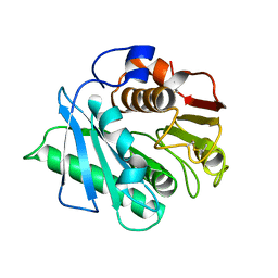
Cyrstal structure of PETase from Rhizobacter gummiphilus
Descriptor: DLH domain-containing protein
Authors:Sagong, H.-Y, Kim, K.-J.
Deposit date:2021-01-26
Release date:2021-07-07
Last modified:2023-11-29
Method:X-RAY DIFFRACTION (2.35 Å)
Cite:Implications for the PET decomposition mechanism through similarity and dissimilarity between PETases from Rhizobacter gummiphilus and Ideonella sakaiensis.
J Hazard Mater, 416, 2021
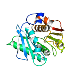
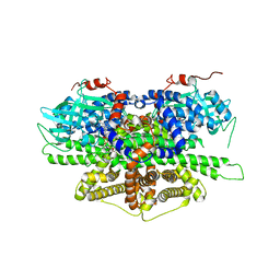
7DZU
DownloadVisualize
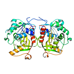
BU of 7dzu by Molmil
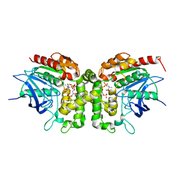
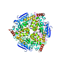
Cyrstal structure of PETase K169A mutant from Rhizobacter gummiphilus
Descriptor: DLH domain-containing protein
Authors:Sagong, H.-Y, Kim, K.-J.
Deposit date:2021-01-26
Release date:2021-07-07
Last modified:2024-10-23
Method:X-RAY DIFFRACTION (2.4 Å)
Cite:Implications for the PET decomposition mechanism through similarity and dissimilarity between PETases from Rhizobacter gummiphilus and Ideonella sakaiensis.
J Hazard Mater, 416, 2021
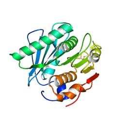
6IOH
DownloadVisualize
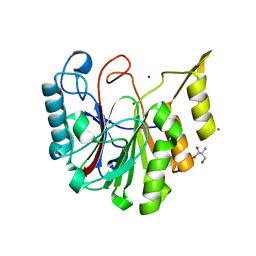
BU of 6ioh by Molmil
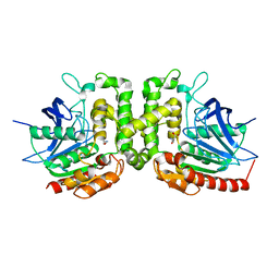
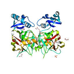
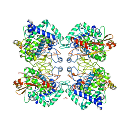
Crystal structure of Homoserine O-acetyltransferase in complex with Homoserine from Mycobacterium smegmatis ATCC 19420
Descriptor: Homoserine O-acetyltransferase, L-HOMOSERINE
Authors:Sagong, H.-Y, Kim, K.-J.
Deposit date:2018-10-30
Release date:2019-09-04
Last modified:2023-11-22
Method:X-RAY DIFFRACTION (2 Å)
Cite:Crystal structure and biochemical characterization of O-acetylhomoserine acetyltransferase from Mycobacterium smegmatis ATCC 19420.
Biochem.Biophys.Res.Commun., 517, 2019
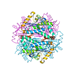
7DZV
DownloadVisualize
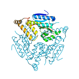
BU of 7dzv by Molmil
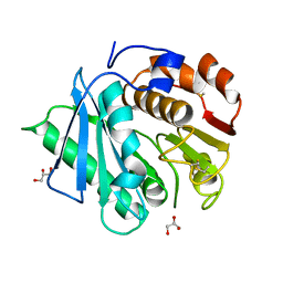
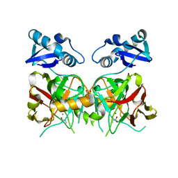
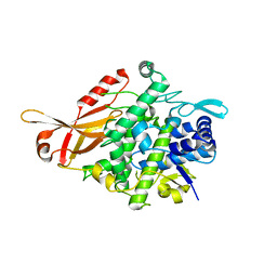
Cyrstal structure of PETase E186A mutant from Rhizobacter gummiphilus
Descriptor: DLH domain-containing protein, GLYCEROL
Authors:Sagong, H.-Y, Kim, K.-J.
Deposit date:2021-01-26
Release date:2021-07-07
Last modified:2024-10-30
Method:X-RAY DIFFRACTION (1.6 Å)
Cite:Implications for the PET decomposition mechanism through similarity and dissimilarity between PETases from Rhizobacter gummiphilus and Ideonella sakaiensis.
J Hazard Mater, 416, 2021
6JWK
DownloadVisualize
BU of 6jwk by Molmil
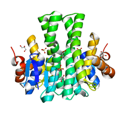
Crystal structure of maleylpyruvate isomerase from Pseudomonas aeruginosa PAO1
Descriptor: GLYCEROL, Probable glutathione S-transferase, SULFATE ION
Authors:Hong, H, Seo, H, Kim, K.-J.
Deposit date:2019-04-20
Release date:2019-05-29
Last modified:2023-11-22
Method:X-RAY DIFFRACTION (1.86 Å)
Cite:Structure and biochemical studies of a pseudomonad maleylpyruvate isomerase from Pseudomonas aeruginosa PAO1.
Biochem.Biophys.Res.Commun., 514, 2019
6JX2
DownloadVisualize
BU of 6jx2 by Molmil
Crystal structure of Ketol-acid reductoisomerase from Corynebacterium glutamicum
Descriptor: 1,2-ETHANEDIOL, Ketol-acid reductoisomerase (NADP(+)), MAGNESIUM ION, ...
Authors:Lee, D, Hong, J, Kim, K.-J.
Deposit date:2019-04-22
Release date:2019-08-07
Last modified:2023-11-22
Method:X-RAY DIFFRACTION (2.6 Å)
Cite:Crystal Structure and Biochemical Characterization of Ketol-Acid Reductoisomerase fromCorynebacterium glutamicum.
J.Agric.Food Chem., 67, 2019
6IOI
DownloadVisualize
BU of 6ioi by Molmil
Crystal structure of Homoserine O-acetyltransferase in complex with CoA from Mycobacterium smegmatis ATCC 19420
Descriptor: COENZYME A, Homoserine O-acetyltransferase
Authors:Sagong, H.-Y, Kim, K.-J.
Deposit date:2018-10-30
Release date:2019-09-04
Last modified:2023-11-22
Method:X-RAY DIFFRACTION (1.6 Å)
Cite:Crystal structure and biochemical characterization of O-acetylhomoserine acetyltransferase from Mycobacterium smegmatis ATCC 19420.
Biochem.Biophys.Res.Commun., 517, 2019
6JVV
DownloadVisualize
BU of 6jvv by Molmil
Crystal structure of maleylpyruvate hydrolase from Sphingobium.sp SYK-6
Descriptor: DI(HYDROXYETHYL)ETHER, GLYCEROL, SULFATE ION, ...
Authors:Hong, H, Kim, K.-J.
Deposit date:2019-04-17
Release date:2019-05-29
Last modified:2023-11-22
Method:X-RAY DIFFRACTION (1.51 Å)
Cite:Structural insights into a maleylpyruvate hydrolase from sphingobium sp. SYK-6, a bacterium degrading lignin-derived aryls.
Biochem.Biophys.Res.Commun., 514, 2019
6JVW
DownloadVisualize
BU of 6jvw by Molmil
Crystal structure of maleylpyruvate hydrolase from Sphingobium sp. SYK-6 in complex with manganese (II) ion and pyruvate
Descriptor: GLYCEROL, MANGANESE (II) ION, PYRUVIC ACID, ...
Authors:Hong, H, Kim, K.-J.
Deposit date:2019-04-17
Release date:2019-05-29
Last modified:2023-11-22
Method:X-RAY DIFFRACTION (2.25 Å)
Cite:Structural insights into a maleylpyruvate hydrolase from sphingobium sp. SYK-6, a bacterium degrading lignin-derived aryls.
Biochem.Biophys.Res.Commun., 514, 2019
6J5X
DownloadVisualize
BU of 6j5x by Molmil
Crystal structure of fumarylpyruvate hydrolase from Corynebacterium glutamicum in complex with Mn2+ and pyruvate
Descriptor: MANGANESE (II) ION, PYRUVIC ACID, Predicted 2-keto-4-pentenoate hydratase/2-oxohepta-3-ene-1,7-dioic acid hydratase, ...
Authors:Hong, H, Seo, H, Kim, K.-J, Park, W.
Deposit date:2019-01-12
Release date:2019-12-18
Last modified:2023-11-22
Method:X-RAY DIFFRACTION (1.79 Å)
Cite:Sequence, structure and function-based classification of the broadly conserved FAH superfamily reveals two distinct fumarylpyruvate hydrolase subfamilies.
Environ.Microbiol., 22, 2020
7CBE
DownloadVisualize
BU of 7cbe by Molmil
Crystal structure of Homoserine O-succinyltransferase from Escherichia coli K-12
Descriptor: 1,2-ETHANEDIOL, GLYCEROL, Homoserine O-succinyltransferase, ...
Authors:Sagong, H.-Y, Kim, K.-J.
Deposit date:2020-06-12
Release date:2021-06-16
Last modified:2023-11-29
Method:X-RAY DIFFRACTION (1.7 Å)
Cite:Crystal structure of Homoserine O-succinyltransferase from Escherichia coli K-12
To Be Published
7FBG
DownloadVisualize
BU of 7fbg by Molmil
Kinetic and structural analysis by Peptidoglycan editing factor from Bacillus cereus ATCC 14579
Descriptor: 2-AMINO-2-HYDROXYMETHYL-PROPANE-1,3-DIOL, CALCIUM ION, Purine nucleoside phosphorylase, ...
Authors:Seok, J, Kim, K.-J.
Deposit date:2021-07-10
Release date:2022-07-13
Last modified:2023-11-29
Method:X-RAY DIFFRACTION (1.6 Å)
Cite:Kinetic and structural analysis by Peptidoglycan editing factor from Bacillus cereus ATCC 14579
to be published
6IJK
DownloadVisualize
BU of 6ijk by Molmil
Enoyl-CoA hydratase/isomerase family protein from Cupriavidus necator H16
Descriptor: Enoyl-CoA hydratase
Authors:Seo, H, Kim, K.-J.
Deposit date:2018-10-10
Release date:2019-08-21
Last modified:2023-11-22
Method:X-RAY DIFFRACTION (2 Å)
Cite:Crystal structure of a novel type isomerase of enoyl-CoA hydratase/isomerase family protein from Cupriavidus necator H16
Biotechnol. Bioprocess Eng., 24, 2019
6J2U
DownloadVisualize
BU of 6j2u by Molmil
Crystal structure of Tyrosinase caddy protein(MelC1)with Tyrosinase (MelC2)from Streptomyces avermitilis in complex with Zinc ion
Descriptor: Tyrosinase, Tyrosinase co-factor protein, ZINC ION
Authors:Lee, S.-H, Hong, H, Kim, K.-J.
Deposit date:2019-01-03
Release date:2020-02-12
Last modified:2023-11-22
Method:X-RAY DIFFRACTION (1.3 Å)
Cite:Crystal structure of Tyrosinase caddy protein(MelC1)with tyrosinase (MelC2)from Streptomyces avermitilis in complex with Zinc ion
To Be Published
6KUS
DownloadVisualize
BU of 6kus by Molmil
Crystal strcuture of PETase S121E, D186H, S242T, N246D mutant from Ideonella sakaiensis
Descriptor: Poly(ethylene terephthalate) hydrolase
Authors:Joo, S, Kim, K.-J.
Deposit date:2019-09-02
Release date:2020-09-02
Last modified:2024-10-09
Method:X-RAY DIFFRACTION (1.8 Å)
Cite:Crystal strcuture of PETase A248D, R280K mutant from Ideonella sakaiensis
To Be Published
6KUQ
DownloadVisualize
BU of 6kuq by Molmil
Crystal strcuture of PETase A248D, R280K mutant from Ideonella sakaiensis
Descriptor: Poly(ethylene terephthalate) hydrolase
Authors:Joo, S, Kim, K.-J.
Deposit date:2019-09-02
Release date:2020-09-02
Last modified:2024-10-16
Method:X-RAY DIFFRACTION (1.91 Å)
Cite:Crystal strcuture of PETase A248D, R280K mutant from Ideonella sakaiensis
To Be Published
6L3O
DownloadVisualize
BU of 6l3o by Molmil
Crystal strcuture of Feruloyl-CoA hydratase lyase(FCHL) from Pseudomonas putida KT2440
Descriptor: Hydroxycinnamoyl-CoA hydratase-lyase
Authors:Seok, J, Seo, H, Kim, K.-J.
Deposit date:2019-10-12
Release date:2020-10-14
Last modified:2023-11-22
Method:X-RAY DIFFRACTION (1.65 Å)
Cite:Kinetic and structural analysis for bioproduction of vanillin by feruloyl-CoA hydratase/lyase from Pseudomonas putida KT2440
to be published
6KUO
DownloadVisualize
BU of 6kuo by Molmil
Crystal strcuture of PETase N246D mutant from Ideonella sakaiensis
Descriptor: Poly(ethylene terephthalate) hydrolase
Authors:Joo, S, Kim, K.-J.
Deposit date:2019-09-02
Release date:2020-09-02
Last modified:2024-10-16
Method:X-RAY DIFFRACTION (1.9 Å)
Cite:Crystal strcuture of PETase A248D, R280K mutant from Ideonella sakaiensis
To Be Published
5Z7R
DownloadVisualize
BU of 5z7r by Molmil
Crystal structure of crotonase from Clostridium acetobutylicum
Descriptor: Short-chain-enoyl-CoA hydratase
Authors:Kim, E.-J, Kim, Y.-J, Kim, K.-J.
Deposit date:2018-01-30
Release date:2018-02-14
Last modified:2023-11-22
Method:X-RAY DIFFRACTION (2.2 Å)
Cite:Structural insights into substrate specificity of crotonase from the n-butanol producing bacterium Clostridium acetobutylicum.
Biochem. Biophys. Res. Commun., 451, 2014
5ZRD
DownloadVisualize
BU of 5zrd by Molmil
Tyrosinase from Burkholderia thailandensis (BtTYR) at low pH condition
Descriptor: CITRIC ACID, COPPER (II) ION, GLYCEROL, ...
Authors:Lee, S, Son, H.-F, Kim, K.-J.
Deposit date:2018-04-24
Release date:2018-10-31
Last modified:2023-11-22
Method:X-RAY DIFFRACTION (2.3 Å)
Cite:Structural Basis for Highly Efficient Production of Catechol Derivatives at Acidic pH by Tyrosinase from Burkholderia thailandensis
Acs Catalysis, 8, 2018
5ZRE
DownloadVisualize
BU of 5zre by Molmil
Tyrosinase from Burkholderia thailandensis (BtTYR) at high pH condition
Descriptor: COPPER (II) ION, GLYCEROL, OXYGEN ATOM, ...
Authors:Lee, S, Son, H.-F, Kim, K.-J.
Deposit date:2018-04-24
Release date:2018-10-31
Last modified:2024-03-27
Method:X-RAY DIFFRACTION (2.5 Å)
Cite:Structural Basis for Highly Efficient Production of Catechol Derivatives at Acidic pH by Tyrosinase from Burkholderia thailandensis
Acs Catalysis, 8, 2018
5YS9
DownloadVisualize
BU of 5ys9 by Molmil
Crystal structure of acyl-coA oxidase3 from Yarrowia lipolytica
Descriptor: Acyl-coenzyme A oxidase 3, FLAVIN-ADENINE DINUCLEOTIDE
Authors:Kim, S, Kim, K.-J.
Deposit date:2017-11-13
Release date:2018-02-28
Last modified:2023-11-22
Method:X-RAY DIFFRACTION (2.5 Å)
Cite:Crystal Structure of Acyl-CoA Oxidase 3 fromYarrowia lipolyticawith Specificity for Short-Chain Acyl-CoA.
J. Microbiol. Biotechnol., 28, 2018
5Y9D
DownloadVisualize
BU of 5y9d by Molmil
Crystal structure of acyl-coA oxidase1 from Yarrowia lipolytica
Descriptor: Acyl-coenzyme A oxidase 1, FLAVIN-ADENINE DINUCLEOTIDE
Authors:Kim, S, Kim, K.-J.
Deposit date:2017-08-24
Release date:2018-01-03
Last modified:2023-11-22
Method:X-RAY DIFFRACTION (2.5 Å)
Cite:Structural insight into the substrate specificity of acyl-CoA oxidase1 from Yarrowia lipolytica for short-chain dicarboxylyl-CoAs.
Biochem. Biophys. Res. Commun., 495, 2018
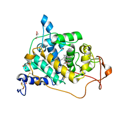
6A6Q
DownloadVisualize
BU of 6a6q by Molmil
Crystal structure of a lignin peroxidase isozyme H8 variant that is stable at very acidic pH
Descriptor: CALCIUM ION, GLYCEROL, HEME B/C, ...
Authors:Seo, H, Kim, K.-J, Pham, L.T.M.
Deposit date:2018-06-29
Release date:2019-01-23
Last modified:2023-11-22
Method:X-RAY DIFFRACTION (1.67 Å)
Cite:In silico-designed lignin peroxidase fromPhanerochaete chrysosporiumshows enhanced acid stability for depolymerization of lignin.
Biotechnol Biofuels, 11, 2018
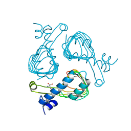
6AG8
DownloadVisualize
BU of 6ag8 by Molmil
Crystal structure of Maltose O-acetyltransferase from E. coli
Descriptor: 2-AMINO-2-HYDROXYMETHYL-PROPANE-1,3-DIOL, Maltose O-acetyltransferase
Authors:Joo, S, Kim, K.-J.
Deposit date:2018-08-09
Release date:2019-08-28
Last modified:2023-11-22
Method:X-RAY DIFFRACTION (1.34 Å)
Cite:Metabolic engineering of Escherichia coli for production of non-natural acetins from glycerol
To Be Published

226707

PDB entries from 2024-10-30

PDB statisticsPDBj update infoContact PDBjnumon