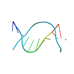
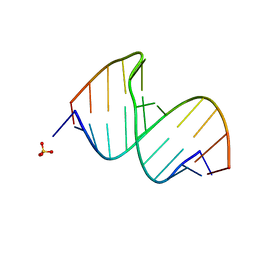
1D8G
 
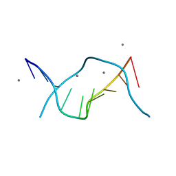 | | ULTRAHIGH RESOLUTION CRYSTAL STRUCTURE OF B-DNA DECAMER D(CCAGTACTGG) | | 分子名称: | 5'-D(*CP*CP*AP*GP*TP*AP*CP*TP*GP*GP*)-3', CALCIUM ION | | 著者 | Kielkopf, C.L, Ding, S, Kuhn, P, Rees, D.C. | | 登録日 | 1999-10-23 | | 公開日 | 2000-02-21 | | 最終更新日 | 2024-02-07 | | 実験手法 | X-RAY DIFFRACTION (0.74 Å) | | 主引用文献 | Conformational flexibility of B-DNA at 0.74 A resolution: d(CCAGTACTGG)(2).
J.Mol.Biol., 296, 2000
|
|
1CVX
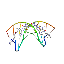 | | CRYSTAL STRUCTURE OF POLYAMIDE DIMER (IMPYHPPYBETADP)2 BOUND TO B-DNA DECAMER CCAGATCTGG | | 分子名称: | 5'-D(*CP*CP*AP*GP*AP*TP*CP*TP*GP*G)-3', HYDROXYPYRROLE-IMIDAZOLE-PYRROLE POLYAMIDE | | 著者 | Kielkopf, C.L, Bremer, R.E, White, S, Baird, E.E, Dervan, P.B, Rees, D.C. | | 登録日 | 1999-08-24 | | 公開日 | 2000-01-15 | | 最終更新日 | 2024-02-07 | | 実験手法 | X-RAY DIFFRACTION (2.27 Å) | | 主引用文献 | Structural effects of DNA sequence on T.A recognition by hydroxypyrrole/pyrrole pairs in the minor groove.
J.Mol.Biol., 295, 2000
|
|
1CVY
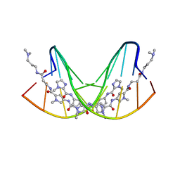 | | CRYSTAL STRUCTURE OF POLYAMIDE DIMER (IMPYPYPYBETADP)2 BOUND TO CCAGATCTGG | | 分子名称: | 5'-D(*CP*CP*AP*GP*AP*TP*CP*TP*GP*G)-3', IMIDAZOLE-PYRROLE POLYAMIDE | | 著者 | Kielkopf, C.L, Bremer, R.E, White, S, Baird, E.E, Dervan, P.B, Rees, D.C. | | 登録日 | 1999-08-24 | | 公開日 | 2000-01-15 | | 最終更新日 | 2024-02-07 | | 実験手法 | X-RAY DIFFRACTION (2.15 Å) | | 主引用文献 | Structural effects of DNA sequence on T.A recognition by hydroxypyrrole/pyrrole pairs in the minor groove.
J.Mol.Biol., 295, 2000
|
|
365D
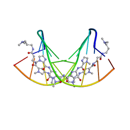 | | STRUCTURAL BASIS FOR G C RECOGNITION IN THE DNA MINOR GROOVE | | 分子名称: | DNA (5'-D(*CP*CP*AP*GP*GP*(CBR)P*CP*TP*GP*G)-3'), IMIDAZOLE-PYRROLE POLYAMIDE | | 著者 | Kielkopf, C.L, Baird, E.E, Dervan, P.B, Rees, D.C. | | 登録日 | 1997-12-17 | | 公開日 | 1998-02-05 | | 最終更新日 | 2024-02-21 | | 実験手法 | X-RAY DIFFRACTION (2 Å) | | 主引用文献 | Structural basis for G.C recognition in the DNA minor groove.
Nat.Struct.Biol., 5, 1998
|
|
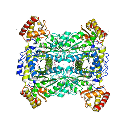
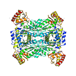
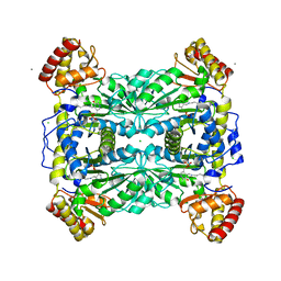
1JG8
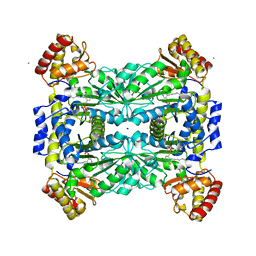 | | Crystal Structure of Threonine Aldolase (Low-specificity) | | 分子名称: | CALCIUM ION, L-allo-threonine aldolase, SODIUM ION | | 著者 | Kielkopf, C.L, Bonanno, J, Ray, S, Burley, S.K, New York SGX Research Center for Structural Genomics (NYSGXRC) | | 登録日 | 2001-06-23 | | 公開日 | 2001-07-04 | | 最終更新日 | 2021-02-03 | | 実験手法 | X-RAY DIFFRACTION (1.8 Å) | | 主引用文献 | Crystal Structure of Low-specificity Threonine Aldolase, a Key Enzyme in Glycine Biosynthesis
To be published
|
|
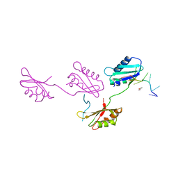
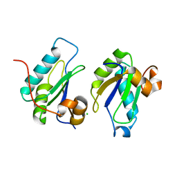
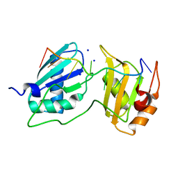
1JMT
 | | X-ray Structure of a Core U2AF65/U2AF35 Heterodimer | | 分子名称: | HEXANE-1,6-DIOL, SPLICING FACTOR U2AF 35 KDA SUBUNIT, SPLICING FACTOR U2AF 65 KDA SUBUNIT | | 著者 | Kielkopf, C.L, Rodionova, N.A, Green, M.R, Burley, S.K. | | 登録日 | 2001-07-19 | | 公開日 | 2001-09-19 | | 最終更新日 | 2024-02-07 | | 実験手法 | X-RAY DIFFRACTION (2.2 Å) | | 主引用文献 | A novel peptide recognition mode revealed by the X-ray structure of a core U2AF35/U2AF65 heterodimer.
Cell(Cambridge,Mass.), 106, 2001
|
|
1LW4
 | |
1LW5
 | |
408D
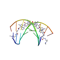 | | STRUCTURAL BASIS FOR RECOGNITION OF A-T AND T-A BASE PAIRS IN THE MINOR GROOVE OF B-DNA | | 分子名称: | DNA (5'-D(*CP*CP*AP*GP*TP*AP*CP*TP*GP*G)-3'), IMIDAZOLE-PYRROLE POLYAMIDE | | 著者 | Kielkopf, C.L, White, S, Szewczyk, J.W, Turner, J.M, Baird, E.E, Dervan, P.B, Rees, D.C. | | 登録日 | 1998-06-24 | | 公開日 | 1998-10-19 | | 最終更新日 | 2024-02-28 | | 実験手法 | X-RAY DIFFRACTION (2.1 Å) | | 主引用文献 | A structural basis for recognition of A.T and T.A base pairs in the minor groove of B-DNA.
Science, 282, 1998
|
|
454D
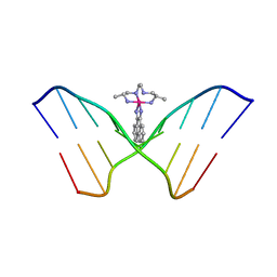 | | INTERCALATION AND MAJOR GROOVE RECOGNITION IN THE 1.2 A RESOLUTION CRYSTAL STRUCTURE OF RH[ME2TRIEN]PHI BOUND TO 5'-G(5IU)TGCAAC-3' | | 分子名称: | 5'-D(*GP*(5IU)P*TP*GP*CP*AP*AP*C)-3', DELTA-ALPHA-RH[2R,9R-DIAMINO-4,7-DIAZADECANE]9,10-PHENANTHRENEQUINONE DIIMINE | | 著者 | Kielkopf, C.L, Erkkila, K.E, Hudson, B.P, Barton, J.K, Rees, D.C. | | 登録日 | 1999-03-03 | | 公開日 | 2000-02-21 | | 最終更新日 | 2024-04-03 | | 実験手法 | X-RAY DIFFRACTION (1.2 Å) | | 主引用文献 | Structure of a photoactive rhodium complex intercalated into DNA.
Nat.Struct.Biol., 7, 2000
|
|
1M6S
 | |
2G4B
 | |
4OZ0
 | |
2HZC
 | |
1XCB
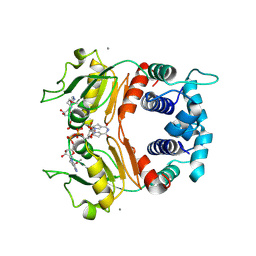 | | X-ray Structure of a Rex-Family Repressor/NADH Complex from Thermus Aquaticus | | 分子名称: | CALCIUM ION, NICOTINAMIDE-ADENINE-DINUCLEOTIDE, Redox-sensing transcriptional repressor rex | | 著者 | Sickmier, E.A, Brekasis, D, Paranawithana, S, Bonanno, J.B, Burley, S.K, Paget, M.S, Kielkopf, C.L, New York SGX Research Center for Structural Genomics (NYSGXRC) | | 登録日 | 2004-09-01 | | 公開日 | 2004-09-28 | | 最終更新日 | 2021-02-03 | | 実験手法 | X-RAY DIFFRACTION (2.9 Å) | | 主引用文献 | X-Ray Structure of a Rex-Family Repressor/NADH Complex: Insights into the Mechanism of Redox Sensing
Structure, 13, 2005
|
|
2OKS
 | | X-ray Structure of a DNA Repair Substrate Containing an Alkyl Interstrand Crosslink at 1.65 Resolution | | 分子名称: | 5'-D(*CP*CP*AP*AP*(C34)P*GP*TP*TP*GP*G)-3', CALCIUM ION | | 著者 | Swenson, M.C, Paranawithana, S.R, Miller, P.S, Kielkopf, C.L. | | 登録日 | 2007-01-17 | | 公開日 | 2007-02-13 | | 最終更新日 | 2023-08-30 | | 実験手法 | X-RAY DIFFRACTION (1.65 Å) | | 主引用文献 | Structure of a DNA repair substrate containing an alkyl interstrand cross-link at 1.65 a resolution.
Biochemistry, 46, 2007
|
|
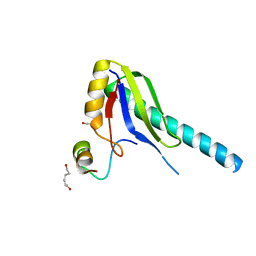
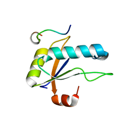
4FXX
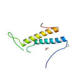 | | Structure of SF1 coiled-coil domain | | 分子名称: | IMIDAZOLE, MALONATE ION, Splicing factor 1 | | 著者 | Gupta, A, Bauer, W.J, Wang, W, Kielkopf, C.L. | | 登録日 | 2012-07-03 | | 公開日 | 2013-01-16 | | 最終更新日 | 2024-02-28 | | 実験手法 | X-RAY DIFFRACTION (2.4801 Å) | | 主引用文献 | Structure of Phosphorylated SF1 Bound to U2AF(65) in an Essential Splicing Factor Complex.
Structure, 21, 2013
|
|
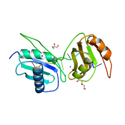
4FXW
 | | Structure of phosphorylated SF1 complex with U2AF65-UHM domain | | 分子名称: | SULFATE ION, Splicing factor 1, Splicing factor U2AF 65 kDa subunit | | 著者 | Wang, W, Bauer, W.J, Wedekind, J.E, Kielkopf, C.L. | | 登録日 | 2012-07-03 | | 公開日 | 2013-01-16 | | 最終更新日 | 2017-11-15 | | 実験手法 | X-RAY DIFFRACTION (2.29 Å) | | 主引用文献 | Structure of Phosphorylated SF1 Bound to U2AF(65) in an Essential Splicing Factor Complex.
Structure, 21, 2013
|
|
6XLW
 | | Crystal structure of U2AF65 bound to AdML splice site sequence | | 分子名称: | DI(HYDROXYETHYL)ETHER, DNA/RNA (5'-R(P*UP*UP*(UD)P*UP*U)-D(P*(BRU))-R(P*CP*C)-3'), GLYCEROL, ... | | 著者 | Maji, D, Jenkins, J.L, Kielkopf, C.L. | | 登録日 | 2020-06-29 | | 公開日 | 2020-10-07 | | 最終更新日 | 2024-05-22 | | 実験手法 | X-RAY DIFFRACTION (1.5 Å) | | 主引用文献 | Representative cancer-associated U2AF2 mutations alter RNA interactions and splicing.
J.Biol.Chem., 295, 2020
|
|
5W0H
 | |
5W0G
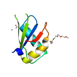 | | Structure of U2AF65 (U2AF2) RRM1 at 1.07 resolution | | 分子名称: | 2-(2-{2-[2-(2-METHOXY-ETHOXY)-ETHOXY]-ETHOXY}-ETHOXY)-ETHANOL, ACETATE ION, Splicing factor U2AF 65 kDa subunit, ... | | 著者 | Agrawal, A.A, Jenkins, J.L, Kielkopf, C.L. | | 登録日 | 2017-05-30 | | 公開日 | 2018-04-11 | | 最終更新日 | 2023-10-04 | | 実験手法 | X-RAY DIFFRACTION (1.07 Å) | | 主引用文献 | Cancer-Associated Mutations Mapped on High-Resolution Structures of the U2AF2 RNA Recognition Motifs.
Biochemistry, 56, 2017
|
|
7S3A
 | |
7SN6
 | | U2AF65 UHM BOUND TO SF3B155 ULM5 | | 分子名称: | ISOPROPYL ALCOHOL, Splicing factor 3B subunit 1, Splicing factor U2AF 65 kDa subunit | | 著者 | Loerch, S, Jenkins, J.L, Kielkopf, C.L. | | 登録日 | 2021-10-27 | | 公開日 | 2022-07-27 | | 最終更新日 | 2023-10-18 | | 実験手法 | X-RAY DIFFRACTION (1.8 Å) | | 主引用文献 | A UHM-ULM interface with unusual structural features contributes to U2AF2 and SF3B1 association for pre-mRNA splicing.
J.Biol.Chem., 298, 2022
|
|
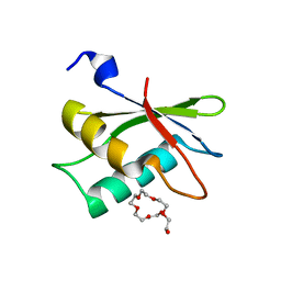
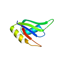
3BS9
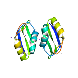 | | X-ray structure of human TIA-1 RRM2 | | 分子名称: | IODIDE ION, Nucleolysin TIA-1 isoform p40 | | 著者 | Kumar, A.O, Kielkopf, C.L. | | 登録日 | 2007-12-22 | | 公開日 | 2008-01-15 | | 最終更新日 | 2023-08-30 | | 実験手法 | X-RAY DIFFRACTION (1.95 Å) | | 主引用文献 | Structure of the central RNA recognition motif of human TIA-1 at 1.95A resolution.
Biochem.Biophys.Res.Commun., 367, 2008
|
|
3CGR
 | |


