Loading
PDBj
MenuPDBj@FacebookPDBj@X(formerly Twitter)PDBj@BlueSkyPDBj@YouTubewwPDB FoundationwwPDB
RCSB PDBPDBeBMRBAdv. SearchSearch help
PDB: 12 results

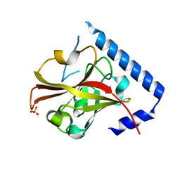
3TB7
DownloadVisualize
BU of 3tb7 by Molmil
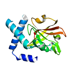
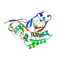
The type I crystal structure of Streptococcus agalactiae sortase C1
Descriptor: Sortase family protein
Authors:Khare, B.
Deposit date:2011-08-05
Release date:2011-10-26
Last modified:2024-02-28
Method:X-RAY DIFFRACTION (2.45 Å)
Cite:The Crystal Structure Analysis of Group B Streptococcus Sortase C1: A Model for the "Lid" Movement upon Substrate Binding.
J.Mol.Biol., 414, 2011
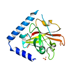
3TBE
DownloadVisualize
BU of 3tbe by Molmil
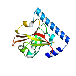
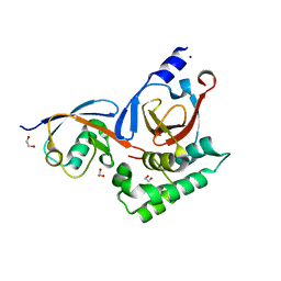
The crystal structure of the complex of Streptococcus agalactiae sortase C1 and MTSET
Descriptor: 2-(TRIMETHYLAMMONIUM)ETHYL THIOL, CHLORIDE ION, SULFATE ION, ...
Authors:Khare, B.
Deposit date:2011-08-05
Release date:2011-10-26
Last modified:2024-11-20
Method:X-RAY DIFFRACTION (2.85 Å)
Cite:The Crystal Structure Analysis of Group B Streptococcus Sortase C1: A Model for the "Lid" Movement upon Substrate Binding.
J.Mol.Biol., 414, 2011
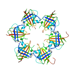
7LCG
DownloadVisualize
BU of 7lcg by Molmil
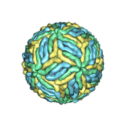
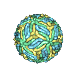
The mature Usutu SAAR-1776, Model A
Descriptor: (7S)-4-HYDROXY-N,N,N-TRIMETHYL-9-OXO-7-[(PALMITOYLOXY)METHYL]-3,5,8-TRIOXA-4-PHOSPHAHEXACOSAN-1-AMINIUM 4-OXIDE, 2-acetamido-2-deoxy-beta-D-glucopyranose, Envelope protein E, ...
Authors:Khare, B, Klose, T, Fang, Q, Kuhn, R.
Deposit date:2021-01-11
Release date:2021-09-01
Last modified:2024-10-30
Method:ELECTRON MICROSCOPY (2.42 Å)
Cite:Structure of Usutu virus SAAR-1776 displays fusion loop asymmetry.
Proc.Natl.Acad.Sci.USA, 118, 2021
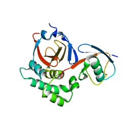
7LCH
DownloadVisualize
BU of 7lch by Molmil
The mature Usutu SAAR-1776, Model B
Descriptor: (7S)-4-HYDROXY-N,N,N-TRIMETHYL-9-OXO-7-[(PALMITOYLOXY)METHYL]-3,5,8-TRIOXA-4-PHOSPHAHEXACOSAN-1-AMINIUM 4-OXIDE, 2-acetamido-2-deoxy-beta-D-glucopyranose, Envelope protein E, ...
Authors:Khare, B, Klose, T, Fang, Q, Kuhn, R.
Deposit date:2021-01-11
Release date:2021-09-01
Last modified:2024-11-06
Method:ELECTRON MICROSCOPY (2.35 Å)
Cite:Structure of Usutu virus SAAR-1776 displays fusion loop asymmetry.
Proc.Natl.Acad.Sci.USA, 118, 2021
3RBJ
DownloadVisualize
BU of 3rbj by Molmil
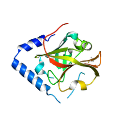
Crystal Structure of the lid-mutant of Streptococcus agalactiae Sortase C1
Descriptor: Sortase family protein
Authors:Khare, B, Narayana, S.V.L.
Deposit date:2011-03-29
Release date:2011-09-07
Last modified:2024-02-21
Method:X-RAY DIFFRACTION (2.3 Å)
Cite:Structural differences between the Streptococcus agalactiae housekeeping and pilus-specific sortases: SrtA and SrtC1.
Plos One, 6, 2011
3RBK
DownloadVisualize
BU of 3rbk by Molmil
The Type II Crystal Structure of Streptococcus agalactiae Sortase C1
Descriptor: SULFATE ION, Sortase family protein
Authors:Khare, B, Narayana, S.V.L.
Deposit date:2011-03-29
Release date:2011-09-07
Last modified:2024-02-21
Method:X-RAY DIFFRACTION (2.9 Å)
Cite:Structural differences between the Streptococcus agalactiae housekeeping and pilus-specific sortases: SrtA and SrtC1.
Plos One, 6, 2011
3RBI
DownloadVisualize
BU of 3rbi by Molmil
The Type III Crystal Structure of Streptococcus agalactiae Sortase C1
Descriptor: SULFATE ION, Sortase family protein
Authors:Khare, B, Narayana, S.V.L.
Deposit date:2011-03-29
Release date:2011-09-07
Last modified:2024-02-21
Method:X-RAY DIFFRACTION (3 Å)
Cite:Structural differences between the Streptococcus agalactiae housekeeping and pilus-specific sortases: SrtA and SrtC1.
Plos One, 6, 2011
3RCC
DownloadVisualize
BU of 3rcc by Molmil
Crystal Structure of the Streptococcus agalactiae Sortase A
Descriptor: Sortase SrtA, ZINC ION
Authors:Khare, B, Narayana, S.V.L.
Deposit date:2011-03-30
Release date:2011-09-07
Last modified:2024-02-21
Method:X-RAY DIFFRACTION (3.1 Å)
Cite:Structural differences between the Streptococcus agalactiae housekeeping and pilus-specific sortases: SrtA and SrtC1.
Plos One, 6, 2011
5V5I
DownloadVisualize
BU of 5v5i by Molmil
OTU protease of Crimean Congo Hemorrhagic Fever Virus bound to ubiquitin variant CC.1
Descriptor: RNA-directed RNA polymerase L, Ubiquitin variant CC.1
Authors:Khare, B, Mark, B.L.
Deposit date:2017-03-14
Release date:2017-05-10
Last modified:2020-01-08
Method:X-RAY DIFFRACTION (2.201 Å)
Cite:OTU protease of Crimean Congo Hemorrhagic Fever Virus bound to ubiquitin variant CC.1
To be published
5V5H
DownloadVisualize
BU of 5v5h by Molmil
OTU protease of Crimean Congo Hemorrhagic Fever Virus bound to ubiquitin variant CC.2
Descriptor: 4-(2-HYDROXYETHYL)-1-PIPERAZINE ETHANESULFONIC ACID, RNA-directed RNA polymerase L, SODIUM ION, ...
Authors:Khare, B, Mark, B.L.
Deposit date:2017-03-14
Release date:2017-05-10
Last modified:2024-11-20
Method:X-RAY DIFFRACTION (1.5 Å)
Cite:Potent and selective inhibition of pathogenic viruses by engineered ubiquitin variants.
PLoS Pathog., 13, 2017
5V5G
DownloadVisualize
BU of 5v5g by Molmil
OTU protease of Crimean Congo Hemorrhagic Fever Virus bound to ubiquitin variant CC.4
Descriptor: 1,2-ETHANEDIOL, RNA-directed RNA polymerase L, SODIUM ION, ...
Authors:Khare, B, Mark, B.L.
Deposit date:2017-03-14
Release date:2017-05-10
Last modified:2023-10-04
Method:X-RAY DIFFRACTION (2.1 Å)
Cite:Potent and selective inhibition of pathogenic viruses by engineered ubiquitin variants.
PLoS Pathog., 13, 2017
1XEY
DownloadVisualize
BU of 1xey by Molmil
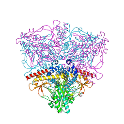
Crystal structure of the complex of Escherichia coli GADA with glutarate at 2.05 A resolution
Descriptor: ACETATE ION, GLUTARIC ACID, Glutamate decarboxylase alpha, ...
Authors:Dutyshev, D.I, Darii, E.L, Fomenkova, N.P, Pechik, I.V, Polyakov, K.M, Nikonov, S.V, Andreeva, N.S, Sukhareva, B.S.
Deposit date:2004-09-13
Release date:2004-10-05
Last modified:2023-08-23
Method:X-RAY DIFFRACTION (2.05 Å)
Cite:Structure of Escherichia coli glutamate decarboxylase (GADalpha) in complex with glutarate at 2.05 angstroms resolution.
Acta Crystallogr.,Sect.D, 61, 2005

227561

PDB entries from 2024-11-20

PDB statisticsPDBj update infoContact PDBjnumon