1S0E
 
 | | Crystal structure of botulinum neurotoxin type B at pH 6.0 | | 分子名称: | Botulinum neurotoxin type B, CALCIUM ION, ZINC ION | | 著者 | Eswaramoorthy, S, Kumaran, D, Keller, J, Swaminathan, S. | | 登録日 | 2003-12-30 | | 公開日 | 2004-03-16 | | 最終更新日 | 2023-08-23 | | 実験手法 | X-RAY DIFFRACTION (1.9 Å) | | 主引用文献 | Role of metals in the biological activity of Clostridium botulinum neurotoxins
Biochemistry, 43, 2004
|
|
1S0B
 | | Crystal structure of botulinum neurotoxin type B at pH 4.0 | | 分子名称: | Botulinum neurotoxin type B, CALCIUM ION | | 著者 | Eswaramoorthy, S, Kumaran, D, Keller, J, Swaminathan, S. | | 登録日 | 2003-12-30 | | 公開日 | 2004-03-16 | | 最終更新日 | 2023-08-23 | | 実験手法 | X-RAY DIFFRACTION (2 Å) | | 主引用文献 | Role of metals in the biological activity of Clostridium botulinum neurotoxins
Biochemistry, 43, 2004
|
|
1S0D
 | | Crystal structure of botulinum neurotoxin type B at pH 5.5 | | 分子名称: | Botulinum neurotoxin type B, CALCIUM ION, ZINC ION | | 著者 | Eswaramoorthy, S, Kumaran, D, Keller, J, Swaminathan, S. | | 登録日 | 2003-12-30 | | 公開日 | 2004-03-16 | | 最終更新日 | 2023-08-23 | | 実験手法 | X-RAY DIFFRACTION (2.2 Å) | | 主引用文献 | Role of metals in the biological activity of Clostridium botulinum neurotoxins
Biochemistry, 43, 2004
|
|
1S0G
 | |
1S0F
 | | Crystal structure of botulinum neurotoxin type B at pH 7.0 | | 分子名称: | Botulinum neurotoxin type B, CALCIUM ION, ZINC ION | | 著者 | Eswaramoorthy, S, Kumaran, D, Keller, J, Swaminathan, S. | | 登録日 | 2003-12-30 | | 公開日 | 2004-03-16 | | 最終更新日 | 2023-08-23 | | 実験手法 | X-RAY DIFFRACTION (2.3 Å) | | 主引用文献 | Role of metals in the biological activity of Clostridium botulinum neurotoxins
Biochemistry, 43, 2004
|
|
5LSJ
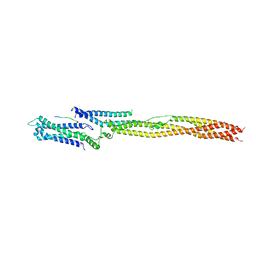 | | CRYSTAL STRUCTURE OF THE HUMAN KINETOCHORE MIS12-CENP-C delta-HEAD2 COMPLEX | | 分子名称: | Centromere protein C, Kinetochore-associated protein DSN1 homolog, Kinetochore-associated protein NSL1 homolog, ... | | 著者 | Vetter, I.R, Petrovic, A, Keller, J, Liu, Y. | | 登録日 | 2016-09-02 | | 公開日 | 2016-11-16 | | 最終更新日 | 2024-01-17 | | 実験手法 | X-RAY DIFFRACTION (3.25 Å) | | 主引用文献 | Structure of the MIS12 Complex and Molecular Basis of Its Interaction with CENP-C at Human Kinetochores.
Cell, 167, 2016
|
|
5LSI
 | | CRYSTAL STRUCTURE OF THE KINETOCHORE MIS12 COMPLEX HEAD2 SUBDOMAIN CONTAINING DSN1 AND NSL1 FRAGMENTS | | 分子名称: | Kinetochore-associated protein DSN1 homolog, Kinetochore-associated protein NSL1 homolog, SULFATE ION | | 著者 | Vetter, I.R, Petrovic, A, Keller, J, Liu, Y. | | 登録日 | 2016-09-02 | | 公開日 | 2016-11-16 | | 最終更新日 | 2024-01-17 | | 実験手法 | X-RAY DIFFRACTION (2.002 Å) | | 主引用文献 | Structure of the MIS12 Complex and Molecular Basis of Its Interaction with CENP-C at Human Kinetochores.
Cell, 167, 2016
|
|
5LSK
 | | CRYSTAL STRUCTURE OF THE HUMAN KINETOCHORE MIS12-CENP-C COMPLEX | | 分子名称: | Centromere protein C, Kinetochore-associated protein DSN1 homolog, Kinetochore-associated protein NSL1 homolog, ... | | 著者 | Vetter, I.R, Petrovic, A, Keller, J, Liu, Y. | | 登録日 | 2016-09-02 | | 公開日 | 2016-11-16 | | 最終更新日 | 2017-10-11 | | 実験手法 | X-RAY DIFFRACTION (3.502 Å) | | 主引用文献 | Structure of the MIS12 Complex and Molecular Basis of Its Interaction with CENP-C at Human Kinetochores.
Cell, 167, 2016
|
|


