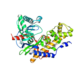
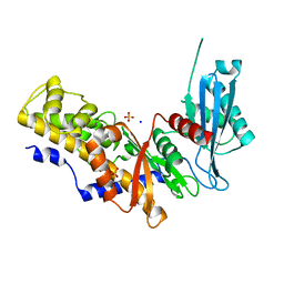
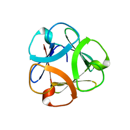
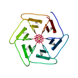
6LF1
 
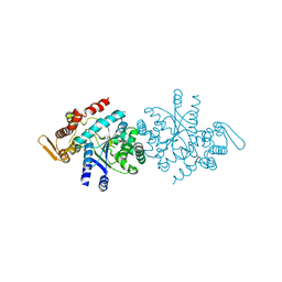
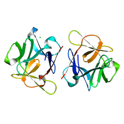 | | SeviL, a GM1b/asialo-GM1 binding lectin | | 分子名称: | CHLORIDE ION, SeviL | | 著者 | Kamata, K, Ozeki, Y, Park, S.-Y, Tame, J.R.H. | | 登録日 | 2019-11-28 | | 公開日 | 2020-12-02 | | 最終更新日 | 2023-11-22 | | 実験手法 | X-RAY DIFFRACTION (1.7 Å) | | 主引用文献 | The structure of SeviL, a GM1b/asialo-GM1 binding R-type lectin from the mussel Mytilisepta virgata.
Sci Rep, 10, 2020
|
|
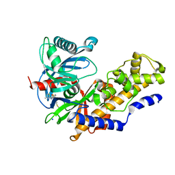
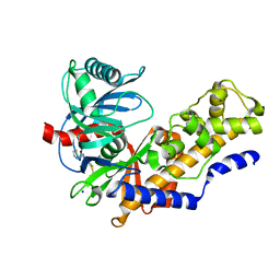
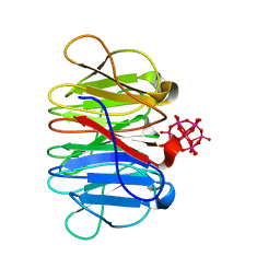
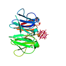
6LF2
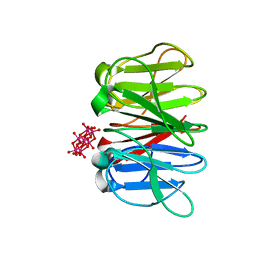
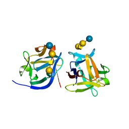 | | SeviL bound to asialo-GM1 saccharide | | 分子名称: | SeviL, beta-D-galactopyranose-(1-3)-2-acetamido-2-deoxy-beta-D-galactopyranose-(1-4)-beta-D-galactopyranose-(1-4)-beta-D-glucopyranose | | 著者 | Kamata, K, Ozeki, Y, Park, S.-Y, Tame, J.R.H. | | 登録日 | 2019-11-28 | | 公開日 | 2020-12-02 | | 最終更新日 | 2023-11-22 | | 実験手法 | X-RAY DIFFRACTION (1.6 Å) | | 主引用文献 | The structure of SeviL, a GM1b/asialo-GM1 binding R-type lectin from the mussel Mytilisepta virgata.
Sci Rep, 10, 2020
|
|
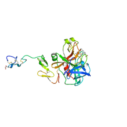
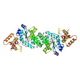
3FR0
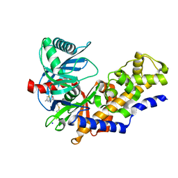 | | Human glucokinase in complex with 2-amino benzamide activator | | 分子名称: | 2-amino-N-(4-methyl-1,3-thiazol-2-yl)-5-[(4-methyl-4H-1,2,4-triazol-3-yl)sulfanyl]benzamide, Glucokinase, SODIUM ION, ... | | 著者 | Kamata, K. | | 登録日 | 2009-01-08 | | 公開日 | 2009-02-17 | | 最終更新日 | 2023-11-01 | | 実験手法 | X-RAY DIFFRACTION (2.7 Å) | | 主引用文献 | Identification of novel and potent 2-amino benzamide derivatives as allosteric glucokinase activators
Bioorg.Med.Chem.Lett., 19, 2009
|
|
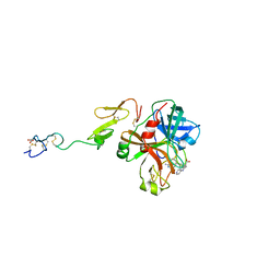
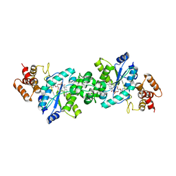
3H1V
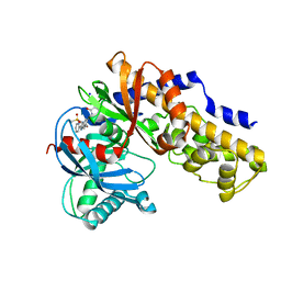 | | Human glucokinase in complex with a synthetic activator | | 分子名称: | 1-({5-[4-(methylsulfonyl)phenoxy]-2-pyridin-2-yl-1H-benzimidazol-6-yl}methyl)pyrrolidine-2,5-dione, Glucokinase, SODIUM ION, ... | | 著者 | Kamata, K, Takahashi, K. | | 登録日 | 2009-04-14 | | 公開日 | 2009-10-06 | | 最終更新日 | 2024-03-20 | | 実験手法 | X-RAY DIFFRACTION (2.11 Å) | | 主引用文献 | The design and optimization of a series of 2-(pyridin-2-yl)-1H-benzimidazole compounds as allosteric glucokinase activators.
Bioorg.Med.Chem., 17, 2009
|
|
3GOI
 | | Human glucokinase in complex with a synthetic activator | | 分子名称: | 2-(methylamino)-N-(4-methyl-1,3-thiazol-2-yl)-5-[(4-methyl-4H-1,2,4-triazol-3-yl)sulfanyl]benzamide, Glucokinase, alpha-D-glucopyranose | | 著者 | Kamata, K, Mitsuya, M. | | 登録日 | 2009-03-19 | | 公開日 | 2009-04-28 | | 最終更新日 | 2024-03-20 | | 実験手法 | X-RAY DIFFRACTION (2.52 Å) | | 主引用文献 | Discovery of novel 3,6-disubstituted 2-pyridinecarboxamide derivatives as GK activators
Bioorg.Med.Chem.Lett., 19, 2009
|
|
3A0I
 | | Human glucokinase in complex with a synthetic activator | | 分子名称: | 3-[(4-fluorophenyl)sulfanyl]-N-(4-methyl-1,3-thiazol-2-yl)-6-[(4-methyl-4H-1,2,4-triazol-3-yl)sulfanyl]pyridine-2-carboxamide, Glucokinase, SODIUM ION, ... | | 著者 | Kamata, K, Mitsuya, M. | | 登録日 | 2009-03-19 | | 公開日 | 2009-04-28 | | 最終更新日 | 2023-11-01 | | 実験手法 | X-RAY DIFFRACTION (2.2 Å) | | 主引用文献 | Discovery of novel 3,6-disubstituted 2-pyridinecarboxamide derivatives as GK activators
Bioorg.Med.Chem.Lett., 19, 2009
|
|
1XKA
 | |
1XKB
 | |
1V4T
 | | Crystal structure of human glucokinase | | 分子名称: | SODIUM ION, SULFATE ION, glucokinase isoform 2 | | 著者 | Kamata, K, Mitsuya, M, Nishimura, T, Eiki, J, Nagata, Y. | | 登録日 | 2003-11-19 | | 公開日 | 2004-03-30 | | 最終更新日 | 2023-11-08 | | 実験手法 | X-RAY DIFFRACTION (3.4 Å) | | 主引用文献 | Structural basis for allosteric regulation of the monomeric allosteric enzyme human glucokinase
Structure, 12, 2004
|
|
1V4S
 | | Crystal structure of human glucokinase | | 分子名称: | 2-AMINO-4-FLUORO-5-[(1-METHYL-1H-IMIDAZOL-2-YL)SULFANYL]-N-(1,3-THIAZOL-2-YL)BENZAMIDE, SODIUM ION, alpha-D-glucopyranose, ... | | 著者 | Kamata, K, Mitsuya, M, Nishimura, T, Eiki, J, Nagata, Y. | | 登録日 | 2003-11-19 | | 公開日 | 2004-03-30 | | 最終更新日 | 2023-11-08 | | 実験手法 | X-RAY DIFFRACTION (2.3 Å) | | 主引用文献 | Structural basis for allosteric regulation of the monomeric allosteric enzyme human glucokinase
Structure, 12, 2004
|
|
7ZCJ
 | | Crystal structure of Pizza6-TNH-TSH with Silicotungstic Acid (STA) polyoxometalate | | 分子名称: | Keggin (STA), Pizza6-TNH-TSH | | 著者 | Wouters, S.M.L, Kamata, K, Takahashi, K, Vandebroek, L, Parac-Vogt, T.N, Tame, J.R.H, Voet, A.R.D. | | 登録日 | 2022-03-28 | | 公開日 | 2023-04-19 | | 最終更新日 | 2024-02-07 | | 実験手法 | X-RAY DIFFRACTION (1.56 Å) | | 主引用文献 | Mutational study of a symmetry matched protein-polyoxometalate interface
To be published
|
|
8P2U
 | |
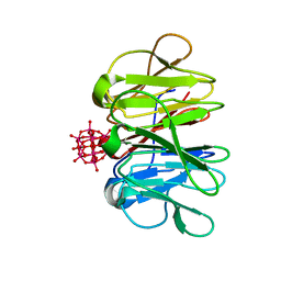
5XG5
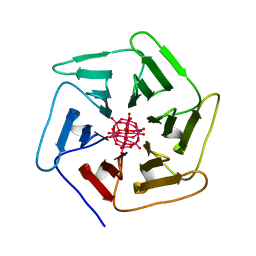
 | | Crystal structure of Mitsuba-1 with bound NAcGal | | 分子名称: | 2-acetamido-2-deoxy-alpha-D-galactopyranose, MITSUBA-1 | | 著者 | Terada, D, Voet, A.R.D, Kamata, K, Zhang, K.Y.J, Tame, J.R.H. | | 登録日 | 2017-04-12 | | 公開日 | 2017-07-12 | | 最終更新日 | 2024-03-27 | | 実験手法 | X-RAY DIFFRACTION (1.54 Å) | | 主引用文献 | Computational design of a symmetrical beta-trefoil lectin with cancer cell binding activity.
Sci Rep, 7, 2017
|
|
7ZPZ
 | | Crystal structure of Pizza6-TSR-TSH with Silicotungstic Acid (STA) polyoxometalate | | 分子名称: | Keggin (STA), Pizza6-TSR-TSH | | 著者 | Wouters, S.M.L, Kamata, K, Takahashi, K, Vandebroek, L, Parac-Vogt, T.N, Tame, J.R.H, Voet, A.R.D. | | 登録日 | 2022-04-29 | | 公開日 | 2023-05-10 | | 最終更新日 | 2024-02-07 | | 実験手法 | X-RAY DIFFRACTION (1.57 Å) | | 主引用文献 | Mutational study of a symmetry matched protein-polyoxometalate interface
To be published
|
|
7ZQ2
 | | Crystal structure of Pizza6-RSH-TSH with Silicotungstic Acid (STA) polyoxometalate | | 分子名称: | Keggin (STA), Pizza6-RSH-TSH | | 著者 | Wouters, S.M.L, Kamata, K, Takahashi, K, Vandebroek, L, Parac-Vogt, T.N, Tame, J.R.H, Voet, A.R.D. | | 登録日 | 2022-04-29 | | 公開日 | 2023-05-10 | | 最終更新日 | 2024-02-07 | | 実験手法 | X-RAY DIFFRACTION (1.6 Å) | | 主引用文献 | Mutational study of a symmetry matched protein-polyoxometalate interface
To be published
|
|
7ZQG
 | | Crystal structure of Pizza6-KSH-TSH with Silicotungstic Acid (STA) polyoxometalate | | 分子名称: | Keggin (STA), Pizza6-KSH-TSH | | 著者 | Wouters, S.M.L, Kamata, K, Takahashi, K, Vandebroek, L, Parac-Vogt, T.N, Tame, J.R.H, Voet, A.R.D. | | 登録日 | 2022-04-29 | | 公開日 | 2023-05-10 | | 最終更新日 | 2024-02-07 | | 実験手法 | X-RAY DIFFRACTION (1.8 Å) | | 主引用文献 | Mutational study of a symmetry matched protein-polyoxometalate interface
To be published
|
|
7ZPW
 | | Crystal structure of Pizza6-TSK-TSH with Silicotungstic Acid (STA) polyoxometalate | | 分子名称: | Keggin (STA), Pizza6-TSK-TSH | | 著者 | Wouters, S.M.L, Kamata, K, Takahashi, K, Vandebroek, L, Parac-Vogt, T.N, Tame, J.R.H, Voet, A.R.D. | | 登録日 | 2022-04-29 | | 公開日 | 2023-05-10 | | 最終更新日 | 2024-02-07 | | 実験手法 | X-RAY DIFFRACTION (1.8 Å) | | 主引用文献 | Mutational study of a symmetry matched protein-polyoxometalate interface.
To be published
|
|
7ZPH
 | | Crystal structure of Pizza6-TNK-RSH with Silicotungstic Acid (STA) polyoxometalate | | 分子名称: | Keggin (STA), Pizza6-TNK-RSH | | 著者 | Wouters, S.M.L, Kamata, K, Takahashi, K, Vandebroek, L, Parac-Vogt, T.N, Tame, J.R.H, Voet, A.R.D. | | 登録日 | 2022-04-27 | | 公開日 | 2023-05-10 | | 最終更新日 | 2024-02-07 | | 実験手法 | X-RAY DIFFRACTION (1.66 Å) | | 主引用文献 | Mutational study of a symmetry matched protein-polyoxometalate interface
To be published
|
|
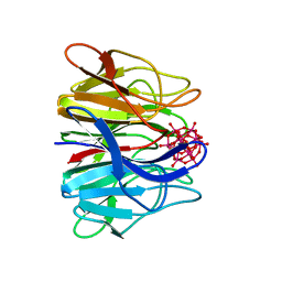
1VBN
 | | Escherichia coli tyrosyl-tRNA synthetase mutant complexed with Tyr-AMS | | 分子名称: | 5'-O-[N-(L-TYROSYL)SULFAMOYL]ADENOSINE, Tyrosyl-tRNA synthetase | | 著者 | Kobayashi, T, Sakamoto, K, Takimura, T, Kamata, K, Sekine, R, Nishimura, S, Yokoyama, S, RIKEN Structural Genomics/Proteomics Initiative (RSGI) | | 登録日 | 2004-02-27 | | 公開日 | 2005-01-25 | | 最終更新日 | 2023-10-25 | | 実験手法 | X-RAY DIFFRACTION (2.7 Å) | | 主引用文献 | Structural basis of nonnatural amino acid recognition by an engineered aminoacyl-tRNA synthetase for genetic code expansion
Proc.Natl.Acad.Sci.USA, 102, 2005
|
|
1VBM
 | | Crystal structure of the Escherichia coli tyrosyl-tRNA synthetase complexed with Tyr-AMS | | 分子名称: | 5'-O-[N-(L-TYROSYL)SULFAMOYL]ADENOSINE, SULFATE ION, Tyrosyl-tRNA synthetase | | 著者 | Kobayashi, T, Sakamoto, K, Takimura, T, Kamata, K, Sekine, R, Nishimura, S, Yokoyama, S, RIKEN Structural Genomics/Proteomics Initiative (RSGI) | | 登録日 | 2004-02-27 | | 公開日 | 2005-01-25 | | 最終更新日 | 2023-10-25 | | 実験手法 | X-RAY DIFFRACTION (2.7 Å) | | 主引用文献 | Structural snapshots of the KMSKS loop rearrangement for amino acid activation by bacterial tyrosyl-tRNA synthetase.
J.Mol.Biol., 346, 2005
|
|
7FG6
 | |
3A8W
 | | Crystal Structure of PKCiota kinase domain | | 分子名称: | ADENOSINE-5'-TRIPHOSPHATE, Protein kinase C iota type, SULFATE ION | | 著者 | Takimura, T, Kamata, K. | | 登録日 | 2009-10-11 | | 公開日 | 2010-05-05 | | 最終更新日 | 2017-10-11 | | 実験手法 | X-RAY DIFFRACTION (2.1 Å) | | 主引用文献 | Structures of the PKC-iota kinase domain in its ATP-bound and apo forms reveal defined structures of residues 533-551 in the C-terminal tail and their roles in ATP binding
Acta Crystallogr.,Sect.D, 66, 2010
|
|
3A8X
 | | Crystal Structure of PKCiota kinase domain | | 分子名称: | Protein kinase C iota type, SULFATE ION | | 著者 | Takimura, T, Kamata, K. | | 登録日 | 2009-10-11 | | 公開日 | 2010-05-05 | | 最終更新日 | 2017-10-11 | | 実験手法 | X-RAY DIFFRACTION (2 Å) | | 主引用文献 | Structures of the PKC-iota kinase domain in its ATP-bound and apo forms reveal defined structures of residues 533-551 in the C-terminal tail and their roles in ATP binding
Acta Crystallogr.,Sect.D, 66, 2010
|
|
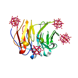
6QSE
 | | Crystal structure of Pizza6S in the presence of Anderson-Evans (TEW) | | 分子名称: | 6-tungstotellurate(VI), Pizza6S | | 著者 | Noguchi, H, Vandebroek, L, Kamata, K, Tame, J.R.H, Van Meervelt, L, Parac-Vogt, T.N, Voet, A.R.D. | | 登録日 | 2019-02-20 | | 公開日 | 2020-03-18 | | 最終更新日 | 2024-01-24 | | 実験手法 | X-RAY DIFFRACTION (1.9 Å) | | 主引用文献 | Hybrid assemblies of a symmetric designer protein and polyoxometalates with matching symmetry.
Chem.Commun.(Camb.), 56, 2020
|
|
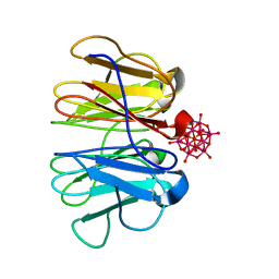
6QSF
 | | Crystal structure of Pizza6S in the presence of Keggin (STA) | | 分子名称: | Keggin (STA), Pizza6S | | 著者 | Noguchi, H, Vandebroek, L, Kamata, K, Tame, J.R.H, Van Meervelt, L, Parac-Vogt, T.N, Voet, A.R.D. | | 登録日 | 2019-02-20 | | 公開日 | 2020-03-18 | | 最終更新日 | 2024-01-24 | | 実験手法 | X-RAY DIFFRACTION (1.5 Å) | | 主引用文献 | Hybrid assemblies of a symmetric designer protein and polyoxometalates with matching symmetry.
Chem.Commun.(Camb.), 56, 2020
|
|


