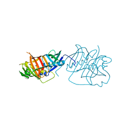
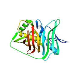
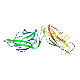
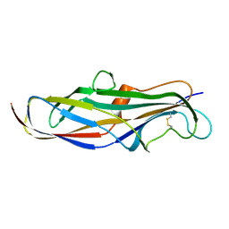
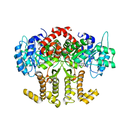
6GTW
 
 | | Crystal structure of the FimH lectin domain from E.coli F18 in complex with trimannose | | 分子名称: | CALCIUM ION, FimH protein, alpha-D-mannopyranose-(1-3)-[alpha-D-mannopyranose-(1-6)]alpha-D-mannopyranose | | 著者 | Jakob, R.P, Sauer, M.M, Luber, T, Canonica, F, Navarra, G, Ernst, B, Unverzagt, C, Maier, T, Glockshuber, R. | | 登録日 | 2018-06-19 | | 公開日 | 2019-01-16 | | 最終更新日 | 2024-01-17 | | 実験手法 | X-RAY DIFFRACTION (2.5 Å) | | 主引用文献 | Binding of the Bacterial Adhesin FimH to Its Natural, Multivalent High-Mannose Type Glycan Targets.
J.Am.Chem.Soc., 141, 2019
|
|
5HST
 | |
5IL5
 | |
5I7Q
 | |
5JCQ
 | |
5IL6
 | |
5J6O
 | |
5JCR
 | |
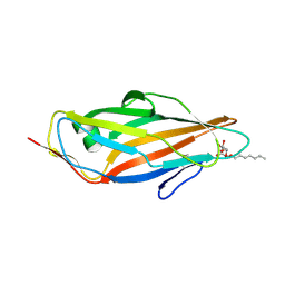
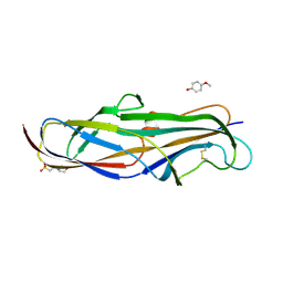
5L4W
 | | Crystal structure of FimH lectin domain in complex with 3-Fluoro-Heptylmannoside | | 分子名称: | Protein FimH, heptyl 3-fluoro-alpha-D-mannopyranoside | | 著者 | Jakob, R.P, Zihlmann, P, Rabbani, S, Maier, T, Ernst, B. | | 登録日 | 2016-05-26 | | 公開日 | 2017-06-21 | | 最終更新日 | 2024-01-10 | | 実験手法 | X-RAY DIFFRACTION (1.9 Å) | | 主引用文献 | High-Affinity Carbohydrate-Lectin Interactions: How Nature Makes it Possible
To Be Published
|
|
5I7P
 | | Crystal structure of Fkbp12-IF(SlyD), a chimeric protein of human Fkbp12 and the insert in flap domain of Ecoli SlyD | | 分子名称: | Peptidyl-prolyl cis-trans isomerase FKBP1A,FKBP-type peptidyl-prolyl cis-trans isomerase SlyD,Peptidyl-prolyl cis-trans isomerase FKBP1A | | 著者 | Jakob, R.P, Knappe, T.A, Dobbek, H, Schmid, F.X. | | 登録日 | 2016-02-18 | | 公開日 | 2017-03-08 | | 最終更新日 | 2024-01-10 | | 実験手法 | X-RAY DIFFRACTION (2.002 Å) | | 主引用文献 | Structural and Functional Analysis of Chaperone Domain Insertion in Fkbp12
To Be Published
|
|
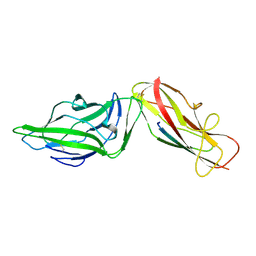
4XO9
 | | Crystal structure of a FimH*DsG complex from E.coli K12 in space group C2 | | 分子名称: | Minor component of type 1 fimbriae, Protein FimH | | 著者 | Jakob, R.P, Eras, J, Glockshuber, R, Maier, T. | | 登録日 | 2015-01-16 | | 公開日 | 2016-01-27 | | 最終更新日 | 2024-01-10 | | 実験手法 | X-RAY DIFFRACTION (1.14 Å) | | 主引用文献 | Catch-bond mechanism of the bacterial adhesin FimH.
Nat Commun, 7, 2016
|
|
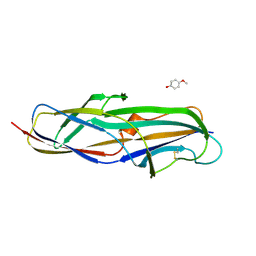
4XO8
 | | Crystal structure of the FimH lectin domain from E.coli K12 in complex with heptyl alpha-D-mannopyrannoside | | 分子名称: | Protein FimH, heptyl alpha-D-mannopyranoside | | 著者 | Jakob, R.P, Eras, J, Navarra, G, Ernst, B, Glockshuber, R, Maier, T. | | 登録日 | 2015-01-16 | | 公開日 | 2016-01-27 | | 最終更新日 | 2024-01-10 | | 実験手法 | X-RAY DIFFRACTION (1.698 Å) | | 主引用文献 | Catch-bond mechanism of the bacterial adhesin FimH.
Nat Commun, 7, 2016
|
|
4XOD
 | | Crystal structure of a FimH*DsG complex from E.coli F18 | | 分子名称: | FimG protein, FimH protein | | 著者 | Jakob, R.P, Sauer, M.M, Glockshuber, R, Maier, T. | | 登録日 | 2015-01-16 | | 公開日 | 2016-01-27 | | 最終更新日 | 2024-01-10 | | 実験手法 | X-RAY DIFFRACTION (1.14 Å) | | 主引用文献 | Catch-bond mechanism of the bacterial adhesin FimH.
Nat Commun, 7, 2016
|
|
4XOC
 | | Crystal structure of the FimH lectin domain from E.coli F18 in complex with heptyl alpha-D-mannopyrannoside | | 分子名称: | FimH protein, heptyl alpha-D-mannopyranoside | | 著者 | Jakob, R.P, Sauer, M.M, Navarra, G, Ernst, B, Glockshuber, R, Maier, T. | | 登録日 | 2015-01-16 | | 公開日 | 2016-01-27 | | 最終更新日 | 2024-01-10 | | 実験手法 | X-RAY DIFFRACTION (1.42 Å) | | 主引用文献 | Catch-bond mechanism of the bacterial adhesin FimH.
Nat Commun, 7, 2016
|
|
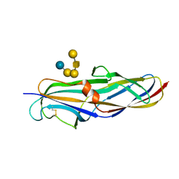
4XOB
 | | Crystal structure of a FimH*DsF complex from E.coli K12 with bound heptyl alpha-D-mannopyrannoside | | 分子名称: | FimF, Protein FimH, SULFATE ION, ... | | 著者 | Jakob, R.P, Eras, J, Navarra, G, Ernst, B, Glockshuber, R, Maier, T. | | 登録日 | 2015-01-16 | | 公開日 | 2016-01-27 | | 最終更新日 | 2024-01-10 | | 実験手法 | X-RAY DIFFRACTION (3.003 Å) | | 主引用文献 | Catch-bond mechanism of the bacterial adhesin FimH.
Nat Commun, 7, 2016
|
|
4XOE
 | | Crystal structure of a FimH*DsG complex from E.coli F18 with bound heptyl alpha-D-mannopyrannoside | | 分子名称: | CACODYLATE ION, FimG protein, FimH protein, ... | | 著者 | Jakob, R.P, Sauer, M.M, Navarra, G, Ernst, B, Glockshuber, R, Maier, T. | | 登録日 | 2015-01-16 | | 公開日 | 2016-01-27 | | 最終更新日 | 2024-01-10 | | 実験手法 | X-RAY DIFFRACTION (2.4 Å) | | 主引用文献 | Catch-bond mechanism of the bacterial adhesin FimH.
Nat Commun, 7, 2016
|
|
4XOA
 | | Crystal structure of a FimH*DsG complex from E.coli K12 in space group P1 | | 分子名称: | FimG, Protein FimH | | 著者 | Jakob, R.P, Eras, J, Glockshuber, R, Maier, T. | | 登録日 | 2015-01-16 | | 公開日 | 2016-01-27 | | 最終更新日 | 2024-01-10 | | 実験手法 | X-RAY DIFFRACTION (2.541 Å) | | 主引用文献 | Catch-bond mechanism of the bacterial adhesin FimH.
Nat Commun, 7, 2016
|
|
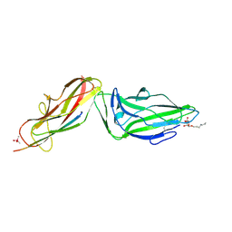
4Z3I
 | | Crystal structure of the lectin domain of PapG from E. coli BI47 in spacegroup P21212 | | 分子名称: | PapG, lectin domain | | 著者 | Jakob, R.P, Navarra, G, Zihlmann, P, Stangier, K, Preston, R.C, Rabbani, S, Maier, T, Ernst, B. | | 登録日 | 2015-03-31 | | 公開日 | 2016-04-13 | | 最終更新日 | 2024-07-17 | | 実験手法 | X-RAY DIFFRACTION (1.739 Å) | | 主引用文献 | Carbohydrate-Lectin Interactions: An Unexpected Contribution to Affinity.
Chembiochem, 18, 2017
|
|
4Z3G
 | | Crystal structure of the lectin domain of PapG from E. coli BI47 in complex with 4-methoxyphenyl beta-D-galabiose in space group P212121 | | 分子名称: | 2-(N-MORPHOLINO)-ETHANESULFONIC ACID, 4-methoxyphenol, PapG, ... | | 著者 | Jakob, R.P, Navarra, G, Zihlmann, P, Stangier, K, Preston, R.C, Rabbani, S, Maier, T, Ernst, B. | | 登録日 | 2015-03-31 | | 公開日 | 2016-04-13 | | 最終更新日 | 2024-07-17 | | 実験手法 | X-RAY DIFFRACTION (1.45 Å) | | 主引用文献 | Carbohydrate-Lectin Interactions: An Unexpected Contribution to Affinity.
Chembiochem, 18, 2017
|
|
4Z3E
 | | Crystal structure of the lectin domain of PapG from E. coli BI47 in complex with SSEA4 in space group P212121 | | 分子名称: | PapG, lectin domain, beta-D-galactopyranose-(1-3)-2-acetamido-2-deoxy-beta-D-galactopyranose-(1-3)-alpha-D-galactopyranose-(1-4)-beta-D-galactopyranose-(1-4)-beta-D-glucopyranose | | 著者 | Jakob, R.P, Navarra, G, Zihlmann, P, Stangier, K, Preston, R.C, Rabbani, S, Maier, T, Ernst, B. | | 登録日 | 2015-03-31 | | 公開日 | 2016-04-13 | | 最終更新日 | 2024-01-10 | | 実験手法 | X-RAY DIFFRACTION (1.8 Å) | | 主引用文献 | Carbohydrate-Lectin Interactions: An Unexpected Contribution to Affinity.
Chembiochem, 18, 2017
|
|
4Z3H
 | | Crystal structure of the lectin domain of PapG from E. coli BI47 in complex with 4-methoxyphenyl beta-D-galabiose in space group P21 | | 分子名称: | 4-methoxyphenol, PapG, lectin domain, ... | | 著者 | Jakob, R.P, Navarra, G, Zihlmann, P, Stangier, K, Preston, R.C, Rabbani, S, Maier, T, Ernst, B. | | 登録日 | 2015-03-31 | | 公開日 | 2016-04-13 | | 最終更新日 | 2024-07-17 | | 実験手法 | X-RAY DIFFRACTION (1.5 Å) | | 主引用文献 | Carbohydrate-Lectin Interactions: An Unexpected Contribution to Affinity.
Chembiochem, 18, 2017
|
|
4Z38
 | |
4Z3F
 | | Crystal structure of the lectin domain of PapG from E. coli BI47 in complex with SSEA4 in space group P21 | | 分子名称: | N-acetyl-alpha-neuraminic acid-(2-3)-beta-D-galactopyranose-(1-3)-2-acetamido-2-deoxy-beta-D-galactopyranose-(1-3)-alpha-D-galactopyranose-(1-4)-beta-D-galactopyranose-(1-4)-beta-D-glucopyranose, PapG, lectin domain, ... | | 著者 | Jakob, R.P, Navarra, G, Zihlmann, P, Stangier, K, Preston, R.C, Rabbani, S, Maier, T, Ernst, B. | | 登録日 | 2015-03-31 | | 公開日 | 2016-04-13 | | 最終更新日 | 2024-07-17 | | 実験手法 | X-RAY DIFFRACTION (1.8 Å) | | 主引用文献 | Carbohydrate-Lectin Interactions: An Unexpected Contribution to Affinity.
Chembiochem, 18, 2017
|
|
4Z3J
 | | Crystal structure of the lectin domain of PapG from E. coli BI47 in space group P1 | | 分子名称: | PapG, lectin domain | | 著者 | Jakob, R.P, Navarra, G, Zihlmann, P, Stangier, K, Preston, R.C, Rabbani, S, Maier, T, Ernst, B. | | 登録日 | 2015-03-31 | | 公開日 | 2016-04-13 | | 最終更新日 | 2024-07-17 | | 実験手法 | X-RAY DIFFRACTION (2.5 Å) | | 主引用文献 | Carbohydrate-Lectin Interactions: An Unexpected Contribution to Affinity.
Chembiochem, 18, 2017
|
|
5OOW
 | | Crystal structure of lobe II from the nucleotide binding domain of DnaK in complex with AMPPCP | | 分子名称: | Chaperone protein DnaK, PHOSPHOMETHYLPHOSPHONIC ACID ADENYLATE ESTER | | 著者 | Jakob, R.P, Bauer, D, Meinhold, S, Stigler, J, Merkel, U, Maier, T, Rief, M, Zoldak, G. | | 登録日 | 2017-08-09 | | 公開日 | 2018-04-25 | | 最終更新日 | 2024-01-17 | | 実験手法 | X-RAY DIFFRACTION (2.9 Å) | | 主引用文献 | A folding nucleus and minimal ATP binding domain of Hsp70 identified by single-molecule force spectroscopy.
Proc. Natl. Acad. Sci. U.S.A., 115, 2018
|
|


