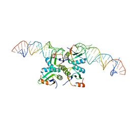
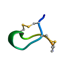
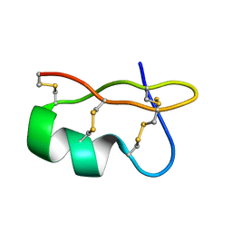
1AUL
 
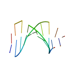 | | SOLUTION STRUCTURE OF A HIGHLY STABLE DNA DUPLEX CONJUGATED TO A MINOR GROOVE BINDER, NMR, MINIMIZED AVERAGE STRUCTURE | | 分子名称: | DNA (5'-D(*CP*AP*GP*AP*TP*AP*AP*TP*CP*A)-3'), DNA (5'-D(P*THXP*GP*AP*TP*TP*AP*TP*CP*TP*G)-3') | | 著者 | Kumar, S, Reed, M.W, Gamper Junior, H.B, Gorn, V.V, Lukhtanov, E.A, Foti, M, West, J, Meyer Junior, R.B, Schweitzer, B.I. | | 登録日 | 1997-08-29 | | 公開日 | 1997-12-24 | | 最終更新日 | 2024-05-22 | | 実験手法 | SOLUTION NMR | | 主引用文献 | Solution structure of a highly stable DNA duplex conjugated to a minor groove binder.
Nucleic Acids Res., 26, 1998
|
|
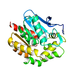
1VEF
 | | Acetylornithine aminotransferase from Thermus thermophilus HB8 | | 分子名称: | Acetylornithine/acetyl-lysine aminotransferase, PYRIDOXAL-5'-PHOSPHATE | | 著者 | Matsumura, M, Goto, M, Omi, R, Miyahara, I, Hirotsu, K, RIKEN Structural Genomics/Proteomics Initiative (RSGI) | | 登録日 | 2004-03-30 | | 公開日 | 2005-08-02 | | 最終更新日 | 2023-10-25 | | 実験手法 | X-RAY DIFFRACTION (1.35 Å) | | 主引用文献 | Three-Dimensional Strutcure of Acetylornithine aminotransferase from Thermus thermophilus HB8
To be Published
|
|
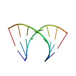
1VEX
 | | F-spondin TSR domain 4 | | 分子名称: | F-spondin | | 著者 | Paakkonen, K, Tossavainen, H, Permi, P, Kilpelainen, I, Guntert, P. | | 登録日 | 2004-04-06 | | 公開日 | 2005-04-19 | | 最終更新日 | 2023-12-27 | | 実験手法 | SOLUTION NMR | | 主引用文献 | Solution structures of the first and fourth TSR domains of F-spondin
Proteins, 64, 2006
|
|
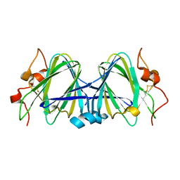
3LLT
 | | Crystal structure of PF14_0431, kinase domain. | | 分子名称: | ACETATE ION, PHOSPHOAMINOPHOSPHONIC ACID-ADENYLATE ESTER, SULFATE ION, ... | | 著者 | Wernimont, A.K, Tempel, W, Lin, Y.H, Loppnau, P, MacKenzie, F, Sullivan, H, Weadge, J, Kozieradzki, I, Cossar, D, Sinesterra, G, Vedadi, M, Arrowsmith, C.H, Edwards, A.M, Bountra, C, Weigelt, J, Bochkarev, A, Hui, R, Qiu, W, Hutchinson, A, Structural Genomics Consortium (SGC) | | 登録日 | 2010-01-29 | | 公開日 | 2010-08-04 | | 最終更新日 | 2023-09-06 | | 実験手法 | X-RAY DIFFRACTION (2.5 Å) | | 主引用文献 | Crystal structure of PF14_0431, kinase domain.
TO BE PUBLISHED
|
|
1VDZ
 | | Crystal structure of A-type ATPase catalytic subunit A from Pyrococcus horikoshii OT3 | | 分子名称: | (4S)-2-METHYL-2,4-PENTANEDIOL, A-type ATPase subunit A | | 著者 | Maegawa, Y, Morita, H, Yao, M, Watanabe, N, Tanaka, I. | | 登録日 | 2004-03-26 | | 公開日 | 2005-06-21 | | 最終更新日 | 2023-12-27 | | 実験手法 | X-RAY DIFFRACTION (2.55 Å) | | 主引用文献 | Crystal structure of A-type ATPase catalytic subunit A from Pyrococcus horikoshii OT3
To be Published
|
|
4AQI
 | | Structure of human S100A15 bound to zinc and calcium | | 分子名称: | CALCIUM ION, CHLORIDE ION, PROTEIN S100-A7A, ... | | 著者 | Murray, J.I, Tonkin, M.L, Whiting, A.L, Peng, F, Farnell, B, Hof, F, Boulanger, M.J. | | 登録日 | 2012-04-17 | | 公開日 | 2012-10-17 | | 最終更新日 | 2023-12-20 | | 実験手法 | X-RAY DIFFRACTION (1.7 Å) | | 主引用文献 | Structural Characterization of S100A15 Reveals a Novel Zinc Coordination Site Among S100 Proteins and Altered Surface Chemistry with Functional Implications for Receptor Binding.
Bmc Struct.Biol., 12, 2012
|
|
3LVQ
 | | The crystal structure of ASAP3 in complex with Arf6 in transition state | | 分子名称: | ALUMINUM FLUORIDE, Arf-GAP with SH3 domain, ANK repeat and PH domain-containing protein 3, ... | | 著者 | Ismail, S.A, Vetter, I.R, Sot, B, Wittinghofer, A. | | 登録日 | 2010-02-22 | | 公開日 | 2010-06-09 | | 最終更新日 | 2023-11-01 | | 実験手法 | X-RAY DIFFRACTION (3.38 Å) | | 主引用文献 | The structure of an Arf-ArfGAP complex reveals a Ca2+ regulatory mechanism
Cell(Cambridge,Mass.), 141, 2010
|
|
3LVW
 | | Glutathione-inhibited ScGCL | | 分子名称: | GLUTATHIONE, Glutamate--cysteine ligase, TRIETHYLENE GLYCOL | | 著者 | Biterova, E.I, Barycki, J.J. | | 登録日 | 2010-02-22 | | 公開日 | 2010-03-16 | | 最終更新日 | 2023-09-06 | | 実験手法 | X-RAY DIFFRACTION (2.5 Å) | | 主引用文献 | Structural basis for feedback and pharmacological inhibition of Saccharomyces cerevisiae glutamate cysteine ligase.
J.Biol.Chem., 285, 2010
|
|
1W0T
 | | hTRF1 DNA-binding domain in complex with telomeric DNA. | | 分子名称: | 5'-D(*CP*TP*GP*TP*TP*AP*GP*GP*GP*TP *TP*AP*GP*GP*GP*TP*TP*AP*G)-3', 5'-D(*TP*CP*TP*AP*AP*CP*CP*CP*TP*AP *AP*CP*CP*CP*TP*AP*AP*CP*A)-3', TELOMERIC REPEAT BINDING FACTOR 1 | | 著者 | Court, R.I, Chapman, L.M, Fairall, L, Rhodes, D. | | 登録日 | 2004-06-11 | | 公開日 | 2004-12-22 | | 最終更新日 | 2024-05-08 | | 実験手法 | X-RAY DIFFRACTION (2 Å) | | 主引用文献 | How the Human Telomeric Proteins Trf1 and Trf2 Recognize Telomeric DNA: A View from High-Resolution Crystal Structures
Embo Rep., 6, 2005
|
|
7DLZ
 | | Crystal Structure of Methyltransferase Ribozyme | | 分子名称: | RNA (45-MER), U1 small nuclear ribonucleoprotein A | | 著者 | Gan, J.H, Gao, Y.Q, Jiang, H.Y, Chen, D.R, Murchie, A.I.H. | | 登録日 | 2020-11-30 | | 公開日 | 2021-10-27 | | 最終更新日 | 2023-11-29 | | 実験手法 | X-RAY DIFFRACTION (3.002 Å) | | 主引用文献 | The identification and characterization of a selected SAM-dependent methyltransferase ribozyme that is present in natural sequences
Nat Catal, 4, 2021
|
|
1BEE
 | | HALOALKANE DEHALOGENASE MUTANT WITH TRP 175 REPLACED BY TYR | | 分子名称: | HALOALKANE DEHALOGENASE | | 著者 | Ridder, I.S, Vos, G.J, Rozeboom, H.J, Kalk, K.H, Dijkstra, B.W. | | 登録日 | 1998-05-13 | | 公開日 | 1998-11-11 | | 最終更新日 | 2024-05-22 | | 実験手法 | X-RAY DIFFRACTION (2.6 Å) | | 主引用文献 | Kinetic analysis and X-ray structure of haloalkane dehalogenase with a modified halide-binding site.
Biochemistry, 37, 1998
|
|
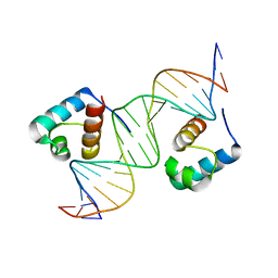
402D
 | | 5'-R(*CP*GP*CP*CP*AP*GP*CP*G)-3' | | 分子名称: | RNA(5'-R(*CP*GP*CP*CP*AP*GP*CP*G)-3') | | 著者 | Jang, S.B, Hung, L.-W, Chi, Y.-I, Holbrook, E.L, Carter, R, Holbrook, S.R. | | 登録日 | 1998-06-04 | | 公開日 | 1998-06-16 | | 最終更新日 | 2024-02-28 | | 実験手法 | X-RAY DIFFRACTION (2.3 Å) | | 主引用文献 | Structure of an RNA internal loop consisting of tandem C-A+ base pairs.
Biochemistry, 37, 1998
|
|
1WLT
 | | Crystal Structure of dTDP-4-dehydrorhamnose 3,5-epimerase homologue from Sulfolobus tokodaii | | 分子名称: | 176aa long hypothetical dTDP-4-dehydrorhamnose 3,5-epimerase | | 著者 | Watanabe, N, Sakai, N, Zilian, Z, Kawarabayasi, Y, Tanaka, I. | | 登録日 | 2004-06-29 | | 公開日 | 2005-07-26 | | 最終更新日 | 2023-10-25 | | 実験手法 | X-RAY DIFFRACTION (1.9 Å) | | 主引用文献 | Crystal Structure of dTDP-4-dehydrorhamnose 3,5-epimerase homologue
to be published
|
|
1WMI
 | | Crystal structure of archaeal RelE-RelB complex from Pyrococcus horikoshii OT3 | | 分子名称: | hypothetical protein PHS013, hypothetical protein PHS014 | | 著者 | Takagi, H, Kakuta, Y, Kamachi, R, Yao, M, Tanaka, I, Kimura, M. | | 登録日 | 2004-07-09 | | 公開日 | 2005-03-15 | | 最終更新日 | 2024-03-13 | | 実験手法 | X-RAY DIFFRACTION (2.3 Å) | | 主引用文献 | Crystal structure of archaeal toxin-antitoxin RelE-RelB complex with implications for toxin activity and antitoxin effects
Nat.Struct.Mol.Biol., 12, 2005
|
|
1B45
 | | ALPHA-CNIA CONOTOXIN FROM CONUS CONSORS, NMR, 43 STRUCTURES | | 分子名称: | ALPHA-CNIA | | 著者 | Favreau, P, Krimm, I, Le Gall, F, Bobenrieth, M.J, Lamthanh, H, Bouet, F, Servent, D, Molgo, J, Menez, A, Letourneux, Y, Lancelin, J.M. | | 登録日 | 1999-01-05 | | 公開日 | 1999-07-09 | | 最終更新日 | 2022-02-16 | | 実験手法 | SOLUTION NMR | | 主引用文献 | Biochemical characterization and nuclear magnetic resonance structure of novel alpha-conotoxins isolated from the venom of Conus consors.
Biochemistry, 38, 1999
|
|
4IZK
 | |
4AQJ
 | | Structure of human S100A7 D24G bound to zinc and calcium | | 分子名称: | CALCIUM ION, CHLORIDE ION, PROTEIN S100-A7, ... | | 著者 | Murray, J.I, Tonkin, M.L, Whiting, A.L, Peng, F, Farnell, B, Hof, F, Boulanger, M.J. | | 登録日 | 2012-04-17 | | 公開日 | 2012-10-17 | | 実験手法 | X-RAY DIFFRACTION (1.6 Å) | | 主引用文献 | Structural Characterization of S100A15 Reveals a Novel Zinc Coordination Site Among S100 Proteins and Altered Surface Chemistry with Functional Implications for Receptor Binding.
Bmc Struct.Biol., 12, 2012
|
|
3LDJ
 | | Crystal structure of aprotinin in complex with sucrose octasulfate: unusual interactions and implication for heparin binding | | 分子名称: | 1,3,4,6-tetra-O-sulfo-beta-D-fructofuranose-(2-1)-2,3,4,6-tetra-O-sulfonato-alpha-D-glucopyranose, ACETATE ION, Pancreatic trypsin inhibitor | | 著者 | Yang, I.S, Kim, T.G, Park, B.S, Kim, K.H. | | 登録日 | 2010-01-13 | | 公開日 | 2010-09-15 | | 最終更新日 | 2020-07-29 | | 実験手法 | X-RAY DIFFRACTION (1.7 Å) | | 主引用文献 | Crystal structures of aprotinin and its complex with sucrose octasulfate reveal multiple modes of interactions with implications for heparin binding.
Biochem.Biophys.Res.Commun., 2010
|
|
1C7W
 | | NMR SOLUTION STRUCTURE OF THE CALCIUM-BOUND C-TERMINAL DOMAIN (W81-S161) OF CALCIUM VECTOR PROTEIN FROM AMPHIOXUS | | 分子名称: | CALCIUM VECTOR PROTEIN | | 著者 | Theret, I, Baladi, S, Cox, J.A, Sakamoto, H, Craescu, C.T. | | 登録日 | 2000-03-27 | | 公開日 | 2000-04-12 | | 最終更新日 | 2023-12-27 | | 実験手法 | SOLUTION NMR | | 主引用文献 | Sequential calcium binding to the regulatory domain of calcium vector protein reveals functional asymmetry and a novel mode of structural rearrangement.
Biochemistry, 39, 2000
|
|
1CCZ
 | | CRYSTAL STRUCTURE OF THE CD2-BINDING DOMAIN OF CD58 (LYMPHOCYTE FUNCTION-ASSOCIATED ANTIGEN 3) AT 1.8-A RESOLUTION | | 分子名称: | 2-acetamido-2-deoxy-beta-D-glucopyranose, PROTEIN (CD58) | | 著者 | Ikemizu, S, Sparks, L.M, Van Der Merwe, P.A, Harlos, K, Stuart, D.I, Jones, E.Y, Davis, S.J. | | 登録日 | 1999-03-02 | | 公開日 | 1999-04-05 | | 最終更新日 | 2023-08-09 | | 実験手法 | X-RAY DIFFRACTION (1.8 Å) | | 主引用文献 | Crystal structure of the CD2-binding domain of CD58 (lymphocyte function-associated antigen 3) at 1.8-A resolution.
Proc.Natl.Acad.Sci.USA, 96, 1999
|
|
7DRE
 | | Cryo-EM structure of DfgA-B at 2.54 angstrom resolution | | 分子名称: | DfgB, Sugar phosphate isomerase/epimerase | | 著者 | Mori, T, Moriya, T, Adachi, N, Senda, T, Abe, I. | | 登録日 | 2020-12-28 | | 公開日 | 2021-12-08 | | 最終更新日 | 2024-06-05 | | 実験手法 | ELECTRON MICROSCOPY (2.54 Å) | | 主引用文献 | C-Glycoside metabolism in the gut and in nature: Identification, characterization, structural analyses and distribution of C-C bond-cleaving enzymes.
Nat Commun, 12, 2021
|
|
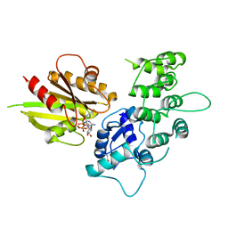
1WNU
 | | Structure of Archaeal Trans-Editing Protein AlaX in complex with L-serine | | 分子名称: | SERINE, ZINC ION, alanyl-tRNA synthetase | | 著者 | Sokabe, M, Okada, A, Nakashima, T, Yao, M, Tanaka, I. | | 登録日 | 2004-08-09 | | 公開日 | 2005-07-26 | | 最終更新日 | 2023-11-15 | | 実験手法 | X-RAY DIFFRACTION (2.8 Å) | | 主引用文献 | Molecular basis of alanine discrimination in editing site
Proc.Natl.Acad.Sci.Usa, 102, 2005
|
|
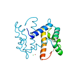
1WPD
 | | Evidence for domain-specific recognition of SK and Kv channels by MTX and HsTx1 scorpion toxins | | 分子名称: | Potassium channel toxin alpha-KTx 6.2,Potassium channel toxin alpha-KTx 6.3 | | 著者 | Regaya, I, Beeton, C, Ferrat, G, Andreotti, N, Chandy, G.K, Darbon, H, De Waard, M, Sabatier, J.M. | | 登録日 | 2004-09-01 | | 公開日 | 2004-10-19 | | 最終更新日 | 2019-12-18 | | 実験手法 | SOLUTION NMR | | 主引用文献 | Evidence for domain-specific recognition of SK and Kv channels by MTX and HsTx1 scorpion toxins
J.Biol.Chem., 279, 2004
|
|
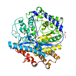
1WU7
 | | Crystal structure of histidyl-tRNA synthetase from Thermoplasma acidophilum | | 分子名称: | Histidyl-tRNA synthetase | | 著者 | Tanaka, Y, Sakai, N, Yao, M, Watanabe, N, Tamura, T, Tanaka, I. | | 登録日 | 2004-12-01 | | 公開日 | 2005-12-06 | | 最終更新日 | 2011-07-13 | | 実験手法 | X-RAY DIFFRACTION (2.4 Å) | | 主引用文献 | Crystal structure of histidyl-tRNA synthetase from Thermoplasma acidophilum
To be Published
|
|
1C7V
 | | NMR SOLUTION STRUCTURE OF THE CALCIUM-BOUND C-TERMINAL DOMAIN (W81-S161) OF CALCIUM VECTOR PROTEIN FROM AMPHIOXUS | | 分子名称: | CALCIUM VECTOR PROTEIN | | 著者 | Theret, I, Baladi, S, Cox, J.A, Sakamoto, H, Craescu, C.T. | | 登録日 | 2000-03-27 | | 公開日 | 2000-04-12 | | 最終更新日 | 2023-12-27 | | 実験手法 | SOLUTION NMR | | 主引用文献 | Sequential calcium binding to the regulatory domain of calcium vector protein reveals functional asymmetry and a novel mode of structural rearrangement.
Biochemistry, 39, 2000
|
|


