7C50
 
 | |
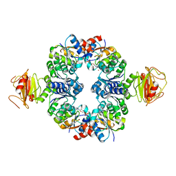
1TZT
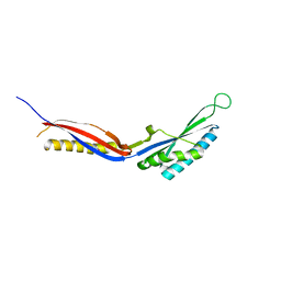
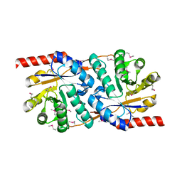
 | | T. maritima NusB, P21 | | 分子名称: | N utilization substance protein B homolog, SULFATE ION | | 著者 | Bonin, I, Robelek, R, Benecke, H, Urlaub, H, Bacher, A, Richter, G, Wahl, M.C. | | 登録日 | 2004-07-12 | | 公開日 | 2004-08-31 | | 最終更新日 | 2023-10-25 | | 実験手法 | X-RAY DIFFRACTION (1.55 Å) | | 主引用文献 | Crystal structures of the antitermination factor NusB from Thermotoga maritima and implications for RNA binding
Biochem.J., 383, 2004
|
|
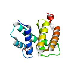
3L2P
 | | Human DNA Ligase III Recognizes DNA Ends by Dynamic Switching Between Two DNA Bound States | | 分子名称: | 5'-D(*GP*CP*CP*AP*GP*TP*CP*CP*GP*AP*CP*GP*AP*CP*GP*CP*AP*TP*CP*CP*CP*G)-3', 5'-D(*GP*TP*CP*GP*GP*AP*CP*TP*G)-3', 5'-D(P*CP*GP*GP*GP*AP*TP*GP*CP*GP*TP*C)-3', ... | | 著者 | Cotner-Gohara, E.A, Kim, I.K, Hammel, M, Tainer, J.A, Tomkinson, A, Ellenberger, T. | | 登録日 | 2009-12-15 | | 公開日 | 2010-07-14 | | 最終更新日 | 2011-07-13 | | 実験手法 | X-RAY DIFFRACTION (3 Å) | | 主引用文献 | Human DNA Ligase III Recognizes DNA Ends by Dynamic Switching between Two DNA-Bound States.
Biochemistry, 49, 2010
|
|
3KOR
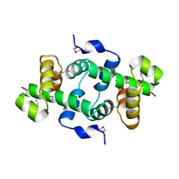 | | Crystal structure of a putative Trp repressor from Staphylococcus aureus | | 分子名称: | Possible Trp repressor | | 著者 | Lam, R, Vodsedalek, J, Lam, K, Romanov, V, Battaile, K.P, Beletskaya, I, Pai, E.F, Chirgadze, N.Y. | | 登録日 | 2009-11-13 | | 公開日 | 2010-11-17 | | 最終更新日 | 2017-11-01 | | 実験手法 | X-RAY DIFFRACTION (1.6 Å) | | 主引用文献 | Crystal structure of a putative Trp repressor from Staphylococcus aureus
To be Published
|
|
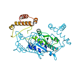
3KJR
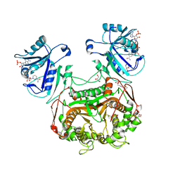 | | Crystal structure of dihydrofolate reductase/thymidylate synthase from Babesia bovis determined using SlipChip based microfluidics | | 分子名称: | 2-[N-CYCLOHEXYLAMINO]ETHANE SULFONIC ACID, Dihydrofolate reductase/thymidylate synthase, GLYCEROL, ... | | 著者 | Li, L, Du, W, Edwards, T.E, Staker, B.L, Phan, I, Stacy, R, Ismagilov, R.F, Accelerated Technologies Center for Gene to 3D Structure (ATCG3D), Seattle Structural Genomics Center for Infectious Disease (SSGCID) | | 登録日 | 2009-11-03 | | 公開日 | 2009-11-17 | | 最終更新日 | 2023-09-06 | | 実験手法 | X-RAY DIFFRACTION (1.95 Å) | | 主引用文献 | Multiparameter screening on SlipChip used for nanoliter protein crystallization combining free interface diffusion and microbatch methods.
J.Am.Chem.Soc., 132, 2010
|
|
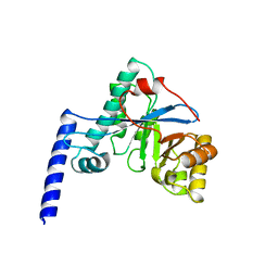
1UAJ
 | | Crystal structure of tRNA(m1G37)methyltransferase: Insight into tRNA recognition | | 分子名称: | tRNA (Guanine-N(1)-)-methyltransferase | | 著者 | Ahn, H.J, Kim, H.-W, Yoon, H.-J, Lee, B.I, Suh, S.W, Yang, J.K. | | 登録日 | 2003-03-11 | | 公開日 | 2003-06-17 | | 最終更新日 | 2023-12-27 | | 実験手法 | X-RAY DIFFRACTION (1.85 Å) | | 主引用文献 | Crystal structure of tRNA(m(1)G37)methyltransferase: insights into tRNA recognition
EMBO J., 22, 2003
|
|
1BK7
 | |
1UCD
 | | Crystal structure of Ribonuclease MC1 from bitter gourd seeds complexed with 5'-UMP | | 分子名称: | Ribonuclease MC, URACIL, URIDINE-5'-MONOPHOSPHATE | | 著者 | Suzuki, A, Numata, T, Yao, M, Kimura, M, Tanaka, I. | | 登録日 | 2003-04-10 | | 公開日 | 2004-05-18 | | 最終更新日 | 2023-10-25 | | 実験手法 | X-RAY DIFFRACTION (1.3 Å) | | 主引用文献 | Structure of RNase MC1 from bitter gourd seeds in complex with 5'UMP
To be published
|
|
1UEL
 | | Solution structure of ubiquitin-like domain of hHR23B complexed with ubiquitin-interacting motif of proteasome subunit S5a | | 分子名称: | 26S proteasome non-ATPase regulatory subunit 4, UV excision repair protein RAD23 homolog B | | 著者 | Fujiwara, K, Tenno, T, Jee, J.G, Sugasawa, K, Ohki, I, Kojima, C, Tochio, H, Hiroaki, H, Hanaoka, H, Shirakawa, M, RIKEN Structural Genomics/Proteomics Initiative (RSGI) | | 登録日 | 2003-05-19 | | 公開日 | 2004-02-10 | | 最終更新日 | 2023-12-27 | | 実験手法 | SOLUTION NMR | | 主引用文献 | Structure of the Ubiquitin-interacting Motif of S5a Bound to the Ubiquitin-like Domain of HR23B
J.Biol.Chem., 279, 2004
|
|
1UG6
 | | Structure of beta-glucosidase at atomic resolution from thermus thermophilus HB8 | | 分子名称: | GLYCEROL, beta-glycosidase | | 著者 | Lokanath, N.K, Shiromizu, I, Miyano, M, Yokoyama, S, Kuramitsu, S, Kunishima, N, RIKEN Structural Genomics/Proteomics Initiative (RSGI) | | 登録日 | 2003-06-12 | | 公開日 | 2003-06-24 | | 最終更新日 | 2023-10-25 | | 実験手法 | X-RAY DIFFRACTION (0.99 Å) | | 主引用文献 | Structure of Beta-Glucosidase at Atomic Resolution from Thermus Thermophilus Hb8
To be Published
|
|
1BV4
 | | APO-MANNOSE-BINDING PROTEIN-C | | 分子名称: | PROTEIN (MANNOSE-BINDING PROTEIN-C) | | 著者 | Ng, K.K.-S, Weis, W.I. | | 登録日 | 1998-09-22 | | 公開日 | 1999-01-13 | | 最終更新日 | 2023-08-09 | | 実験手法 | X-RAY DIFFRACTION (1.85 Å) | | 主引用文献 | Ca2+-dependent structural changes in C-type mannose-binding proteins.
Biochemistry, 37, 1998
|
|
1TWD
 | | Crystal Structure of the Putative Copper Homeostasis Protein (CutC) from Shigella flexneri, Northeast Structural Genomics Target SfR33 | | 分子名称: | Copper homeostasis protein cutC | | 著者 | Forouhar, F, Lee, I, Vorobiev, S.M, Ma, L.-C, Shastry, R, Conover, K, Xiao, R, Acton, T.B, Montelione, G.T, Tong, L, Hunt, J.F, Northeast Structural Genomics Consortium (NESG) | | 登録日 | 2004-06-30 | | 公開日 | 2004-07-20 | | 最終更新日 | 2017-10-11 | | 実験手法 | X-RAY DIFFRACTION (1.7 Å) | | 主引用文献 | Crystal Structure of the Putative Copper Homeostasis Protein (CutC) from Shigella flexneri, Northeast Structural Genomics Target SfR33
To be Published
|
|
1TZ5
 | | [pNPY19-23]-hPP bound to DPC Micelles | | 分子名称: | Pancreatic prohormone,neuropeptide Y,Pancreatic prohormone | | 著者 | Lerch, M, Kamimori, H, Folkers, G, Aguilar, M.I, Beck-Sickinger, A.G, Zerbe, O. | | 登録日 | 2004-07-09 | | 公開日 | 2005-07-05 | | 最終更新日 | 2020-01-01 | | 実験手法 | SOLUTION NMR | | 主引用文献 | Strongly Altered Receptor Binding Properties in PP and NPY Chimeras Are Accompanied by Changes
in Structure and Membrane Binding
Biochemistry, 44, 2005
|
|
1BY0
 | |
1C08
 | | CRYSTAL STRUCTURE OF HYHEL-10 FV-HEN LYSOZYME COMPLEX | | 分子名称: | ANTI-HEN EGG WHITE LYSOZYME ANTIBODY (HYHEL-10), LYSOZYME | | 著者 | Shiroishi, M, Kondo, H, Matsushima, M, Tsumoto, K, Kumagai, I. | | 登録日 | 1999-07-15 | | 公開日 | 2000-07-19 | | 最終更新日 | 2023-05-31 | | 実験手法 | X-RAY DIFFRACTION (2.3 Å) | | 主引用文献 | Crystal structure of anti-Hen egg white lysozyme antibody (HyHEL-10) Fv-antigen complex. Local structural changes in the protein antigen and water-mediated interactions of Fv-antigen and light chain-heavy chain interfaces.
J.Biol.Chem., 274, 1999
|
|
1TXQ
 | |
3L4B
 | | Crystal Structure of an Octomeric Two-Subunit TrkA K+ Channel Ring Gating Assembly, TM1088A:TM1088B, from Thermotoga maritima | | 分子名称: | ADENOSINE MONOPHOSPHATE, TrkA K+ Channel protein TM1088A, TrkA K+ Channel protein TM1088B, ... | | 著者 | Deller, M.C, Johnson, H.A, Miller, M, Spraggon, G, Wilson, I.A, Lesley, S.A, Joint Center for Structural Genomics (JCSG) | | 登録日 | 2009-12-18 | | 公開日 | 2010-02-16 | | 最終更新日 | 2023-09-06 | | 実験手法 | X-RAY DIFFRACTION (3.45 Å) | | 主引用文献 | Crystal Structure of an Octomeric Two-Subunit TrkA K+ Channel Ring Gating Assembly, TM1088A:TM1088B, from Thermotoga maritima
To be Published
|
|
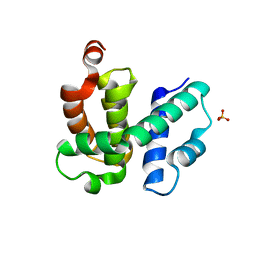
1TZX
 | | T. maritima NusB, P3221 | | 分子名称: | CITRIC ACID, N utilization substance protein B homolog | | 著者 | Bonin, I, Robelek, R, Benecke, H, Urlaub, H, Bacher, A, Richter, G, Wahl, M.C. | | 登録日 | 2004-07-12 | | 公開日 | 2004-08-31 | | 最終更新日 | 2023-10-25 | | 実験手法 | X-RAY DIFFRACTION (1.72 Å) | | 主引用文献 | Crystal structures of the antitermination factor NusB from Thermotoga maritima and implications for RNA binding
Biochem.J., 383, 2004
|
|
1UAM
 | | Crystal structure of tRNA(m1G37)methyltransferase: Insight into tRNA recognition | | 分子名称: | PHOSPHATE ION, S-ADENOSYL-L-HOMOCYSTEINE, tRNA (Guanine-N(1)-)-methyltransferase | | 著者 | Ahn, H.J, Kim, H.-W, Yoon, H.-J, Lee, B.I, Suh, S.W, Yang, J.K. | | 登録日 | 2003-03-11 | | 公開日 | 2003-06-17 | | 最終更新日 | 2023-12-27 | | 実験手法 | X-RAY DIFFRACTION (2.2 Å) | | 主引用文献 | Crystal structure of tRNA(m(1)G37)methyltransferase: insights into tRNA recognition
EMBO J., 22, 2003
|
|
4HU3
 | | Crystal structure of EAL domain of the E. coli DosP - monomeric form | | 分子名称: | Oxygen sensor protein DosP | | 著者 | Tarnawski, M, Barends, T.R.M, Hartmann, E, Schlichting, I. | | 登録日 | 2012-11-02 | | 公開日 | 2013-05-29 | | 最終更新日 | 2024-02-28 | | 実験手法 | X-RAY DIFFRACTION (3.301 Å) | | 主引用文献 | Structures of the catalytic EAL domain of the Escherichia coli direct oxygen sensor.
Acta Crystallogr.,Sect.D, 69, 2013
|
|
3KJ6
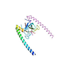
 | | Crystal structure of a Methylated beta2 Adrenergic Receptor-Fab complex | | 分子名称: | Beta-2 adrenergic receptor, Fab heavy chain, Fab light chain, ... | | 著者 | Bokoch, M.P, Zou, Y, Rasmussen, S.G.F, Liu, C.W, Nygaard, R, Rosenbaum, D.M, Fung, J.J, Choi, H.-J, Thian, F.S, Kobilka, T.S, Puglisi, J.D, Weis, W.I, Pardo, L, Prosser, S, Mueller, L, Kobilka, B.K. | | 登録日 | 2009-11-02 | | 公開日 | 2010-02-16 | | 最終更新日 | 2021-10-13 | | 実験手法 | X-RAY DIFFRACTION (3.4 Å) | | 主引用文献 | Ligand-specific regulation of the extracellular surface of a G-protein-coupled receptor.
Nature, 463, 2010
|
|
7CC7
 | |
1BMR
 | | ALPHA-LIKE TOXIN LQH III FROM SCORPION LEIURUS QUINQUESTRIATUS HEBRAEUS, NMR, 25 STRUCTURES | | 分子名称: | LQH III ALPHA-LIKE TOXIN | | 著者 | Krimm, I, Gilles, N, Sautiere, P, Stankiewicz, M, Pelhate, M, Gordon, D, Lancelin, J.-M. | | 登録日 | 1998-07-24 | | 公開日 | 1999-02-16 | | 最終更新日 | 2022-02-16 | | 実験手法 | SOLUTION NMR | | 主引用文献 | NMR structures and activity of a novel alpha-like toxin from the scorpion Leiurus quinquestriatus hebraeus.
J.Mol.Biol., 285, 1999
|
|
1BX7
 | | HIRUSTASIN FROM HIRUDO MEDICINALIS AT 1.2 ANGSTROMS | | 分子名称: | HIRUSTASIN, SULFATE ION | | 著者 | Uson, I, Sheldrick, G.M, De La Fortelle, E, Bricogne, G, Di Marco, S, Priestle, J.P, Gruetter, M.G, Mittl, P.R.E. | | 登録日 | 1998-10-14 | | 公開日 | 1999-04-27 | | 最終更新日 | 2024-06-05 | | 実験手法 | X-RAY DIFFRACTION (1.2 Å) | | 主引用文献 | The 1.2 A crystal structure of hirustasin reveals the intrinsic flexibility of a family of highly disulphide-bridged inhibitors.
Structure Fold.Des., 7, 1999
|
|
1U3O
 | | Solution structure of rat Kalirin N-terminal SH3 domain | | 分子名称: | Huntingtin-associated protein-interacting protein | | 著者 | Schiller, M.R, Chakrabarti, K, King, G.F, Schiller, N.I, Eipper, B.A, Maciejewski, M.W. | | 登録日 | 2004-07-22 | | 公開日 | 2005-07-26 | | 最終更新日 | 2024-05-22 | | 実験手法 | SOLUTION NMR | | 主引用文献 | Regulation of RhoGEF Activity by Intramolecular and Intermolecular SH3 Domain Interactions.
J.Biol.Chem., 281, 2006
|
|


