Loading
PDBj
MenuPDBj@FacebookPDBj@TwitterPDBj@YouTubewwPDB FoundationwwPDB
RCSB PDBPDBeBMRBAdv. SearchSearch help
PDB: 256 results

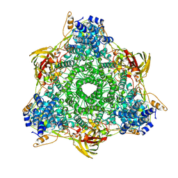
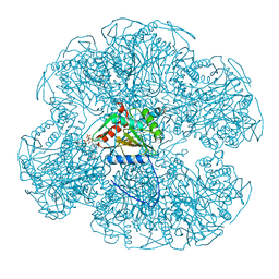
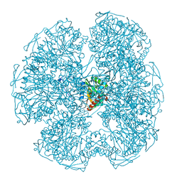
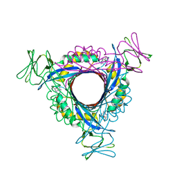
1Y63
DownloadVisualize
BU of 1y63 by Molmil
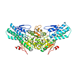
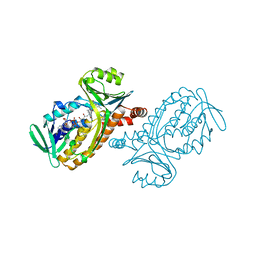
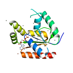
Initial crystal structural analysis of a probable kinase from Leishmania major Friedlin
Descriptor: ADENOSINE-5'-DIPHOSPHATE, BROMIDE ION, Lmaj004144AAA protein, ...
Authors:Robien, M.A, Hol, W.G.J, Structural Genomics of Pathogenic Protozoa Consortium (SGPP)
Deposit date:2004-12-03
Release date:2005-01-04
Last modified:2024-02-14
Method:X-RAY DIFFRACTION (1.7 Å)
Cite:Crystal structural analysis of a probable kinase from Leishmania major Friedlin
To be Published
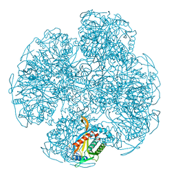
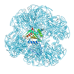
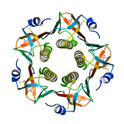
1Y13
DownloadVisualize
BU of 1y13 by Molmil
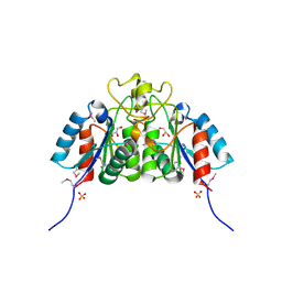
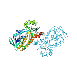
Structural Analysis of Plasmodium falciparum 6-pyruvoyl tetrahydropterin synthase (PTPS)
Descriptor: 6-pyruvoyl tetrahydropterin synthase, BIOPTERIN, ZINC ION
Authors:Bosch, J, Hol, W.G.J, Structural Genomics of Pathogenic Protozoa Consortium (SGPP)
Deposit date:2004-11-17
Release date:2005-01-04
Last modified:2024-10-30
Method:X-RAY DIFFRACTION (2.2 Å)
Cite:Structural Analysis of Plasmodium falciparum 6-pyruvoyl tetrahydropterin synthase (PTPS)
To be Published
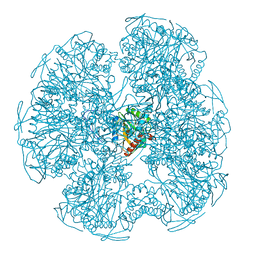
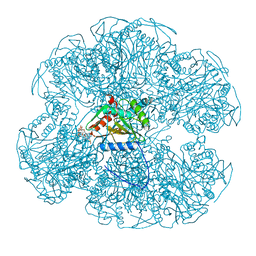
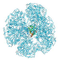
1ZLJ
DownloadVisualize
BU of 1zlj by Molmil
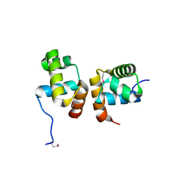
Crystal Structure of the Mycobacterium tuberculosis Hypoxic Response Regulator DosR C-terminal Domain
Descriptor: Dormancy Survival Regulator
Authors:Wisedchaisri, G, Wu, M, Rice, A.E, Roberts, D.M, Sherman, D.R, Hol, W.G.J.
Deposit date:2005-05-06
Release date:2006-01-31
Last modified:2011-07-13
Method:X-RAY DIFFRACTION (2 Å)
Cite:Structures of Mycobacterium tuberculosis DosR and DosR-DNA complex involved in gene activation during adaptation to hypoxic latency.
J.Mol.Biol., 354, 2005
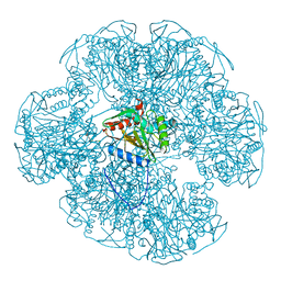
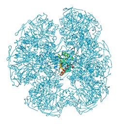
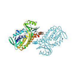
2A0K
DownloadVisualize
BU of 2a0k by Molmil
Crystal structure of Nucleoside 2-deoxyribosyltransferase from Trypanosoma brucei at 1.8 A resolution
Descriptor: GLYCEROL, Nucleoside 2-deoxyribosyltransferase, SULFATE ION
Authors:Bosch, J, Robien, M.A, Hol, W.G.J, Structural Genomics of Pathogenic Protozoa Consortium (SGPP)
Deposit date:2005-06-16
Release date:2005-07-26
Last modified:2024-10-09
Method:X-RAY DIFFRACTION (1.8 Å)
Cite:Using fragment cocktail crystallography to assist inhibitor design of Trypanosoma brucei nucleoside 2-deoxyribosyltransferase.
J.Med.Chem., 49, 2006
2A0U
DownloadVisualize
BU of 2a0u by Molmil
Crystal structure of the eukaryotic initiation factor 2B from Leishmania major at 2.1 A resolution
Descriptor: SULFATE ION, initiation factor 2B
Authors:Bosch, J, Hol, W.G.J, Structural Genomics of Pathogenic Protozoa Consortium (SGPP)
Deposit date:2005-06-17
Release date:2005-08-02
Last modified:2011-07-13
Method:X-RAY DIFFRACTION (2.1 Å)
Cite:Crystal structure of the eukaryotic initiation factor 2B from Leishmania major at 2.1 A resolution
To be Published
1HC1
DownloadVisualize
BU of 1hc1 by Molmil
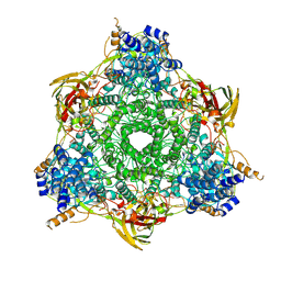
CRYSTAL STRUCTURE OF HEXAMERIC HAEMOCYANIN FROM PANULIRUS INTERRUPTUS REFINED AT 3.2 ANGSTROMS RESOLUTION
Descriptor: ARTHROPODAN HEMOCYANIN, COPPER (II) ION
Authors:Volbeda, A, Hol, W.G.J.
Deposit date:1991-05-15
Release date:1994-01-31
Last modified:2024-06-05
Method:X-RAY DIFFRACTION (3.2 Å)
Cite:Crystal structure of hexameric haemocyanin from Panulirus interruptus refined at 3.2 A resolution.
J.Mol.Biol., 209, 1989
1HCY
DownloadVisualize
BU of 1hcy by Molmil
CRYSTAL STRUCTURE OF HEXAMERIC HAEMOCYANIN FROM PANULIRUS INTERRUPTUS REFINED AT 3.2 ANGSTROMS RESOLUTION
Descriptor: 2-acetamido-2-deoxy-beta-D-glucopyranose-(1-4)-2-acetamido-2-deoxy-beta-D-glucopyranose, ARTHROPODAN HEMOCYANIN, COPPER (II) ION
Authors:Volbeda, A, Hol, W.G.J.
Deposit date:1991-05-15
Release date:1994-01-31
Last modified:2024-10-23
Method:X-RAY DIFFRACTION (3.2 Å)
Cite:Crystal structure of hexameric haemocyanin from Panulirus interruptus refined at 3.2 A resolution.
J.Mol.Biol., 209, 1989
1EAF
DownloadVisualize
BU of 1eaf by Molmil
ATOMIC STRUCTURE OF THE CUBIC CORE OF THE PYRUVATE DEHYDROGENASE MULTIENZYME COMPLEX
Descriptor: DIHYDROLIPOYL-TRANSACETYLASE, SULFITE ION
Authors:Mattevi, A, Hol, W.G.J.
Deposit date:1992-12-16
Release date:1993-10-31
Last modified:2024-02-07
Method:X-RAY DIFFRACTION (2.6 Å)
Cite:Crystallographic analysis of substrate binding and catalysis in dihydrolipoyl transacetylase (E2p).
Biochemistry, 32, 1993
1EAA
DownloadVisualize
BU of 1eaa by Molmil
ATOMIC STRUCTURE OF THE CUBIC CORE OF THE PYRUVATE DEHYDROGENASE MULTIENZYME COMPLEX
Descriptor: DIHYDROLIPOYL-TRANSACETYLASE
Authors:Mattevi, A, Hol, W.G.J.
Deposit date:1992-12-16
Release date:1993-10-31
Last modified:2024-02-07
Method:X-RAY DIFFRACTION (2.6 Å)
Cite:Crystallographic analysis of substrate binding and catalysis in dihydrolipoyl transacetylase (E2p).
Biochemistry, 32, 1993
1EAE
DownloadVisualize
BU of 1eae by Molmil
ATOMIC STRUCTURE OF THE CUBIC CORE OF THE PYRUVATE DEHYDROGENASE MULTIENZYME COMPLEX
Descriptor: 6,8-DIMERCAPTO-OCTANOIC ACID AMIDE, DIHYDROLIPOYL-TRANSACETYLASE
Authors:Mattevi, A, Hol, W.G.J.
Deposit date:1992-12-16
Release date:1993-10-31
Last modified:2024-02-07
Method:X-RAY DIFFRACTION (2.6 Å)
Cite:Crystallographic analysis of substrate binding and catalysis in dihydrolipoyl transacetylase (E2p).
Biochemistry, 32, 1993
1EAC
DownloadVisualize
BU of 1eac by Molmil
ATOMIC STRUCTURE OF THE CUBIC CORE OF THE PYRUVATE DEHYDROGENASE MULTIENZYME COMPLEX
Descriptor: 2,3-DIHYDROXY-1,4-DITHIOBUTANE, DIHYDROLIPOYL-TRANSACETYLASE, OXIDIZED COENZYME A
Authors:Mattevi, A, Hol, W.G.J.
Deposit date:1992-12-16
Release date:1993-10-31
Last modified:2024-02-07
Method:X-RAY DIFFRACTION (2.6 Å)
Cite:Crystallographic analysis of substrate binding and catalysis in dihydrolipoyl transacetylase (E2p).
Biochemistry, 32, 1993
1EAB
DownloadVisualize
BU of 1eab by Molmil
ATOMIC STRUCTURE OF THE CUBIC CORE OF THE PYRUVATE DEHYDROGENASE MULTIENZYME COMPLEX
Descriptor: 6,8-DIMERCAPTO-OCTANOIC ACID AMIDE, COENZYME A, DIHYDROLIPOYL-TRANSACETYLASE
Authors:Mattevi, A, Hol, W.G.J.
Deposit date:1992-12-16
Release date:1993-10-31
Last modified:2024-02-07
Method:X-RAY DIFFRACTION (2.6 Å)
Cite:Crystallographic analysis of substrate binding and catalysis in dihydrolipoyl transacetylase (E2p).
Biochemistry, 32, 1993
1EAD
DownloadVisualize
BU of 1ead by Molmil
ATOMIC STRUCTURE OF THE CUBIC CORE OF THE PYRUVATE DEHYDROGENASE MULTIENZYME COMPLEX
Descriptor: DIHYDROLIPOYL-TRANSACETYLASE, OXIDIZED COENZYME A
Authors:Mattevi, A, Hol, W.G.J.
Deposit date:1992-12-16
Release date:1993-10-31
Last modified:2024-02-07
Method:X-RAY DIFFRACTION (2.6 Å)
Cite:Crystallographic analysis of substrate binding and catalysis in dihydrolipoyl transacetylase (E2p).
Biochemistry, 32, 1993
1DPD
DownloadVisualize
BU of 1dpd by Molmil
CRYSTALLOGRAPHIC AND ENZYMATIC INVESTIGATIONS ON THE ROLE OF SER558, HIS610 AND ASN614 IN THE CATALYTIC MECHANISM OF AZOTOBACTER VINELANDII DIHYDROLIPOAMIDE ACETYLTRANSFERASE (E2P)
Descriptor: DIHYDROLIPOYL-TRANSACETYLASE
Authors:Hendle, J, Hol, W.G.J.
Deposit date:1995-02-03
Release date:1995-04-20
Last modified:2024-02-07
Method:X-RAY DIFFRACTION (2.7 Å)
Cite:Crystallographic and enzymatic investigations on the role of Ser558, His610, and Asn614 in the catalytic mechanism of Azotobacter vinelandii dihydrolipoamide acetyltransferase (E2p).
Biochemistry, 34, 1995
1DPC
DownloadVisualize
BU of 1dpc by Molmil
CRYSTALLOGRAPHIC AND ENZYMATIC INVESTIGATIONS ON THE ROLE OF SER558, HIS610 AND ASN614 IN THE CATALYTIC MECHANISM OF AZOTOBACTER VINELANDII DIHYDROLIPOAMIDE ACETYLTRANSFERASE (E2P)
Descriptor: DIHYDROLIPOYL-TRANSACETYLASE
Authors:Hendle, J, Hol, W.G.J.
Deposit date:1995-02-03
Release date:1995-04-20
Last modified:2024-02-07
Method:X-RAY DIFFRACTION (2.6 Å)
Cite:Crystallographic and enzymatic investigations on the role of Ser558, His610, and Asn614 in the catalytic mechanism of Azotobacter vinelandii dihydrolipoamide acetyltransferase (E2p).
Biochemistry, 34, 1995
1EFI
DownloadVisualize
BU of 1efi by Molmil
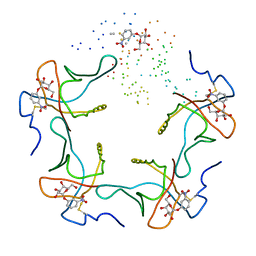
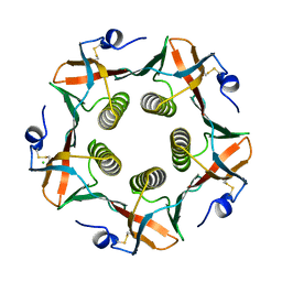
HEAT-LABILE ENTEROTOXIN B-PENTAMER COMPLEXED WITH PARA-AMINOPHENYL-ALPHA-D-GALACTOPYRANOSIDE
Descriptor: 4-aminophenyl alpha-D-galactopyranoside, PROTEIN (HEAT-LABILE ENTEROTOXIN B CHAIN)
Authors:Merritt, E.A, Hol, W.G.J.
Deposit date:2000-02-08
Release date:2000-02-23
Last modified:2024-10-30
Method:X-RAY DIFFRACTION (1.6 Å)
Cite:Exploration of the GM1 receptor-binding site of heat-labile enterotoxin and cholera toxin by phenyl-ring-containing galactose derivatives.
Acta Crystallogr.,Sect.D, 57, 2001
1DPB
DownloadVisualize
BU of 1dpb by Molmil
CRYSTALLOGRAPHIC AND ENZYMATIC INVESTIGATIONS ON THE ROLE OF SER558, HIS610 AND ASN614 IN THE CATALYTIC MECHANISM OF AZOTOBACTER VINELANDII DIHYDROLIPOAMIDE ACETYLTRANSFERASE (E2P)
Descriptor: DIHYDROLIPOYL-TRANSACETYLASE
Authors:Hendle, J, Hol, W.G.J.
Deposit date:1995-02-03
Release date:1995-04-20
Last modified:2024-02-07
Method:X-RAY DIFFRACTION (2.5 Å)
Cite:Crystallographic and enzymatic investigations on the role of Ser558, His610, and Asn614 in the catalytic mechanism of Azotobacter vinelandii dihydrolipoamide acetyltransferase (E2p).
Biochemistry, 34, 1995
1PBB
DownloadVisualize
BU of 1pbb by Molmil
CRYSTAL STRUCTURES OF WILD-TYPE P-HYDROXYBENZOATE HYDROXYLASE COMPLEXED WITH 4-AMINOBENZOATE, 2,4-DIHYDROXYBENZOATE AND 2-HYDROXY-4-AMINOBENZOATE AND OF THE TRY222ALA MUTANT, COMPLEXED WITH 2-HYDROXY-4-AMINOBENZOATE. EVIDENCE FOR A PROTON CHANNEL AND A NEW BINDING MODE OF THE FLAVIN RING
Descriptor: 2,4-DIHYDROXYBENZOIC ACID, FLAVIN-ADENINE DINUCLEOTIDE, P-HYDROXYBENZOATE HYDROXYLASE
Authors:Schreuder, H.A, Mattevi, A, Hol, W.G.J.
Deposit date:1994-07-06
Release date:1994-09-30
Last modified:2024-02-14
Method:X-RAY DIFFRACTION (2.5 Å)
Cite:Crystal structures of wild-type p-hydroxybenzoate hydroxylase complexed with 4-aminobenzoate,2,4-dihydroxybenzoate, and 2-hydroxy-4-aminobenzoate and of the Tyr222Ala mutant complexed with 2-hydroxy-4-aminobenzoate. Evidence for a proton channel and a new binding mode of the flavin ring
Biochemistry, 33, 1994
1EEI
DownloadVisualize
BU of 1eei by Molmil
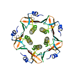
CHOLERA TOXIN B-PENTAMER COMPLEXED WITH METANITROPHENYL-ALPHA-D-GALACTOSE
Descriptor: 3-nitrophenyl alpha-D-galactopyranoside, PROTEIN (CHOLERA TOXIN B)
Authors:Merritt, E.A, Hol, W.G.J.
Deposit date:2000-01-31
Release date:2000-02-16
Last modified:2023-08-09
Method:X-RAY DIFFRACTION (2 Å)
Cite:Exploration of the GM1 receptor-binding site of heat-labile enterotoxin and cholera toxin by phenyl-ring-containing galactose derivatives.
Acta Crystallogr.,Sect.D, 57, 2001
1PBD
DownloadVisualize
BU of 1pbd by Molmil
CRYSTAL STRUCTURES OF WILD-TYPE P-HYDROXYBENZOATE HYDROXYLASE COMPLEXED WITH 4-AMINOBENZOATE, 2,4-DIHYDROXYBENZOATE AND 2-HYDROXY-4-AMINOBENZOATE AND OF THE TRY222ALA MUTANT, COMPLEXED WITH 2-HYDROXY-4-AMINOBENZOATE. EVIDENCE FOR A PROTON CHANNEL AND A NEW BINDING MODE OF THE FLAVIN RING
Descriptor: 4-AMINOBENZOIC ACID, FLAVIN-ADENINE DINUCLEOTIDE, P-HYDROXYBENZOATE HYDROXYLASE
Authors:Schreuder, H.A, Mattevi, A, Hol, W.G.J.
Deposit date:1994-07-06
Release date:1994-09-30
Last modified:2024-02-14
Method:X-RAY DIFFRACTION (2.3 Å)
Cite:Crystal structures of wild-type p-hydroxybenzoate hydroxylase complexed with 4-aminobenzoate,2,4-dihydroxybenzoate, and 2-hydroxy-4-aminobenzoate and of the Tyr222Ala mutant complexed with 2-hydroxy-4-aminobenzoate. Evidence for a proton channel and a new binding mode of the flavin ring
Biochemistry, 33, 1994
1PBE
DownloadVisualize
BU of 1pbe by Molmil
CRYSTAL STRUCTURE OF THE P-HYDROXYBENZOATE HYDROXYLASE-SUBSTRATE COMPLEX REFINED AT 1.9 ANGSTROMS RESOLUTION. ANALYSIS OF THE ENZYME-SUBSTRATE AND ENZYME-PRODUCT COMPLEXES
Descriptor: FLAVIN-ADENINE DINUCLEOTIDE, P-HYDROXYBENZOATE HYDROXYLASE, P-HYDROXYBENZOIC ACID
Authors:Schreuder, H.A, Hol, W.G.J, Drenth, J.
Deposit date:1994-07-06
Release date:1994-09-30
Last modified:2024-02-14
Method:X-RAY DIFFRACTION (1.9 Å)
Cite:Crystal structure of the p-hydroxybenzoate hydroxylase-substrate complex refined at 1.9 A resolution. Analysis of the enzyme-substrate and enzyme-product complexes.
J.Mol.Biol., 208, 1989
1CHQ
DownloadVisualize
BU of 1chq by Molmil
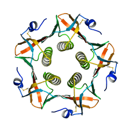
SURPRISING LEADS FOR A CHOLERA TOXIN RECEPTOR BINDING ANTAGONIST; CRYSTALLOGRAPHIC STUDIES OF CTB MUTANTS
Descriptor: CHOLERA TOXIN B PENTAMER
Authors:Merritt, E.A, Hol, W.G.J.
Deposit date:1995-02-15
Release date:1996-03-08
Last modified:2021-11-03
Method:X-RAY DIFFRACTION (2.1 Å)
Cite:Surprising leads for a cholera toxin receptor-binding antagonist: crystallographic studies of CTB mutants.
Structure, 3, 1995
1CHP
DownloadVisualize
BU of 1chp by Molmil
SURPRISING LEADS FOR A CHOLERA TOXIN RECEPTOR BINDING ANTAGONIST; CRYSTALLOGRAPHIC STUDIES OF CTB MUTANTS
Descriptor: CHLORIDE ION, CHOLERA TOXIN B PENTAMER
Authors:Merritt, E.A, Hol, W.G.J.
Deposit date:1995-02-15
Release date:1996-03-08
Last modified:2021-11-03
Method:X-RAY DIFFRACTION (2 Å)
Cite:Surprising leads for a cholera toxin receptor-binding antagonist: crystallographic studies of CTB mutants.
Structure, 3, 1995
1EEF
DownloadVisualize
BU of 1eef by Molmil
HEAT-LABILE ENTEROTOXIN B-PENTAMER COMPLEXED WITH BOUND LIGAND PEPG
Descriptor: 2-PHENETHYL-2,3-DIHYDRO-PHTHALAZINE-1,4-DIONE, PROTEIN (HEAT-LABILE ENTEROTOXIN B CHAIN), alpha-D-galactopyranose
Authors:Merritt, E.A, Hol, W.G.J.
Deposit date:2000-01-31
Release date:2000-02-16
Last modified:2024-10-16
Method:X-RAY DIFFRACTION (1.8 Å)
Cite:Exploration of the GM1 receptor-binding site of heat-labile enterotoxin and cholera toxin by phenyl-ring-containing galactose derivatives.
Acta Crystallogr.,Sect.D, 57, 2001
1U8R
DownloadVisualize
BU of 1u8r by Molmil
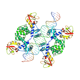
Crystal Structure of an IdeR-DNA Complex Reveals a Conformational Change in Activated IdeR for Base-specific Interactions
Descriptor: COBALT (II) ION, Iron-dependent repressor ideR, SODIUM ION, ...
Authors:Wisedchaisri, G, Holmes, R.K, Hol, W.G.J.
Deposit date:2004-08-06
Release date:2004-10-05
Last modified:2023-08-23
Method:X-RAY DIFFRACTION (2.75 Å)
Cite:Crystal Structure of an IdeR-DNA Complex Reveals a Conformational Change in Activated IdeR for Base-specific Interactions.
J.Mol.Biol., 342, 2004

226707

數據於2024-10-30公開中

PDB statisticsPDBj update infoContact PDBjnumon