Loading
PDBj
メニューPDBj@FacebookPDBj@TwitterPDBj@YouTubewwPDB FoundationwwPDB
RCSB PDBPDBeBMRBAdv. SearchSearch help
PDB: 148 件

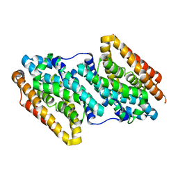
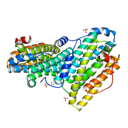
6QO8
DownloadVisualize
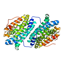
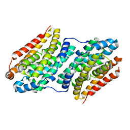
BU of 6qo8 by Molmil
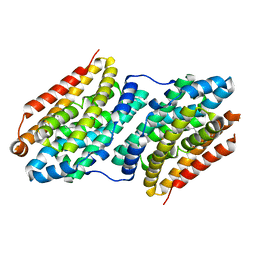
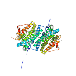
Crystal structure of ribonucleotide reductase NrdF from Bacillus anthracis anaerobically soaked with ferrous ions
分子名称: 2-[BIS-(2-HYDROXY-ETHYL)-AMINO]-2-HYDROXYMETHYL-PROPANE-1,3-DIOL, FE (II) ION, Ribonucleoside-diphosphate reductase subunit beta, ...
著者Grave, K, Hogbom, M.
登録日2019-02-12
公開日2019-08-21
最終更新日2024-01-24
実験手法X-RAY DIFFRACTION (1.31921768 Å)
主引用文献Redox-induced structural changes in the di-iron and di-manganese forms of Bacillus anthracis ribonucleotide reductase subunit NrdF suggest a mechanism for gating of radical access.
J.Biol.Inorg.Chem., 24, 2019
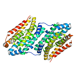
6QO9
DownloadVisualize
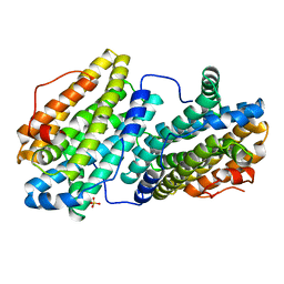
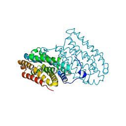
BU of 6qo9 by Molmil
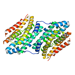
Crystal structure of ribonucleotide reductase NrdF from Bacillus anthracis soaked with manganese ions
分子名称: 2-[BIS-(2-HYDROXY-ETHYL)-AMINO]-2-HYDROXYMETHYL-PROPANE-1,3-DIOL, MANGANESE (II) ION, Ribonucleoside-diphosphate reductase subunit beta, ...
著者Grave, K, Hogbom, M.
登録日2019-02-12
公開日2019-08-21
最終更新日2024-01-24
実験手法X-RAY DIFFRACTION (1.299 Å)
主引用文献Redox-induced structural changes in the di-iron and di-manganese forms of Bacillus anthracis ribonucleotide reductase subunit NrdF suggest a mechanism for gating of radical access.
J.Biol.Inorg.Chem., 24, 2019
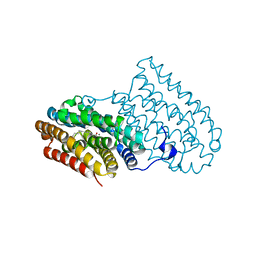
6QO7
DownloadVisualize
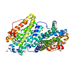
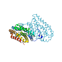
BU of 6qo7 by Molmil
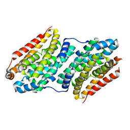
Crystal structure of ribonucleotide reductase NrdF from Bacillus anthracis aerobically soaked with ferrous ions (photo-reduced)
分子名称: CHLORIDE ION, FE (II) ION, Ribonucleoside-diphosphate reductase subunit beta
著者Grave, K, Hogbom, M.
登録日2019-02-12
公開日2019-08-21
最終更新日2024-05-15
実験手法X-RAY DIFFRACTION (1.628 Å)
主引用文献Redox-induced structural changes in the di-iron and di-manganese forms of Bacillus anthracis ribonucleotide reductase subunit NrdF suggest a mechanism for gating of radical access.
J.Biol.Inorg.Chem., 24, 2019
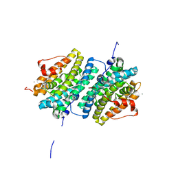
6QO5
DownloadVisualize
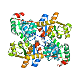
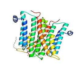
BU of 6qo5 by Molmil
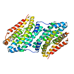
Crystal structure of apo (metal-free) ribonucleotide reductase NrdF from Bacillus anthracis
分子名称: CHLORIDE ION, Ribonucleoside-diphosphate reductase subunit beta, SULFATE ION
著者Grave, K, Hogbom, M.
登録日2019-02-12
公開日2019-08-21
最終更新日2024-01-24
実験手法X-RAY DIFFRACTION (1.511 Å)
主引用文献Redox-induced structural changes in the di-iron and di-manganese forms of Bacillus anthracis ribonucleotide reductase subunit NrdF suggest a mechanism for gating of radical access.
J.Biol.Inorg.Chem., 24, 2019
6QOB
DownloadVisualize
BU of 6qob by Molmil
Crystal structure of ribonucleotide reductase NrdF from Bacillus anthracis with partially oxidised di-iron metallocofactor
分子名称: 2-[BIS-(2-HYDROXY-ETHYL)-AMINO]-2-HYDROXYMETHYL-PROPANE-1,3-DIOL, FE (III) ION, Ribonucleoside-diphosphate reductase subunit beta, ...
著者Grave, K, Hogbom, M.
登録日2019-02-12
公開日2019-08-21
最終更新日2024-01-24
実験手法X-RAY DIFFRACTION (1.457 Å)
主引用文献Redox-induced structural changes in the di-iron and di-manganese forms of Bacillus anthracis ribonucleotide reductase subunit NrdF suggest a mechanism for gating of radical access.
J.Biol.Inorg.Chem., 24, 2019
2D1F
DownloadVisualize
BU of 2d1f by Molmil
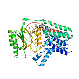
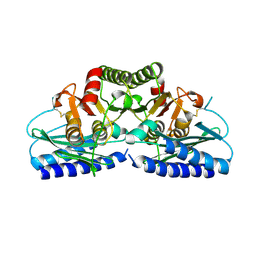
Structure of Mycobacterium tuberculosis threonine synthase
分子名称: PYRIDOXAL-5'-PHOSPHATE, Threonine synthase
著者Covarrubias, A.S, Bergfors, T, Mannerstedt, K, Oscarson, S, Jones, T.A, Mowbray, S.L, Hogbom, M.
登録日2005-08-20
公開日2006-09-05
最終更新日2011-07-13
実験手法X-RAY DIFFRACTION (2.5 Å)
主引用文献Structural, biochemical, and in vivo investigations of the threonine synthase from Mycobacterium tuberculosis.
J.Mol.Biol., 381, 2008
6Q3A
DownloadVisualize
BU of 6q3a by Molmil
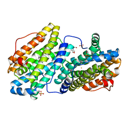
Apo form of Apolipoprotein N-acyltransferase (Lnt)
分子名称: (2R)-2,3-dihydroxypropyl (9Z)-octadec-9-enoate, Apolipoprotein N-acyltransferase, GLYCEROL
著者Wiseman, B, Hogbom, M.
登録日2018-12-03
公開日2019-12-18
最終更新日2024-01-24
実験手法X-RAY DIFFRACTION (3.1 Å)
主引用文献Conformational changes in Apolipoprotein N-acyltransferase (Lnt).
Sci Rep, 10, 2020
6F6C
DownloadVisualize
BU of 6f6c by Molmil
R2-like ligand-binding oxidase V72A mutant with aerobically reconstituted Mn/Fe cofactor
分子名称: FE (III) ION, MANGANESE (II) ION, MANGANESE (III) ION, ...
著者Griese, J.J, Hogbom, M.
登録日2017-12-05
公開日2018-07-04
最終更新日2024-01-17
実験手法X-RAY DIFFRACTION (1.766 Å)
主引用文献Ether cross-link formation in the R2-like ligand-binding oxidase.
J. Biol. Inorg. Chem., 23, 2018
6F6K
DownloadVisualize
BU of 6f6k by Molmil
R2-like ligand-binding oxidase V72L mutant with anaerobically reconstituted Mn/Fe cofactor
分子名称: FE (II) ION, LAURIC ACID, MANGANESE (II) ION, ...
著者Griese, J.J, Hogbom, M.
登録日2017-12-05
公開日2018-07-04
最終更新日2024-01-17
実験手法X-RAY DIFFRACTION (1.982 Å)
主引用文献Ether cross-link formation in the R2-like ligand-binding oxidase.
J. Biol. Inorg. Chem., 23, 2018
6F6H
DownloadVisualize
BU of 6f6h by Molmil
R2-like ligand-binding oxidase V72L mutant with aerobically reconstituted Mn/Fe cofactor
分子名称: FE (III) ION, MANGANESE (II) ION, MANGANESE (III) ION, ...
著者Griese, J.J, Hogbom, M.
登録日2017-12-05
公開日2018-07-04
最終更新日2024-01-17
実験手法X-RAY DIFFRACTION (1.764 Å)
主引用文献Ether cross-link formation in the R2-like ligand-binding oxidase.
J. Biol. Inorg. Chem., 23, 2018
6F6G
DownloadVisualize
BU of 6f6g by Molmil
R2-like ligand-binding oxidase V72I mutant with anaerobically reconstituted Mn/Fe cofactor
分子名称: FE (II) ION, MANGANESE (II) ION, PALMITIC ACID, ...
著者Griese, J.J, Hogbom, M.
登録日2017-12-05
公開日2018-07-04
最終更新日2024-01-17
実験手法X-RAY DIFFRACTION (1.991 Å)
主引用文献Ether cross-link formation in the R2-like ligand-binding oxidase.
J. Biol. Inorg. Chem., 23, 2018
6F6E
DownloadVisualize
BU of 6f6e by Molmil
R2-like ligand-binding oxidase V72A mutant with anaerobically reconstituted Mn/Fe cofactor
分子名称: FE (II) ION, MANGANESE (II) ION, PALMITIC ACID, ...
著者Griese, J.J, Hogbom, M.
登録日2017-12-05
公開日2018-07-04
最終更新日2024-01-17
実験手法X-RAY DIFFRACTION (1.627 Å)
主引用文献Ether cross-link formation in the R2-like ligand-binding oxidase.
J. Biol. Inorg. Chem., 23, 2018
6F6F
DownloadVisualize
BU of 6f6f by Molmil
R2-like ligand-binding oxidase V72I mutant with aerobically reconstituted Mn/Fe cofactor
分子名称: FE (III) ION, MANGANESE (II) ION, MANGANESE (III) ION, ...
著者Griese, J.J, Hogbom, M.
登録日2017-12-05
公開日2018-07-04
最終更新日2024-01-17
実験手法X-RAY DIFFRACTION (1.788 Å)
主引用文献Ether cross-link formation in the R2-like ligand-binding oxidase.
J. Biol. Inorg. Chem., 23, 2018
6GP2
DownloadVisualize
BU of 6gp2 by Molmil
Ribonucleotide Reductase class Ie R2 from Mesoplasma florum, DOPA-active form
分子名称: CALCIUM ION, Ribonucleoside-diphosphate reductase beta chain
著者Srinivas, V, Lebrette, H, Lundin, D, Kutin, Y, Sahlin, M, Lerche, M, Enrich, J, Branca, R.M.M, Cox, N, Sjoberg, B.M, Hogbom, M.
登録日2018-06-05
公開日2018-08-22
最終更新日2024-01-17
実験手法X-RAY DIFFRACTION (1.48 Å)
主引用文献Metal-free ribonucleotide reduction powered by a DOPA radical in Mycoplasma pathogens.
Nature, 563, 2018
6GP3
DownloadVisualize
BU of 6gp3 by Molmil
Ribonucleotide Reductase class Ie R2 from Mesoplasma florum, inactive form
分子名称: CALCIUM ION, Ribonucleoside-diphosphate reductase beta chain
著者Srinivas, V, Lebrette, H, Lundin, D, Kutin, Y, Sahlin, M, Lerche, M, Enrich, J, Branca, R.M.M, Cox, N, Sjoberg, B.M, Hogbom, M.
登録日2018-06-05
公開日2018-08-22
最終更新日2024-01-17
実験手法X-RAY DIFFRACTION (1.23 Å)
主引用文献Metal-free ribonucleotide reduction powered by a DOPA radical in Mycoplasma pathogens.
Nature, 563, 2018
6H5A
DownloadVisualize
BU of 6h5a by Molmil
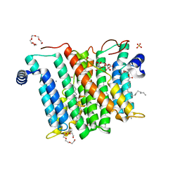
Crystal structure of Mycobacterium tuberculosis phosphatidylinositol phosphate synthase (PgsA1) in complex with manganese and citrate
分子名称: (2R)-2,3-dihydroxypropyl (9Z)-octadec-9-enoate, CDP-diacylglycerol--inositol 3-phosphatidyltransferase, CITRATE ANION, ...
著者Grave, K, Hogbom, M.
登録日2018-07-24
公開日2019-05-15
最終更新日2024-01-17
実験手法X-RAY DIFFRACTION (1.88 Å)
主引用文献Structure ofMycobacterium tuberculosisphosphatidylinositol phosphate synthase reveals mechanism of substrate binding and metal catalysis.
Commun Biol, 2, 2019
6H59
DownloadVisualize
BU of 6h59 by Molmil
Crystal structure of Mycobacterium tuberculosis phosphatidylinositol phosphate synthase (PgsA1) with CDP-DAG bound
分子名称: (2S)-2,3-dihydroxypropyl (9Z)-octadec-9-enoate, CDP-diacylglycerol--inositol 3-phosphatidyltransferase, MAGNESIUM ION, ...
著者Grave, K, Hogbom, M.
登録日2018-07-24
公開日2019-05-15
最終更新日2024-01-17
実験手法X-RAY DIFFRACTION (1.8 Å)
主引用文献Structure ofMycobacterium tuberculosisphosphatidylinositol phosphate synthase reveals mechanism of substrate binding and metal catalysis.
Commun Biol, 2, 2019
6F6M
DownloadVisualize
BU of 6f6m by Molmil
R2-like ligand-binding oxidase Y162F mutant with anaerobically reconstituted Mn/Fe cofactor
分子名称: FE (II) ION, MANGANESE (II) ION, PALMITIC ACID, ...
著者Griese, J.J, Hogbom, M.
登録日2017-12-05
公開日2018-12-19
最終更新日2024-01-17
実験手法X-RAY DIFFRACTION (1.393 Å)
主引用文献Assembly of a heterodinuclear Mn/Fe cofactor is coupled to tyrosine-valine ether cross-link formation in the R2-like ligand-binding oxidase.
J. Biol. Inorg. Chem., 24, 2019
6F6L
DownloadVisualize
BU of 6f6l by Molmil
R2-like ligand-binding oxidase Y162F mutant with aerobically reconstituted Mn/Fe cofactor
分子名称: FE (III) ION, MANGANESE (II) ION, MANGANESE (III) ION, ...
著者Griese, J.J, Hogbom, M.
登録日2017-12-05
公開日2018-12-19
最終更新日2024-01-17
実験手法X-RAY DIFFRACTION (1.904 Å)
主引用文献Assembly of a heterodinuclear Mn/Fe cofactor is coupled to tyrosine-valine ether cross-link formation in the R2-like ligand-binding oxidase.
J. Biol. Inorg. Chem., 24, 2019
6F6B
DownloadVisualize
BU of 6f6b by Molmil
R2-like ligand-binding oxidase A171F mutant with anaerobically reconstituted Mn/Fe cofactor
分子名称: FE (II) ION, MANGANESE (II) ION, Ribonucleotide reductase small subunit
著者Griese, J.J, Hogbom, M.
登録日2017-12-05
公開日2018-12-19
最終更新日2024-01-17
実験手法X-RAY DIFFRACTION (2.009 Å)
主引用文献Assembly of a heterodinuclear Mn/Fe cofactor is coupled to tyrosine-valine ether cross-link formation in the R2-like ligand-binding oxidase.
J. Biol. Inorg. Chem., 24, 2019
6F65
DownloadVisualize
BU of 6f65 by Molmil
R2-like ligand-binding oxidase A171F mutant with aerobically reconstituted Mn/Fe cofactor
分子名称: FE (III) ION, MANGANESE (II) ION, MANGANESE (III) ION, ...
著者Griese, J.J, Hogbom, M.
登録日2017-12-05
公開日2018-12-19
最終更新日2024-01-17
実験手法X-RAY DIFFRACTION (1.948 Å)
主引用文献Assembly of a heterodinuclear Mn/Fe cofactor is coupled to tyrosine-valine ether cross-link formation in the R2-like ligand-binding oxidase.
J. Biol. Inorg. Chem., 24, 2019
6H53
DownloadVisualize
BU of 6h53 by Molmil
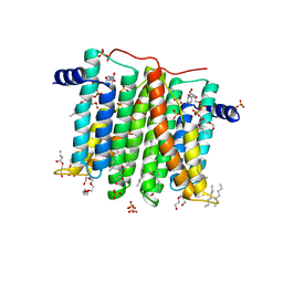
Crystal structure of Mycobacterium tuberculosis phosphatidylinositol phosphate synthase (PgsA1) in apo form
分子名称: CDP-diacylglycerol--inositol 3-phosphatidyltransferase, SULFATE ION, UNKNOWN BRANCHED FRAGMENT OF PHOSPHOLIPID
著者Grave, K, Hogbom, M.
登録日2018-07-23
公開日2019-05-15
最終更新日2024-01-17
実験手法X-RAY DIFFRACTION (2.9 Å)
主引用文献Structure ofMycobacterium tuberculosisphosphatidylinositol phosphate synthase reveals mechanism of substrate binding and metal catalysis.
Commun Biol, 2, 2019
2H4U
DownloadVisualize
BU of 2h4u by Molmil
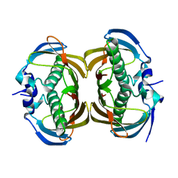
Crystal Structure of Human Thioesterase Superfamily Member 2
分子名称: Thioesterase superfamily member 2
著者Ogg, D.J, Uppenberg, J, Arrowsmith, C, Berglund, H, Edwards, A, Ehn, M, Grasslund, S, Flodin, S, Hammerstrom, M, Hogbom, M, Holmberg-Schiavone, L, Kotenyova, T, Nilsson-Ehle, P, Nordlund, P, Nyman, T, Persson, C, Sagemark, J, Sundstrom, M, Thorsell, A.-G, Weigelt, J, Hallberg, M, Structural Genomics Consortium (SGC)
登録日2006-05-25
公開日2006-06-20
最終更新日2023-08-30
実験手法X-RAY DIFFRACTION (2.2 Å)
主引用文献The crystal structure of human thioesterase superfamily member 2
To be Published
2J91
DownloadVisualize
BU of 2j91 by Molmil
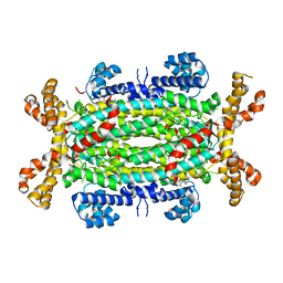
Crystal structure of Human Adenylosuccinate Lyase in complex with AMP
分子名称: ADENOSINE MONOPHOSPHATE, ADENYLOSUCCINATE LYASE, CHLORIDE ION, ...
著者Stenmark, P, Moche, M, Arrowsmith, C, Berglund, H, Busam, R, Collins, R, Edwards, A, Ericsson, U.B, Flodin, S, Flores, A, Graslund, S, Hammarstrom, M, Hallberg, B.M, Holmberg Schiavone, L, Hogbom, M, Johansson, I, Karlberg, T, Kosinska, U, Kotenyova, T, Magnusdottir, A, Nilsson, M.E, Nilsson-Ehle, P, Nyman, T, Ogg, D, Persson, C, Sagemark, J, Sundstrom, M, Uppenberg, J, Uppsten, M, Thorsell, A.G, van Den Berg, S, Wallden, K, Weigelt, J, Nordlund, P.
登録日2006-11-01
公開日2006-11-07
最終更新日2023-12-13
実験手法X-RAY DIFFRACTION (1.8 Å)
主引用文献Crystal Structure of Human Adenylosuccinate Lyase
To be Published
2FVZ
DownloadVisualize
BU of 2fvz by Molmil
Human Inositol Monophosphosphatase 2
分子名称: Inositol monophosphatase 2
著者Ogg, D, Hallberg, B.M, Arrowsmith, C, Berglund, H, Collins, R, Edwards, A, Ehn, M, Flodin, S, Graslund, S, Hammarstrom, M, Hogbom, M, Holmberg-Schiavone, L, Kotenyova, T, Kursula, P, Nilsson-Ehle, P, Nordlund, P, Nyman, T, Persson, C, Sagemark, J, Stenmark, P, Sundstrom, M, Van Den Berg, S, Weigelt, J, Thorsell, A.G, Structural Genomics Consortium (SGC)
登録日2006-01-31
公開日2006-02-21
最終更新日2024-10-23
実験手法X-RAY DIFFRACTION (2.4 Å)
主引用文献Structure of Human Inositol Monophosphatase 2
To be published

226707

件を2024-10-30に公開中

PDB statisticsPDBj update infoContact PDBjnumon