8OVO
 
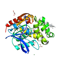
 | | X-ray structure of the SF-iGluSnFR-S72A in complex with L-aspartate | | 分子名称: | ASPARTIC ACID, Putative periplasmic binding transport protein,Green fluorescent protein | | 著者 | Tarnawski, M, Hellweg, L, Bergner, A, Hiblot, J, Leippe, P, Johnsson, K. | | 登録日 | 2023-04-26 | | 公開日 | 2023-05-17 | | 実験手法 | X-RAY DIFFRACTION (1.7 Å) | | 主引用文献 | X-ray structure of the SF-iGluSnFR-S72A in complex with L-aspartate
To Be Published
|
|
8OVP
 | | X-ray structure of the iAspSnFR in complex with L-aspartate | | 分子名称: | ACETATE ION, ASPARTIC ACID, MAGNESIUM ION, ... | | 著者 | Tarnawski, M, Hellweg, L, Bergner, A, Hiblot, J, Leippe, P, Johnsson, K. | | 登録日 | 2023-04-26 | | 公開日 | 2023-05-17 | | 実験手法 | X-RAY DIFFRACTION (1.7 Å) | | 主引用文献 | X-ray structure of the SF-iAspSnFR in complex with L-aspartate
To Be Published
|
|
8B6T
 | | X-ray structure of the interface optimized haloalkane dehalogenase HaloTag7 fusion to the green fluorescent protein GFP (ChemoG5-TMR) labeled with a chloroalkane tetramethylrhodamine fluorophore substrate | | 分子名称: | CHLORIDE ION, Green fluorescent protein,Haloalkane dehalogenase, [9-[2-carboxy-5-[2-[2-(6-chloranylhexoxy)ethoxy]ethylcarbamoyl]phenyl]-6-(dimethylamino)xanthen-3-ylidene]-dimethyl-azanium | | 著者 | Tarnawski, M, Hellweg, L, Hiblot, J. | | 登録日 | 2022-09-27 | | 公開日 | 2023-07-26 | | 最終更新日 | 2023-11-15 | | 実験手法 | X-RAY DIFFRACTION (2 Å) | | 主引用文献 | A general method for the development of multicolor biosensors with large dynamic ranges.
Nat.Chem.Biol., 19, 2023
|
|
8B6S
 | | X-ray structure of the haloalkane dehalogenase HaloTag7 fusion to the green fluorescent protein GFP (ChemoG1) labeled with a chloroalkane tetramethylrhodamine fluorophore substrate | | 分子名称: | CHLORIDE ION, GLYCEROL, Green fluorescent protein,Haloalkane dehalogenase, ... | | 著者 | Tarnawski, M, Hellweg, L, Hiblot, J. | | 登録日 | 2022-09-27 | | 公開日 | 2023-07-26 | | 最終更新日 | 2023-11-15 | | 実験手法 | X-RAY DIFFRACTION (1.8 Å) | | 主引用文献 | A general method for the development of multicolor biosensors with large dynamic ranges.
Nat.Chem.Biol., 19, 2023
|
|
8B6R
 | | X-ray structure of the haloalkane dehalogenase HaloTag7 labeled with a chloroalkane Cyanine3 fluorophore substrate | | 分子名称: | CHLORIDE ION, GLYCEROL, Haloalkane dehalogenase, ... | | 著者 | Tarnawski, M, Hellweg, L, Hiblot, J. | | 登録日 | 2022-09-27 | | 公開日 | 2023-07-26 | | 最終更新日 | 2023-09-06 | | 実験手法 | X-RAY DIFFRACTION (1.5 Å) | | 主引用文献 | A general method for the development of multicolor biosensors with large dynamic ranges.
Nat.Chem.Biol., 19, 2023
|
|
8OVN
 | | X-ray structure of the SF-iGluSnFR-S72A | | 分子名称: | CITRIC ACID, Putative periplasmic binding transport protein,Green fluorescent protein | | 著者 | Tarnawski, M, Hellweg, L, Bergner, A, Hiblot, J, Leippe, P, Johnsson, K. | | 登録日 | 2023-04-26 | | 公開日 | 2023-05-17 | | 実験手法 | X-RAY DIFFRACTION (2.6 Å) | | 主引用文献 | X-ray structure of the SF-iGluSnFR-S72A
To Be Published
|
|


