8HN0
 
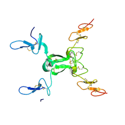 | |
8HNA
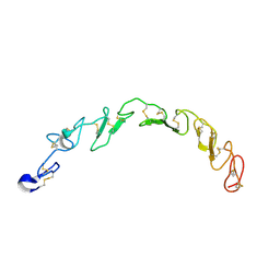 | |
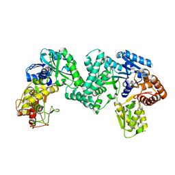
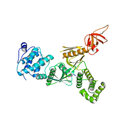
3L8J
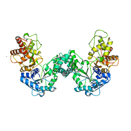
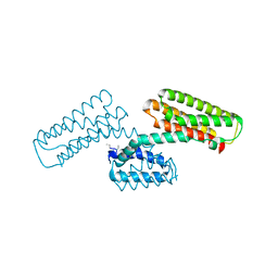 | | Crystal structure of CCM3, a cerebral cavernous malformation protein critical for vascular integrity | | 分子名称: | Programmed cell death protein 10 | | 著者 | Li, X, Zhang, R, Zhang, H, He, Y, Ji, W, Min, W, Boggon, T.J. | | 登録日 | 2009-12-31 | | 公開日 | 2010-05-19 | | 最終更新日 | 2023-09-06 | | 実験手法 | X-RAY DIFFRACTION (3.05 Å) | | 主引用文献 | Crystal structure of CCM3, a cerebral cavernous malformation protein critical for vascular integrity.
J.Biol.Chem., 285, 2010
|
|
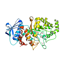
3L8I
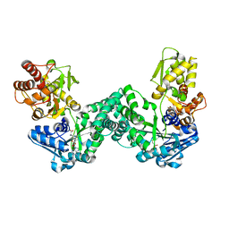
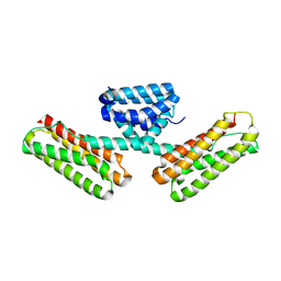 | | Crystal structure of CCM3, a cerebral cavernous malformation protein critical for vascular integrity | | 分子名称: | Programmed cell death protein 10 | | 著者 | Li, X, Zhang, R, Zhang, H, He, Y, Ji, W, Min, W, Boggon, T.J. | | 登録日 | 2009-12-31 | | 公開日 | 2010-05-19 | | 最終更新日 | 2024-02-21 | | 実験手法 | X-RAY DIFFRACTION (2.5 Å) | | 主引用文献 | Crystal structure of CCM3, a cerebral cavernous malformation protein critical for vascular integrity.
J.Biol.Chem., 285, 2010
|
|
5DOO
 | | The structure of PKMT2 from Rickettsia typhi | | 分子名称: | CALCIUM ION, protein lysine methyltransferase 2 | | 著者 | Noinaj, N, Abeykoon, A, He, Y, Yang, D.C, Buchanan, S.K. | | 登録日 | 2015-09-11 | | 公開日 | 2016-08-10 | | 最終更新日 | 2023-09-27 | | 実験手法 | X-RAY DIFFRACTION (3.133 Å) | | 主引用文献 | Structural Insights into Substrate Recognition and Catalysis in Outer Membrane Protein B (OmpB) by Protein-lysine Methyltransferases from Rickettsia.
J.Biol.Chem., 291, 2016
|
|
5DNK
 | | The structure of PKMT1 from Rickettsia prowazekii in complex with AdoHcy | | 分子名称: | S-ADENOSYL-L-HOMOCYSTEINE, protein lysine methyltransferase 1 | | 著者 | Noinaj, N, Abeykoon, A, He, Y, Yang, D.C, Buchanan, S.K. | | 登録日 | 2015-09-10 | | 公開日 | 2016-08-10 | | 最終更新日 | 2024-03-06 | | 実験手法 | X-RAY DIFFRACTION (1.9 Å) | | 主引用文献 | Structural Insights into Substrate Recognition and Catalysis in Outer Membrane Protein B (OmpB) by Protein-lysine Methyltransferases from Rickettsia.
J.Biol.Chem., 291, 2016
|
|
5DPL
 | | The structure of PKMT2 from Rickettsia typhi in complex with AdoHcy | | 分子名称: | S-ADENOSYL-L-HOMOCYSTEINE, protein lysine methyltransferase 2 | | 著者 | Noinaj, N, Abeykoon, A, He, Y, Yang, D.C, Buchanan, S.K. | | 登録日 | 2015-09-12 | | 公開日 | 2016-08-10 | | 最終更新日 | 2024-03-06 | | 実験手法 | X-RAY DIFFRACTION (3.2 Å) | | 主引用文献 | Structural Insights into Substrate Recognition and Catalysis in Outer Membrane Protein B (OmpB) by Protein-lysine Methyltransferases from Rickettsia.
J.Biol.Chem., 291, 2016
|
|
5DPD
 | | The structure of PKMT1 from Rickettsia prowazekii in complex with AdoMet | | 分子名称: | S-ADENOSYLMETHIONINE, protein lysine methyltransferase 1 | | 著者 | Noinaj, N, Abeykoon, A, He, Y, Yang, D.C, Buchanan, S.K. | | 登録日 | 2015-09-12 | | 公開日 | 2016-08-10 | | 最終更新日 | 2024-03-06 | | 実験手法 | X-RAY DIFFRACTION (3 Å) | | 主引用文献 | Structural Insights into Substrate Recognition and Catalysis in Outer Membrane Protein B (OmpB) by Protein-lysine Methyltransferases from Rickettsia.
J.Biol.Chem., 291, 2016
|
|
5DO0
 | | The structure of PKMT1 from Rickettsia prowazekii | | 分子名称: | protein lysine methyltransferase 1 | | 著者 | Noinaj, N, Abeykoon, A, He, Y, Yang, D.C, Buchanan, S.K. | | 登録日 | 2015-09-10 | | 公開日 | 2016-08-10 | | 最終更新日 | 2024-03-06 | | 実験手法 | X-RAY DIFFRACTION (2.6 Å) | | 主引用文献 | Structural Insights into Substrate Recognition and Catalysis in Outer Membrane Protein B (OmpB) by Protein-lysine Methyltransferases from Rickettsia.
J.Biol.Chem., 291, 2016
|
|
7EK6
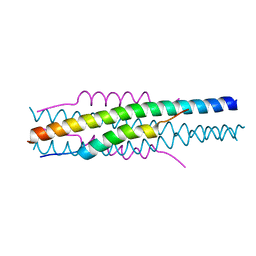 | | Structure of viral peptides IPB19/N52 | | 分子名称: | Spike protein S2 | | 著者 | Yu, D, Qin, B, Cui, S, He, Y. | | 登録日 | 2021-04-04 | | 公開日 | 2021-06-09 | | 最終更新日 | 2023-11-29 | | 実験手法 | X-RAY DIFFRACTION (1.243 Å) | | 主引用文献 | Structure-based design and characterization of novel fusion-inhibitory lipopeptides against SARS-CoV-2 and emerging variants.
Emerg Microbes Infect, 10, 2021
|
|
4LYB
 | | CdS within a lysoyzme single crystal | | 分子名称: | CADMIUM ION, Lysozyme C | | 著者 | Wei, H, House, S, Wu, J, Zhang, J, Wang, Z, He, Y, Gao, Y.-G, Robinson, H, Li, W, Zuo, J.-M, Robertson, I.M, Lu, Y. | | 登録日 | 2013-07-30 | | 公開日 | 2015-02-25 | | 実験手法 | X-RAY DIFFRACTION (1.21 Å) | | 主引用文献 | Enhanced and tunable fluorescent quantum dots within a single crystal of protein
TO BE PUBLISHED
|
|
3KX2
 | |
4I2L
 | | New HIV entry inhibitor MTSFT/T23 complex | | 分子名称: | GP41, inhibitor MTSFT | | 著者 | Yao, X, Chong, H.H, Waltersperger, S, Wang, M.T, He, Y.X, Cui, S. | | 登録日 | 2012-11-22 | | 公開日 | 2014-01-22 | | 最終更新日 | 2023-11-08 | | 実験手法 | X-RAY DIFFRACTION (1.426 Å) | | 主引用文献 | Potent antiviral activity of the novel HIV entry inhibitor MTSFT
To be Published
|
|
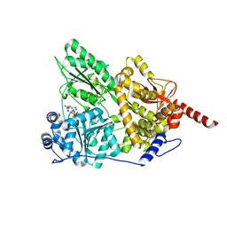
3UJL
 | | Crystal structure of abscisic acid bound PYL2 in complex with type 2C protein phosphatase ABI2 | | 分子名称: | (2Z,4E)-5-[(1S)-1-hydroxy-2,6,6-trimethyl-4-oxocyclohex-2-en-1-yl]-3-methylpenta-2,4-dienoic acid, Abscisic acid receptor PYL2, MAGNESIUM ION, ... | | 著者 | Zhou, X.E, Soon, F.-F, Ng, L.-M, Kovach, A, Tan, M.H.E, Suino-Powell, K.M, He, Y, Xu, Y, Brunzelle, J.S, Li, J, Melcher, K, Xu, H.E. | | 登録日 | 2011-11-07 | | 公開日 | 2012-02-15 | | 最終更新日 | 2012-03-28 | | 実験手法 | X-RAY DIFFRACTION (2.5 Å) | | 主引用文献 | Molecular mimicry regulates ABA signaling by SnRK2 kinases and PP2C phosphatases.
Science, 335, 2012
|
|
4LYC
 | | Cd ions within a lysoyzme single crystal | | 分子名称: | CADMIUM ION, Lysozyme C | | 著者 | Wei, H, House, S, Wu, J, Zhang, J, Wang, Z, He, Y, Gao, Y.-G, Robinson, H, Li, W, Zuo, J.-M, Robertson, I.M, Lu, Y. | | 登録日 | 2013-07-30 | | 公開日 | 2015-02-25 | | 実験手法 | X-RAY DIFFRACTION (1.35 Å) | | 主引用文献 | Enhanced and tunable fluorescent quantum dots within a single crystal of protein
TO BE PUBLISHED
|
|
3UJG
 | | Crystal structure of SnRK2.6 in complex with HAB1 | | 分子名称: | MAGNESIUM ION, Protein phosphatase 2C 16, SULFATE ION, ... | | 著者 | Zhou, X.E, Soon, F.-F, Ng, L.-M, Kovach, A, Tan, M.H.E, Suino-Powell, K.M, He, Y, Xu, Y, Brunzelle, J.S, Li, J, Melcher, K, Xu, H.E. | | 登録日 | 2011-11-07 | | 公開日 | 2012-02-15 | | 最終更新日 | 2024-02-28 | | 実験手法 | X-RAY DIFFRACTION (2.6 Å) | | 主引用文献 | Molecular mimicry regulates ABA signaling by SnRK2 kinases and PP2C phosphatases.
Science, 335, 2012
|
|
3UJK
 | | Crystal structure of protein phosphatase ABI2 | | 分子名称: | MAGNESIUM ION, Protein phosphatase 2C 77 | | 著者 | Zhou, X.E, Soon, F.-F, Ng, L.-M, Kovach, A, Tan, M.H.E, Suino-Powell, K.M, He, Y, Xu, Y, Brunzelle, J.S, Li, J, Melcher, K, Xu, H.E. | | 登録日 | 2011-11-07 | | 公開日 | 2012-02-15 | | 最終更新日 | 2024-02-28 | | 実験手法 | X-RAY DIFFRACTION (1.9 Å) | | 主引用文献 | Molecular mimicry regulates ABA signaling by SnRK2 kinases and PP2C phosphatases.
Science, 335, 2012
|
|
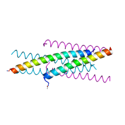
5F3X
 | | Crystal structure of Harmonin NPDZ1 in complex with ANKS4B SAM-PBM | | 分子名称: | Ankyrin repeat and SAM domain-containing protein 4B, CHLORIDE ION, Harmonin | | 著者 | Li, J, He, Y, Lu, Q, Zhang, M. | | 登録日 | 2015-12-03 | | 公開日 | 2016-03-16 | | 最終更新日 | 2023-11-08 | | 実験手法 | X-RAY DIFFRACTION (2.649 Å) | | 主引用文献 | Mechanistic Basis of Organization of the Harmonin/USH1C-Mediated Brush Border Microvilli Tip-Link Complex
Dev.Cell, 36, 2016
|
|
5F3Y
 | | Crystal Structure of Myo7b N-MyTH4-FERM-SH3 in complex with Anks4b CEN | | 分子名称: | Ankyrin repeat and SAM domain-containing protein 4B, Unconventional myosin-VIIb | | 著者 | Li, J, He, Y, Lu, Q, Zhang, M. | | 登録日 | 2015-12-03 | | 公開日 | 2016-03-16 | | 最終更新日 | 2023-11-08 | | 実験手法 | X-RAY DIFFRACTION (3.409 Å) | | 主引用文献 | Mechanistic Basis of Organization of the Harmonin/USH1C-Mediated Brush Border Microvilli Tip-Link Complex
Dev.Cell, 36, 2016
|
|
7DUP
 | | Apo structure of wild type Bt4394, a GH20 family sulfoglycosidase | | 分子名称: | 2-(N-MORPHOLINO)-ETHANESULFONIC ACID, Beta-N-acetylhexosaminidase, CHLORIDE ION, ... | | 著者 | Zhang, Z, He, Y, Jin, Y. | | 登録日 | 2021-01-10 | | 公開日 | 2022-01-19 | | 最終更新日 | 2023-11-29 | | 実験手法 | X-RAY DIFFRACTION (1.62 Å) | | 主引用文献 | Mechanistic and Structural Insights into the Specificity and Biological Functions of Bacterial Sulfoglycosidases
Acs Catalysis, 13, 2023
|
|
7DVA
 | | Structure of wild type Bt4394, a GH20 family sulfoglycosidase, in complex with 6S-GlcNAc | | 分子名称: | 2-acetamido-2-deoxy-6-O-sulfo-beta-D-glucopyranose, Beta-N-acetylhexosaminidase, GLYCEROL | | 著者 | Zhang, Z, He, Y, Jin, Y. | | 登録日 | 2021-01-13 | | 公開日 | 2022-01-19 | | 最終更新日 | 2023-11-29 | | 実験手法 | X-RAY DIFFRACTION (1.55 Å) | | 主引用文献 | Mechanistic and Structural Insights into the Specificity and Biological Functions of Bacterial Sulfoglycosidases
Acs Catalysis, 13, 2023
|
|
7DVB
 | | D335N variant of Bt4394 in complex with 6SO3-NAG-oxazoline intermediate | | 分子名称: | 2-acetamido-2-deoxy-6-O-sulfo-beta-D-glucopyranose, Beta-N-acetylhexosaminidase, [(3~{a}~{R},5~{R},6~{S},7~{R},7~{a}~{R})-2-methyl-6,7-bis(oxidanyl)-5,6,7,7~{a}-tetrahydro-3~{a}~{H}-pyrano[3,2-d][1,3]oxazol-1-ium-5-yl]methyl sulfate | | 著者 | Zhang, Z, He, Y, Jin, Y. | | 登録日 | 2021-01-13 | | 公開日 | 2022-01-19 | | 最終更新日 | 2023-11-29 | | 実験手法 | X-RAY DIFFRACTION (2.05 Å) | | 主引用文献 | Mechanistic and Structural Insights into the Specificity and Biological Functions of Bacterial Sulfoglycosidases
Acs Catalysis, 13, 2023
|
|
5FUR
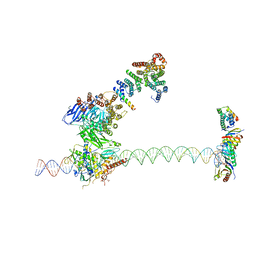 | | Structure of human TFIID-IIA bound to core promoter DNA | | 分子名称: | SUPER CORE PROMOTER, TATA-BOX-BINDING PROTEIN, TRANSCRIPTION INITIATION FACTOR IIA SUBUNIT 1, ... | | 著者 | Louder, R.K, He, Y, Lopez-Blanco, J.R, Fang, J, Chacon, P, Nogales, E. | | 登録日 | 2016-01-29 | | 公開日 | 2016-04-06 | | 最終更新日 | 2017-08-02 | | 実験手法 | ELECTRON MICROSCOPY (8.5 Å) | | 主引用文献 | Structure of Promoter-Bound TFIID and Model of Human Pre-Initiation Complex Assembly.
Nature, 531, 2016
|
|
7XU2
 | | Structure of SARS-CoV-2 Spike Protein with Engineered x3 Disulfide (x3(D427C, V987C) and single Arg S1/S2 cleavage site), Locked-2 Conformation | | 分子名称: | 2-acetamido-2-deoxy-beta-D-glucopyranose, 2-acetamido-2-deoxy-beta-D-glucopyranose-(1-4)-2-acetamido-2-deoxy-beta-D-glucopyranose, BILIVERDINE IX ALPHA, ... | | 著者 | Qu, K, Chen, Q, Ciazynska, K.A, Liu, B, Zhang, X, Wang, J, He, Y, Guan, J, He, J, Liu, T, Zhang, X, Carter, A.P, Xiong, X, Briggs, J.A.G. | | 登録日 | 2022-05-18 | | 公開日 | 2022-07-20 | | 最終更新日 | 2022-08-17 | | 実験手法 | ELECTRON MICROSCOPY (3.2 Å) | | 主引用文献 | Engineered disulfide reveals structural dynamics of locked SARS-CoV-2 spike.
Plos Pathog., 18, 2022
|
|
7XU0
 | | Structure of SARS-CoV-2 Spike Protein with Engineered x3 Disulfide (x3(D427C, V987C) and single Arg S1/S2 cleavage site), Locked-211 Conformation | | 分子名称: | 2-acetamido-2-deoxy-beta-D-glucopyranose, 2-acetamido-2-deoxy-beta-D-glucopyranose-(1-4)-2-acetamido-2-deoxy-beta-D-glucopyranose, BILIVERDINE IX ALPHA, ... | | 著者 | Qu, K, Chen, Q, Ciazynska, K.A, Liu, B, Zhang, X, Wang, J, He, Y, Guan, J, He, J, Liu, T, Zhang, X, Carter, A.P, Xiong, X, Briggs, J.A.G. | | 登録日 | 2022-05-18 | | 公開日 | 2022-07-20 | | 最終更新日 | 2022-08-17 | | 実験手法 | ELECTRON MICROSCOPY (2.9 Å) | | 主引用文献 | Engineered disulfide reveals structural dynamics of locked SARS-CoV-2 spike.
Plos Pathog., 18, 2022
|
|


