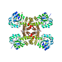
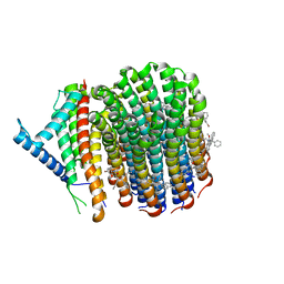
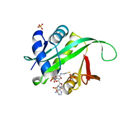
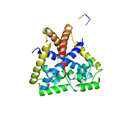
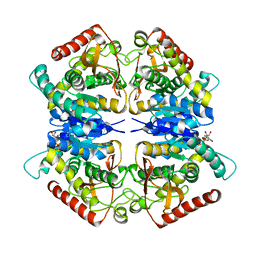
3OGG
 
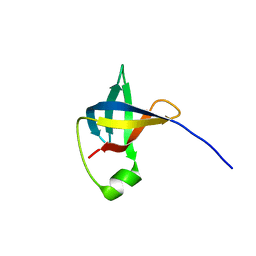
 | | Crystal structure of the receptor binding domain of botulinum neurotoxin D | | 分子名称: | Botulinum neurotoxin type D | | 著者 | Zhang, Y, Gao, X, Qin, L, Buchko, G.W, Robinson, H, Varnum, S.M. | | 登録日 | 2010-08-16 | | 公開日 | 2010-09-01 | | 最終更新日 | 2023-09-06 | | 実験手法 | X-RAY DIFFRACTION (1.651 Å) | | 主引用文献 | Structural analysis of the receptor binding domain of botulinum neurotoxin serotype D.
Biochem.Biophys.Res.Commun., 401, 2010
|
|
3PDU
 | |
3PEF
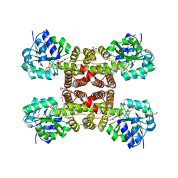 | |
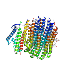
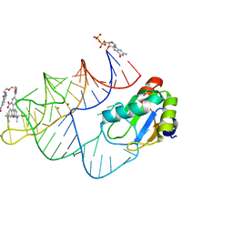
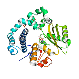
3UUX
 | | Crystal structure of yeast Fis1 in complex with Mdv1 fragment containing N-terminal extension and coiled coil domains | | 分子名称: | Mitochondria fission 1 protein, Mitochondrial division protein 1 | | 著者 | Zhang, Y, Chan, N.C, Gristick, H, Chan, D.C. | | 登録日 | 2011-11-28 | | 公開日 | 2012-02-08 | | 最終更新日 | 2012-04-25 | | 実験手法 | X-RAY DIFFRACTION (3.9 Å) | | 主引用文献 | Crystal structure of mitochondrial fission complex reveals scaffolding function for mitochondrial division 1 (mdv1) coiled coil.
J.Biol.Chem., 287, 2012
|
|
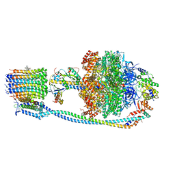
8J58
 | | Cryo-EM structure of Mycobacterium tuberculosis ATP synthase Fo in the apo-form | | 分子名称: | ATP synthase subunit a, ATP synthase subunit c | | 著者 | Zhang, Y, Lai, Y, Liu, F, Rao, Z, Gong, H. | | 登録日 | 2023-04-21 | | 公開日 | 2024-05-01 | | 実験手法 | ELECTRON MICROSCOPY (3.15 Å) | | 主引用文献 | Structure of Mycobacterium tuberculosis ATP synthase
To Be Published
|
|
8JR1
 | | Cryo-EM structure of Mycobacterium tuberculosis ATP synthase Fo in complex with TBAJ-587 | | 分子名称: | (1~{S},2~{S})-1-(6-bromanyl-2-methoxy-quinolin-3-yl)-2-(2,6-dimethoxypyridin-4-yl)-4-(dimethylamino)-1-(2-fluoranyl-3-methoxy-phenyl)butan-2-ol, ATP synthase subunit a, ATP synthase subunit c | | 著者 | Zhang, Y, Lai, Y, Liu, F, Rao, Z, Gong, H. | | 登録日 | 2023-06-15 | | 公開日 | 2024-05-01 | | 実験手法 | ELECTRON MICROSCOPY (3.17 Å) | | 主引用文献 | Structure of Mycobacterium tuberculosis ATP synthase
To Be Published
|
|
8JR0
 | | Cryo-EM structure of Mycobacterium tuberculosis ATP synthase in complex with TBAJ-587 | | 分子名称: | (1~{S},2~{S})-1-(6-bromanyl-2-methoxy-quinolin-3-yl)-2-(2,6-dimethoxypyridin-4-yl)-4-(dimethylamino)-1-(2-fluoranyl-3-methoxy-phenyl)butan-2-ol, ADENOSINE-5'-DIPHOSPHATE, ADENOSINE-5'-TRIPHOSPHATE, ... | | 著者 | Zhang, Y, Lai, Y, Liu, F, Rao, Z, Gong, H. | | 登録日 | 2023-06-15 | | 公開日 | 2024-05-01 | | 実験手法 | ELECTRON MICROSCOPY (2.8 Å) | | 主引用文献 | Structure of Mycobacterium tuberculosis ATP synthase
To Be Published
|
|
8J57
 | | Cryo-EM structure of Mycobacterium tuberculosis ATP synthase Fo in complex with bedaquiline(BDQ) | | 分子名称: | ATP synthase subunit a, ATP synthase subunit c, Bedaquiline | | 著者 | Zhang, Y, Lai, Y, Liu, F, Rao, Z, Gong, H. | | 登録日 | 2023-04-21 | | 公開日 | 2024-05-01 | | 実験手法 | ELECTRON MICROSCOPY (2.85 Å) | | 主引用文献 | Structure of Mycobacterium tuberculosis ATP synthase
To Be Published
|
|
8J0T
 | | Cryo-EM structure of Mycobacterium tuberculosis ATP synthase in the apo-form | | 分子名称: | ADENOSINE-5'-DIPHOSPHATE, ADENOSINE-5'-TRIPHOSPHATE, ATP synthase epsilon chain, ... | | 著者 | Zhang, Y, Lai, Y, Liu, F, Rao, Z, Gong, H. | | 登録日 | 2023-04-11 | | 公開日 | 2024-05-22 | | 実験手法 | ELECTRON MICROSCOPY (2.8 Å) | | 主引用文献 | Structure of Mycobacterium tuberculosis ATP synthase
To Be Published
|
|
8J0S
 | | Cryo-EM structure of Mycobacterium tuberculosis ATP synthase in complex with bedaquiline(BDQ) | | 分子名称: | ADENOSINE-5'-DIPHOSPHATE, ADENOSINE-5'-TRIPHOSPHATE, ATP synthase epsilon chain, ... | | 著者 | Zhang, Y, Lai, Y, Liu, F, Rao, Z, Gong, H. | | 登録日 | 2023-04-11 | | 公開日 | 2024-05-22 | | 実験手法 | ELECTRON MICROSCOPY (2.58 Å) | | 主引用文献 | Structure of Mycobacterium tuberculosis ATP synthase
To Be Published
|
|
6C00
 | |
5GXF
 | |
5GXE
 | |
4R3K
 | |
8JY0
 | | Crystal structure of RhoBAST complexed with TMR-DN | | 分子名称: | 2,4-dinitroaniline, 5-aminocarbonyl-2-[3-(dimethylamino)-6-dimethylazaniumylidene-xanthen-9-yl]benzoate, GUANOSINE-5'-DIPHOSPHATE, ... | | 著者 | Zhang, Y, Xiao, Y, Xu, Z, Fang, X. | | 登録日 | 2023-07-02 | | 公開日 | 2024-05-29 | | 実験手法 | X-RAY DIFFRACTION (2.75 Å) | | 主引用文献 | Structural mechanisms for binding and activation of a contact-quenched fluorophore by RhoBAST.
Nat Commun, 15, 2024
|
|
4R3L
 | |
5GXD
 | |
4EJT
 | |
5ZLC
 | | Binary complex of human DNA Polymerase Mu with MndGTP | | 分子名称: | 2'-DEOXYGUANOSINE-5'-TRIPHOSPHATE, DNA-directed DNA/RNA polymerase mu, MANGANESE (II) ION, ... | | 著者 | Chang, Y.K, Wu, W.J, Tsai, M.D. | | 登録日 | 2018-03-27 | | 公開日 | 2019-05-29 | | 最終更新日 | 2023-11-22 | | 実験手法 | X-RAY DIFFRACTION (2 Å) | | 主引用文献 | Human DNA Polymerase mu Can Use a Noncanonical Mechanism for Multiple Mn2+-Mediated Functions.
J.Am.Chem.Soc., 141, 2019
|
|
2PQN
 | |
3PQE
 | |
3PQF
 | |
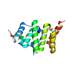
3LZC
 | | Crystal structure of Dph2 from Pyrococcus horikoshii | | 分子名称: | Dph2 | | 著者 | Zhang, Y, Zhu, X, Torelli, A.T, Lee, M, Dzikovski, B, Koralewski, R.M, Wang, E, Freed, J, Krebs, C, Lin, H, Ealick, S.E. | | 登録日 | 2010-03-01 | | 公開日 | 2010-06-23 | | 最終更新日 | 2024-02-21 | | 実験手法 | X-RAY DIFFRACTION (2.261 Å) | | 主引用文献 | Diphthamide biosynthesis requires an organic radical generated by an iron-sulphur enzyme.
Nature, 465, 2010
|
|
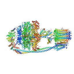
7W3Z
 | | Cryo-EM Structure of Human Gastrin Releasing Peptide Receptor in complex with the agonist Gastrin Releasing Peptide and Gq heterotrimers | | 分子名称: | Gastrin Releasing Peptide PRGNHWAVGHLM(NH2), Guanine nucleotide-binding protein G(I)/G(S)/G(O) subunit gamma-2, Guanine nucleotide-binding protein G(I)/G(S)/G(T) subunit beta-1, ... | | 著者 | Zhan, Y, Peng, S, Zhang, H. | | 登録日 | 2021-11-26 | | 公開日 | 2023-02-22 | | 実験手法 | ELECTRON MICROSCOPY (3 Å) | | 主引用文献 | Structures of human gastrin-releasing peptide receptors bound to antagonist and agonist for cancer and itch therapy.
Proc.Natl.Acad.Sci.USA, 120, 2023
|
|
7W40
 | | Cryo-EM Structure of Human Gastrin Releasing Peptide Receptor in complex with the agonist Bombesin (6-14) [D-Phe6, beta-Ala11, Phe13, Nle14] and Gq heterotrimers | | 分子名称: | Bombesin, Guanine nucleotide-binding protein G(I)/G(S)/G(O) subunit gamma-2, Guanine nucleotide-binding protein G(I)/G(S)/G(T) subunit beta-1, ... | | 著者 | Zhan, Y, Peng, S, Zhang, H. | | 登録日 | 2021-11-26 | | 公開日 | 2023-02-22 | | 実験手法 | ELECTRON MICROSCOPY (3 Å) | | 主引用文献 | Structures of human gastrin-releasing peptide receptors bound to antagonist and agonist for cancer and itch therapy.
Proc.Natl.Acad.Sci.USA, 120, 2023
|
|


