6JPK
 
 | |
6KHE
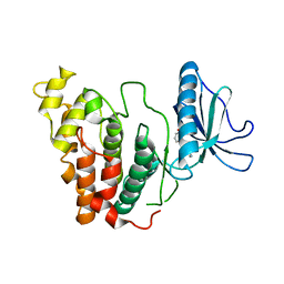
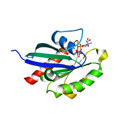
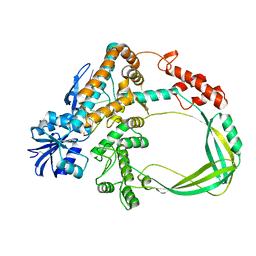
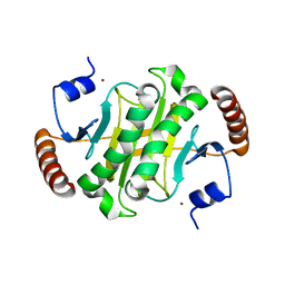
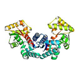
 | | Crystal structure of CLK2 in complex with CX-4945 | | 分子名称: | 5-[(3-chlorophenyl)amino]benzo[c][2,6]naphthyridine-8-carboxylic acid, Dual specificity protein kinase CLK2 | | 著者 | Lee, J.Y, Yun, J.S, Jin, H, Chang, J.H. | | 登録日 | 2019-07-15 | | 公開日 | 2019-10-02 | | 最終更新日 | 2024-03-27 | | 実験手法 | X-RAY DIFFRACTION (2.8 Å) | | 主引用文献 | Structural Basis for the Selective Inhibition of Cdc2-Like Kinases by CX-4945.
Biomed Res Int, 2019, 2019
|
|
6KWS
 | |
6KWT
 | |
6KHF
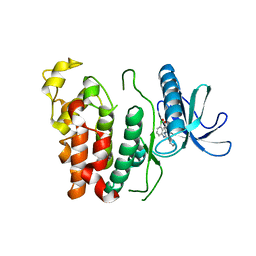
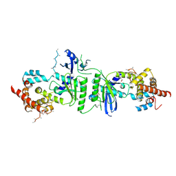
 | | Crystal structure of CLK3 in complex with CX-4945 | | 分子名称: | 5-[(3-chlorophenyl)amino]benzo[c][2,6]naphthyridine-8-carboxylic acid, Dual specificity protein kinase CLK3 | | 著者 | Lee, J.Y, Yun, J.S, Jin, H, Chang, J.H. | | 登録日 | 2019-07-15 | | 公開日 | 2019-10-02 | | 最終更新日 | 2023-11-22 | | 実験手法 | X-RAY DIFFRACTION (2.598 Å) | | 主引用文献 | Structural Basis for the Selective Inhibition of Cdc2-Like Kinases by CX-4945.
Biomed Res Int, 2019, 2019
|
|
6KHD
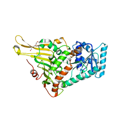
 | | Crystal structure of CLK1 in complex with CX-4945 | | 分子名称: | 5-[(3-chlorophenyl)amino]benzo[c][2,6]naphthyridine-8-carboxylic acid, Dual specificity protein kinase CLK1 | | 著者 | Lee, J.Y, Yun, J.S, Jin, H, Chang, J.H. | | 登録日 | 2019-07-15 | | 公開日 | 2019-10-02 | | 最終更新日 | 2024-03-27 | | 実験手法 | X-RAY DIFFRACTION (2.7 Å) | | 主引用文献 | Structural Basis for the Selective Inhibition of Cdc2-Like Kinases by CX-4945.
Biomed Res Int, 2019, 2019
|
|
5XR4
 | |
5XR7
 | |
5XR6
 | |
5BY2
 | |
2JWE
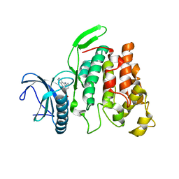
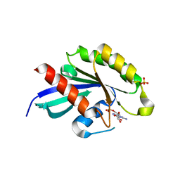
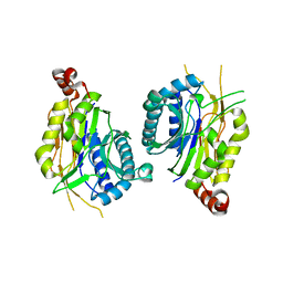
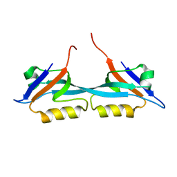 | | Solution structure of the second PDZ domain from human zonula occludens-1: A dimeric form with 3D domain swapping | | 分子名称: | Tight junction protein ZO-1 | | 著者 | Ji, P, Wu, J.W, Zhang, J.H, Yang, Y.S, Wu, J.H, Shi, Y.Y. | | 登録日 | 2007-10-10 | | 公開日 | 2007-10-30 | | 最終更新日 | 2024-05-01 | | 実験手法 | SOLUTION NMR | | 主引用文献 | Solution structure of the second PDZ domain of Zonula Occludens 1
Proteins, 79, 2011
|
|
6K8N
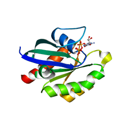
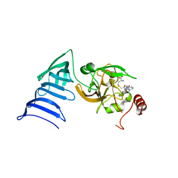
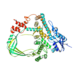 | | Crystal structure of the Sulfolobus solfataricus topoisomerase III | | 分子名称: | ZINC ION, topoisomerase III | | 著者 | Wang, H.Q, Zhang, J.H, Zheng, X, Zheng, Z.F, Dong, Y.H, Huang, L, Gong, Y. | | 登録日 | 2019-06-13 | | 公開日 | 2020-06-24 | | 最終更新日 | 2023-11-22 | | 実験手法 | X-RAY DIFFRACTION (2.1 Å) | | 主引用文献 | Crystal structures of the Sulfolobus solfataricus topoisomerase III reveal that its C-terminal novel zinc finger part is a unique decatenation domain
To Be Published
|
|
6K8O
 | | Crystal structure of the Sulfolobus solfataricus topoisomerase III in complex with DNA | | 分子名称: | DNA (5'-D(*GP*CP*AP*AP*GP*GP*TP*C)-3'), ZINC ION, topoisomerase III | | 著者 | Wang, H.Q, Zhang, J.H, Zheng, X, Zheng, Z.F, Dong, Y.H, Huang, L, Gong, Y. | | 登録日 | 2019-06-13 | | 公開日 | 2020-06-24 | | 実験手法 | X-RAY DIFFRACTION (2.5 Å) | | 主引用文献 | Crystal structures of the Sulfolobus solfataricus topoisomerase III reveal that its C-terminal novel zinc finger part is a unique decatenation domain
To Be Published
|
|
3PQ1
 | | Crystal structure of human mitochondrial poly(A) polymerase (PAPD1) | | 分子名称: | Poly(A) RNA polymerase | | 著者 | Bai, Y, Srivastava, S.K, Chang, J.H, Tong, L. | | 登録日 | 2010-11-25 | | 公開日 | 2011-03-30 | | 最終更新日 | 2017-08-02 | | 実験手法 | X-RAY DIFFRACTION (3.1 Å) | | 主引用文献 | Structural basis for dimerization and activity of human PAPD1, a noncanonical poly(A) polymerase.
Mol.Cell, 41, 2011
|
|
7E6H
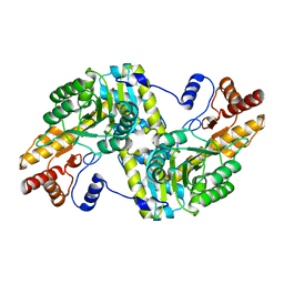
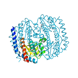
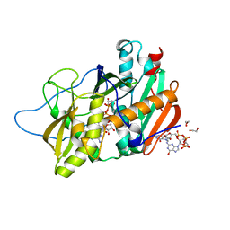
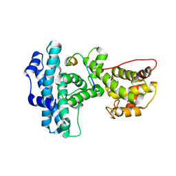
 | | glucose-6-phosphate dehydrogenase from Kluyveromyces lactis | | 分子名称: | 2-(2-(2-(2-(2-(2-ETHOXYETHOXY)ETHOXY)ETHOXY)ETHOXY)ETHOXY)ETHANOL, 2-AMINO-2-HYDROXYMETHYL-PROPANE-1,3-DIOL, Glucose-6-phosphate 1-dehydrogenase | | 著者 | Ha, V.H, Chang, J.H. | | 登録日 | 2021-02-22 | | 公開日 | 2021-04-14 | | 最終更新日 | 2023-11-29 | | 実験手法 | X-RAY DIFFRACTION (2.7 Å) | | 主引用文献 | Structural basis for substrate recognition of glucose-6-phosphate dehydrogenase from Kluyveromyces lactis.
Biochem.Biophys.Res.Commun., 553, 2021
|
|
7E6I
 | |
7BU3
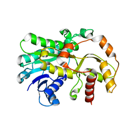
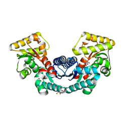
 | | Structure of alcohol dehydrogenase YjgB in complex with NADP from Escherichia coli | | 分子名称: | ASPARTIC ACID, Alcohol dehydrogenase, DI(HYDROXYETHYL)ETHER, ... | | 著者 | Nguyen, G.T, Kim, Y.-G, Ahn, J.-W, Chang, J.H. | | 登録日 | 2020-04-03 | | 公開日 | 2020-05-13 | | 最終更新日 | 2023-11-29 | | 実験手法 | X-RAY DIFFRACTION (2 Å) | | 主引用文献 | Structural Basis for Broad Substrate Selectivity of Alcohol Dehydrogenase YjgB from Escherichia coli .
Molecules, 25, 2020
|
|
6IQ1
 | |
7ELF
 | |
4J7N
 | | Crystal structure of mouse DXO in complex with M7GPPPG cap | | 分子名称: | 1,2-ETHANEDIOL, 7-METHYL-GUANOSINE-5'-TRIPHOSPHATE-5'-GUANOSINE, 9-METHYLGUANINE, ... | | 著者 | Kilic, T, Chang, J.H, Tong, L. | | 登録日 | 2013-02-13 | | 公開日 | 2013-03-27 | | 最終更新日 | 2024-02-28 | | 実験手法 | X-RAY DIFFRACTION (1.5 Å) | | 主引用文献 | A mammalian pre-mRNA 5' end capping quality control mechanism and an unexpected link of capping to pre-mRNA processing.
Mol.Cell, 50, 2013
|
|
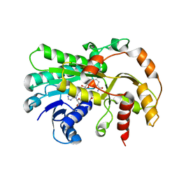
5ELM
 | | Crystal structure of L-aspartate/glutamate specific racemase in complex with L-glutamate | | 分子名称: | Asp/Glu_racemase family protein, GLUTAMIC ACID, GLYCEROL, ... | | 著者 | Ahn, J.W, Chang, J.H, Kim, K.J. | | 登録日 | 2015-11-04 | | 公開日 | 2015-11-18 | | 最終更新日 | 2023-11-08 | | 実験手法 | X-RAY DIFFRACTION (2 Å) | | 主引用文献 | Structural basis for an atypical active site of an l-aspartate/glutamate-specific racemase from Escherichia coli
Febs Lett., 589, 2015
|
|
5ELL
 | |
1N6A
 | | Structure of SET7/9 | | 分子名称: | S-ADENOSYLMETHIONINE, SET domain-containing protein 7 | | 著者 | Kwon, T.W, Chang, J.H, Kwak, E, Lee, C.W, Joachimiak, A, Kim, Y.C, Lee, J, Cho, Y. | | 登録日 | 2002-11-09 | | 公開日 | 2003-02-04 | | 最終更新日 | 2011-07-13 | | 実験手法 | X-RAY DIFFRACTION (1.7 Å) | | 主引用文献 | Mechanism of histone lysine methyl transfer revealed by the structure of SET7/9-AdoMet
EMBO J., 22, 2003
|
|
1N6C
 | | Structure of SET7/9 | | 分子名称: | S-ADENOSYLMETHIONINE, SET domain-containing protein 7 | | 著者 | Kwon, T.W, Chang, J.H, Cho, Y. | | 登録日 | 2002-11-09 | | 公開日 | 2003-02-04 | | 最終更新日 | 2024-03-13 | | 実験手法 | X-RAY DIFFRACTION (2.3 Å) | | 主引用文献 | Mechanism of histone lysine methyl transfer revealed by the structure of SET7/9-AdoMet
EMBO J., 22, 2003
|
|
1N4M
 | | Structure of Rb tumor suppressor bound to the transactivation domain of E2F-2 | | 分子名称: | Retinoblastoma Pocket, Transcription factor E2F2 | | 著者 | Lee, C, Chang, J.H, Lee, H.S, Cho, Y. | | 登録日 | 2002-10-31 | | 公開日 | 2003-01-07 | | 最終更新日 | 2024-02-14 | | 実験手法 | X-RAY DIFFRACTION (2.2 Å) | | 主引用文献 | Structural basis for the recognition of the E2F transactivation domain by the retinoblastoma tumor suppressor
GENES DEV., 16, 2002
|
|


