Loading
PDBj
メニューPDBj@FacebookPDBj@TwitterPDBj@YouTubewwPDB FoundationwwPDB
RCSB PDBPDBeBMRBAdv. SearchSearch help
PDB: 70 件

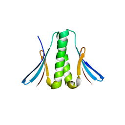
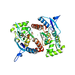
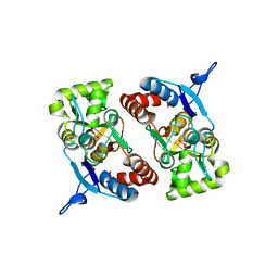
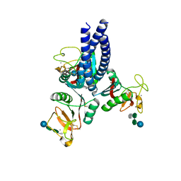
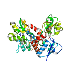
4BDL
DownloadVisualize
BU of 4bdl by Molmil
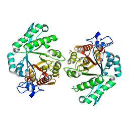
Crystal structure of the GluK2 K531A LBD dimer in complex with glutamate
分子名称: GLUTAMATE RECEPTOR, IONOTROPIC KAINATE 2, GLUTAMIC ACID, ...
著者Nayeem, N, Mayans, O, Green, T.
登録日2012-10-05
公開日2013-04-10
最終更新日2023-12-20
実験手法X-RAY DIFFRACTION (1.75 Å)
主引用文献Correlating Efficacy and Desensitization with Gluk2 Ligand-Binding Domain Movements.
Open Biol., 3, 2013
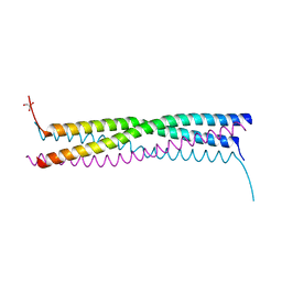
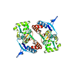
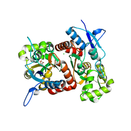
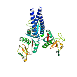
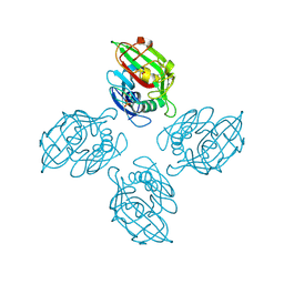
2FQM
DownloadVisualize
BU of 2fqm by Molmil
Crystal structure of the oligomerization domain of the phosphoprotein of vesicular stomatitis virus
分子名称: Phosphoprotein
著者Ding, H, Green, T.J, Lu, S, Luo, M.
登録日2006-01-18
公開日2006-02-07
最終更新日2024-02-14
実験手法X-RAY DIFFRACTION (2.3 Å)
主引用文献Crystal structure of the oligomerization domain of the phosphoprotein of vesicular stomatitis virus
J.Virol., 80, 2006
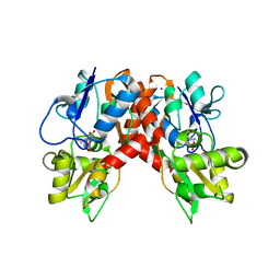
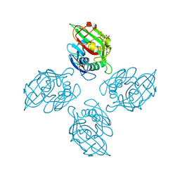
4EIJ
DownloadVisualize
BU of 4eij by Molmil
Structure of the Mumps virus phosphoprotein oligomerization domain
分子名称: GLYCEROL, P protein
著者Cox, R, Green, T.J, Luo, M.
登録日2012-04-05
公開日2013-05-08
最終更新日2024-02-28
実験手法X-RAY DIFFRACTION (2.2001 Å)
主引用文献Structural and functional characterization of the mumps virus phosphoprotein.
J.Virol., 87, 2013
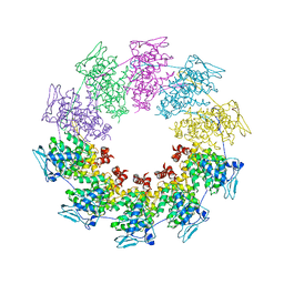
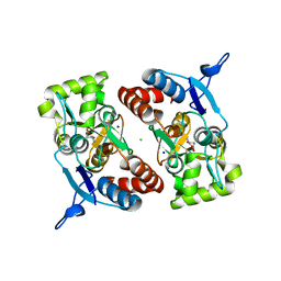
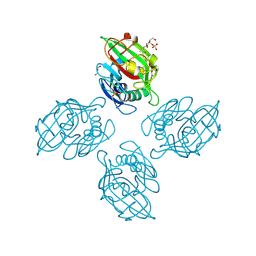
2QVJ
DownloadVisualize
BU of 2qvj by Molmil
Crystal structure of a vesicular stomatitis virus nucleocapsid protein Ser290Trp mutant
分子名称: Nucleocapsid protein
著者Luo, M, Green, T.J, Zhang, X, Tsao, J, Qiu, S.
登録日2007-08-08
公開日2008-01-08
最終更新日2024-02-21
実験手法X-RAY DIFFRACTION (2.8 Å)
主引用文献Role of intermolecular interactions of vesicular stomatitis virus nucleoprotein in RNA encapsidation.
J.Virol., 82, 2008
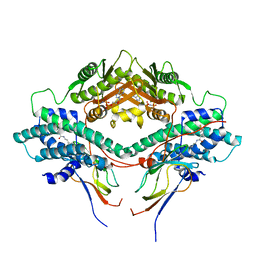
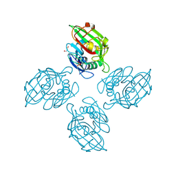
3CRL
DownloadVisualize
BU of 3crl by Molmil
Crystal structure of the PDHK2-L2 complex.
分子名称: Dihydrolipoyllysine-residue acetyltransferase component of pyruvate dehydrogenase complex, mitochondrial, MAGNESIUM ION, ...
著者Popov, K.M, Luo, M, Green, T.J, Grigorian, A, Klyuyeva, A, Tuganova, A.
登録日2008-04-07
公開日2008-04-29
最終更新日2017-10-25
実験手法X-RAY DIFFRACTION (2.61 Å)
主引用文献Structural and functional insights into the molecular mechanisms responsible for the regulation of pyruvate dehydrogenase kinase 2.
J.Biol.Chem., 283, 2008
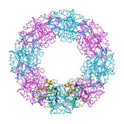
2WYY
DownloadVisualize
BU of 2wyy by Molmil
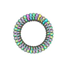
CRYOEM MODEL OF THE VESICULAR STOMATITIS VIRUS
分子名称: NUCLEOPROTEIN, POLY-URIDINE
著者Ge, P, Tsao, J, Green, T.J, Luo, M, Zhou, Z.H.
登録日2009-11-20
公開日2010-02-16
最終更新日2024-05-08
実験手法ELECTRON MICROSCOPY (10.6 Å)
主引用文献Cryo-Em Model of the Bullet-Shaped Vesicular Stomatitis Virus.
Science, 327, 2010
3BXW
DownloadVisualize
BU of 3bxw by Molmil
Crystal Structure of Stabilin-1 Interacting Chitinase-Like Protein, SI-CLP
分子名称: Chitinase domain-containing protein 1, SULFATE ION
著者Meng, G, Green, T.J.
登録日2008-01-15
公開日2009-01-20
最終更新日2024-03-13
実験手法X-RAY DIFFRACTION (2.7 Å)
主引用文献Structure of human stabilin-1 interacting chitinase-like protein (SI-CLP) reveals a saccharide-binding cleft with lower sugar-binding selectivity.
J.Biol.Chem., 285, 2010
1YAE
DownloadVisualize
BU of 1yae by Molmil
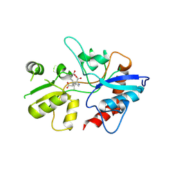
Structure of the Kainate Receptor Subunit GluR6 Agonist Binding Domain Complexed with Domoic Acid
分子名称: (2S,3S,4S)-2-CARBOXY-4-[(1Z,3E,5R)-5-CARBOXY-1-METHYL-1,3-HEXADIENYL]-3-PYRROLIDINEACETIC ACID, 2-acetamido-2-deoxy-beta-D-glucopyranose, Glutamate receptor, ...
著者Nanao, M.H, Green, T, Stern-Bach, Y, Heinemann, S.F, Choe, S.
登録日2004-12-17
公開日2005-02-01
最終更新日2023-08-23
実験手法X-RAY DIFFRACTION (3.11 Å)
主引用文献Structure of the kainate receptor subunit GluR6 agonist-binding domain complexed with domoic acid.
Proc.Natl.Acad.Sci.USA, 102, 2005
2XXV
DownloadVisualize
BU of 2xxv by Molmil
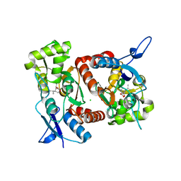
Crystal structure of the GluK2 (GluR6) M770K LBD dimer in complex with kainate
分子名称: 3-(CARBOXYMETHYL)-4-ISOPROPENYLPROLINE, CHLORIDE ION, GLUTAMATE RECEPTOR, ...
著者Nayeem, N, Mayans, O, Green, T.
登録日2010-11-12
公開日2011-02-09
最終更新日2023-12-20
実験手法X-RAY DIFFRACTION (1.7 Å)
主引用文献Conformational Flexibility of the Ligand-Binding Domain Dimer in Kainate Receptor Gating and Desensitization
J.Neurosci., 31, 2011
2XXR
DownloadVisualize
BU of 2xxr by Molmil
Crystal structure of the GluK2 (GluR6) wild-type LBD dimer in complex with glutamate
分子名称: CHLORIDE ION, GLUTAMATE RECEPTOR, IONOTROPIC KAINATE 2, ...
著者Nayeem, N, Mayans, O, Green, T.
登録日2010-11-11
公開日2011-02-09
最終更新日2023-12-20
実験手法X-RAY DIFFRACTION (1.6 Å)
主引用文献Conformational Flexibility of the Ligand-Binding Domain Dimer in Kainate Receptor Gating and Desensitization
J.Neurosci., 31, 2011
2XXY
DownloadVisualize
BU of 2xxy by Molmil
Crystal structure of the GluK2 (GluR6) D776K LBD dimer in complex with kainate
分子名称: 3-(CARBOXYMETHYL)-4-ISOPROPENYLPROLINE, GLUTAMATE RECEPTOR, IONOTROPIC KAINATE 2
著者Nayeem, N, Mayans, O, Green, T.
登録日2010-11-12
公開日2011-02-09
最終更新日2023-12-20
実験手法X-RAY DIFFRACTION (3 Å)
主引用文献Conformational Flexibility of the Ligand-Binding Domain Dimer in Kainate Receptor Gating and Desensitization
J.Neurosci., 31, 2011
2XXT
DownloadVisualize
BU of 2xxt by Molmil
Crystal structure of the GluK2 (GluR6) wild-type LBD dimer in complex with kainate
分子名称: 3-(CARBOXYMETHYL)-4-ISOPROPENYLPROLINE, CHLORIDE ION, GLUTAMATE RECEPTOR, ...
著者Nayeem, N, Mayans, O, Green, T.
登録日2010-11-12
公開日2011-02-09
最終更新日2023-12-20
実験手法X-RAY DIFFRACTION (1.9 Å)
主引用文献Conformational Flexibility of the Ligand-Binding Domain Dimer in Kainate Receptor Gating and Desensitization
J.Neurosci., 31, 2011
2XXU
DownloadVisualize
BU of 2xxu by Molmil
Crystal structure of the GluK2 (GluR6) M770K LBD dimer in complex with glutamate
分子名称: CHLORIDE ION, GLUTAMATE RECEPTOR, IONOTROPIC KAINATE 2, ...
著者Nayeem, N, Mayans, O, Green, T.
登録日2010-11-12
公開日2011-02-09
最終更新日2023-12-20
実験手法X-RAY DIFFRACTION (1.5 Å)
主引用文献Conformational Flexibility of the Ligand-Binding Domain Dimer in Kainate Receptor Gating and Desensitization
J.Neurosci., 31, 2011
2XXX
DownloadVisualize
BU of 2xxx by Molmil
Crystal structure of the GluK2 (GluR6) D776K LBD dimer in complex with glutamate (P21 21 21)
分子名称: GLUTAMATE RECEPTOR, IONOTROPIC KAINATE 2, GLUTAMIC ACID
著者Nayeem, N, Mayans, O, Green, T.
登録日2010-11-12
公開日2011-02-09
最終更新日2023-12-20
実験手法X-RAY DIFFRACTION (2.098 Å)
主引用文献Conformational Flexibility of the Ligand-Binding Domain Dimer in Kainate Receptor Gating and Desensitization
J.Neurosci., 31, 2011
2XXW
DownloadVisualize
BU of 2xxw by Molmil
Crystal structure of the GluK2 (GluR6) D776K LBD dimer in complex with glutamate
分子名称: CHLORIDE ION, GLUTAMATE RECEPTOR, IONOTROPIC KAINATE 2, ...
著者Nayeem, N, Mayans, O, Green, T.
登録日2010-11-12
公開日2011-02-09
最終更新日2023-12-20
実験手法X-RAY DIFFRACTION (2.3 Å)
主引用文献Conformational Flexibility of the Ligand-Binding Domain Dimer in Kainate Receptor Gating and Desensitization
J.Neurosci., 31, 2011
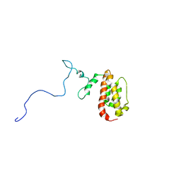
1N4C
DownloadVisualize
BU of 1n4c by Molmil
NMR Structure of the J-Domain and Clathrin Substrate Binding Domain of Bovine Auxilin
分子名称: Auxilin
著者Gruschus, J.M, Han, C.J, Greener, T, Greene, L.E, Ferretti, J.A, Eisenberg, E.
登録日2002-10-30
公開日2003-11-11
最終更新日2024-05-22
実験手法SOLUTION NMR
主引用文献Structure of the functional fragment of auxilin required for catalytic uncoating of clathrin-coated vesicles.
Biochemistry, 43, 2004
1QTJ
DownloadVisualize
BU of 1qtj by Molmil
CRYSTAL STRUCTURE OF LIMULUS POLYPHEMUS SAP
分子名称: PROTEIN (SERUM AMYLOID P COMPONENT)
著者Shrive, A.K, Metcalfe, A.M, Cartwright, J.R, Greenhough, T.J.
登録日1999-06-28
公開日2000-06-28
最終更新日2023-08-16
実験手法X-RAY DIFFRACTION (3 Å)
主引用文献C-reactive protein and SAP-like pentraxin are both present in Limulus polyphemus haemolymph: crystal structure of Limulus SAP.
J.Mol.Biol., 290, 1999
5OXS
DownloadVisualize
BU of 5oxs by Molmil
Crystal structure of human lung surfactant protein D trimeric fragment with bound ligand Salmonella enterica Minnesota R5 oligosaccharide
分子名称: CALCIUM ION, Pulmonary surfactant-associated protein D, alpha-D-glucopyranose-(1-3)-L-glycero-alpha-D-manno-heptopyranose-(1-3)-L-glycero-alpha-D-manno-heptopyranose-(1-5)-4,7-anhydro-3-deoxy-D-gluco-oct-2-ulosonic acid
著者Shrive, A.K, Greenhough, T.J.
登録日2017-09-07
公開日2018-06-20
最終更新日2024-10-16
実験手法X-RAY DIFFRACTION (1.65 Å)
主引用文献Structural definition of hSP-D recognition of Salmonella enterica LPS inner core oligosaccharides reveals alternative binding modes for the same LPS.
PLoS ONE, 13, 2018
5OXR
DownloadVisualize
BU of 5oxr by Molmil
Crystal structure of human lung surfactant protein D trimeric fragment with bound ligand Salmonella enterica Minnesota R7 oligosaccharide
分子名称: CALCIUM ION, L-glycero-alpha-D-manno-heptopyranose-(1-3)-L-glycero-alpha-D-manno-heptopyranose-(1-5)-4,7-anhydro-3-deoxy-D-gluco-oct-2-ulosonic acid, Pulmonary surfactant-associated protein D
著者Shrive, A.K, Greenhough, T.J.
登録日2017-09-07
公開日2018-06-20
最終更新日2024-10-23
実験手法X-RAY DIFFRACTION (1.75 Å)
主引用文献Structural definition of hSP-D recognition of Salmonella enterica LPS inner core oligosaccharides reveals alternative binding modes for the same LPS.
PLoS ONE, 13, 2018
6ZR0
DownloadVisualize
BU of 6zr0 by Molmil
Crystal structure of tetrameric fibrinogen-like recognition domain of FIBCD1 with N-acetylalanine ligand bound
分子名称: ACETIC ACID, CALCIUM ION, Fibrinogen C domain-containing protein 1, ...
著者Shrive, A.K, Greenhough, T.J, Williams, H.M.
登録日2020-07-10
公開日2021-07-21
最終更新日2024-01-31
実験手法X-RAY DIFFRACTION (1.94 Å)
主引用文献Crystal structures of human immune protein FIBCD1 suggest an extended binding site compatible with recognition of pathogen associated carbohydrate motifs
J.Biol.Chem., 2023
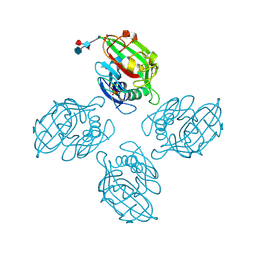
6ZQR
DownloadVisualize
BU of 6zqr by Molmil
Crystal structure of tetrameric fibrinogen-like recognition domain of FIBCD1 with GlcNAc ligand bound
分子名称: 2-acetamido-2-deoxy-beta-D-glucopyranose, ACETIC ACID, CALCIUM ION, ...
著者Shrive, A.K, Greenhough, T.J, Williams, H.M.
登録日2020-07-10
公開日2021-07-21
最終更新日2024-01-31
実験手法X-RAY DIFFRACTION (1.93 Å)
主引用文献Crystal structures of human immune protein FIBCD1 suggest an extended binding site compatible with recognition of pathogen associated carbohydrate motifs
J.Biol.Chem., 2023
6ZQY
DownloadVisualize
BU of 6zqy by Molmil
Crystal structure of tetrameric fibrinogen-like recognition domain of FIBCD1 with Neu5Ac ligand bound
分子名称: ACETIC ACID, CALCIUM ION, Fibrinogen C domain-containing protein 1, ...
著者Shrive, A.K, Greenhough, T.J, Williams, H.M.
登録日2020-07-10
公開日2021-07-21
最終更新日2024-10-09
実験手法X-RAY DIFFRACTION (1.85 Å)
主引用文献Crystal structures of human immune protein FIBCD1 suggest an extended binding site compatible with recognition of pathogen associated carbohydrate motifs
J.Biol.Chem., 2023
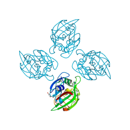
6ZR4
DownloadVisualize
BU of 6zr4 by Molmil
Crystal structure of tetrameric fibrinogen-like recognition domain of FIBCD1
分子名称: ACETIC ACID, CALCIUM ION, Fibrinogen C domain-containing protein 1, ...
著者Shrive, A.K, Greenhough, T.J, Williams, H.M.
登録日2020-07-10
公開日2021-07-21
最終更新日2024-01-31
実験手法X-RAY DIFFRACTION (2 Å)
主引用文献Crystal structures of human immune protein FIBCD1 suggest an extended binding site compatible with recognition of pathogen associated carbohydrate motifs
J.Biol.Chem., 2023
6ZR3
DownloadVisualize
BU of 6zr3 by Molmil
Crystal structure of tetrameric fibrinogen-like recognition domain of FIBCD1 with N-acetyl-galactosamine-4-sulfate ligand bound
分子名称: 2-acetamido-2-deoxy-4-O-sulfo-beta-D-galactopyranose, ACETIC ACID, CALCIUM ION, ...
著者Shrive, A.K, Greenhough, T.J, Williams, H.M.
登録日2020-07-10
公開日2021-07-21
最終更新日2024-01-31
実験手法X-RAY DIFFRACTION (1.97 Å)
主引用文献Crystal structures of human immune protein FIBCD1 suggest an extended binding site compatible with recognition of pathogen associated carbohydrate motifs
J.Biol.Chem., 2023
6ZQX
DownloadVisualize
BU of 6zqx by Molmil
Crystal structure of tetrameric fibrinogen-like recognition domain of FIBCD1 with N,N'-diacetyl chitobiose ligand bound
分子名称: 2-acetamido-2-deoxy-beta-D-glucopyranose, ACETIC ACID, CALCIUM ION, ...
著者Shrive, A.K, Greenhough, T.J, Williams, H.M.
登録日2020-07-10
公開日2021-07-21
最終更新日2024-10-23
実験手法X-RAY DIFFRACTION (1.84 Å)
主引用文献Crystal structures of human immune protein FIBCD1 suggest an extended binding site compatible with recognition of pathogen associated carbohydrate motifs
J.Biol.Chem., 2023

226707

件を2024-10-30に公開中

PDB statisticsPDBj update infoContact PDBjnumon