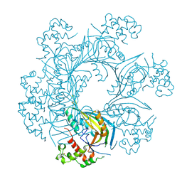
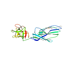
4C4N
 
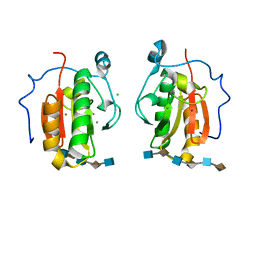 | | Crystal structure of the Sonic Hedgehog-heparin complex | | 分子名称: | 2-O-sulfo-alpha-L-idopyranuronic acid-(1-4)-2-deoxy-6-O-sulfo-2-(sulfoamino)-alpha-D-glucopyranose-(1-4)-2-O-sulfo-alpha-L-idopyranuronic acid-(1-4)-2-deoxy-6-O-sulfo-2-(sulfoamino)-alpha-D-glucopyranose-(1-4)-2-O-sulfo-alpha-L-idopyranuronic acid-(1-4)-2-deoxy-6-O-sulfo-2-(sulfoamino)-alpha-D-glucopyranose, 2-deoxy-6-O-sulfo-2-(sulfoamino)-alpha-D-glucopyranose-(1-4)-2-O-sulfo-alpha-L-idopyranuronic acid-(1-4)-2-deoxy-6-O-sulfo-2-(sulfoamino)-alpha-D-glucopyranose-(1-4)-2-O-sulfo-alpha-L-idopyranuronic acid-(1-4)-2-deoxy-6-O-sulfo-2-(sulfoamino)-alpha-D-glucopyranose-(1-4)-2-O-sulfo-alpha-L-idopyranuronic acid, CALCIUM ION, ... | | 著者 | Whalen, D.M, Malinauskas, T, Gilbert, R.J.C, Siebold, C. | | 登録日 | 2013-09-05 | | 公開日 | 2013-10-02 | | 最終更新日 | 2024-05-08 | | 実験手法 | X-RAY DIFFRACTION (2.36 Å) | | 主引用文献 | Structural Insights Into Proteoglycan-Shaped Hedgehog Signaling.
Proc.Natl.Acad.Sci.USA, 110, 2013
|
|
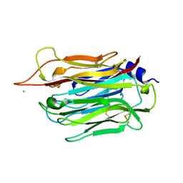
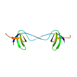
2VSK
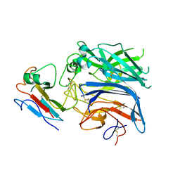 | | Hendra virus attachment glycoprotein in complex with human cell surface receptor ephrinB2 | | 分子名称: | EPHRIN-B2, HEMAGGLUTININ-NEURAMINIDASE | | 著者 | Bowden, T.A, Aricescu, A.R, Gilbert, R.J, Grimes, J.M, Jones, E.Y, Stuart, D.I. | | 登録日 | 2008-04-24 | | 公開日 | 2008-05-20 | | 最終更新日 | 2023-12-13 | | 実験手法 | X-RAY DIFFRACTION (3.3 Å) | | 主引用文献 | Structural Basis of Nipah and Hendra Virus Attachment to Their Cell-Surface Receptor Ephrin-B2
Nat.Struct.Mol.Biol., 15, 2008
|
|
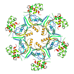
1UW7
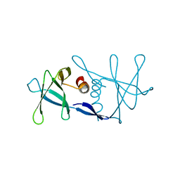 | | Nsp9 protein from SARS-coronavirus. | | 分子名称: | NSP9 | | 著者 | Sutton, G, Fry, E, Carter, L, Sainsbury, S, Walter, T, Nettleship, J, Berrow, N, Owens, R, Gilbert, R, Davidson, A, Siddell, S, Poon, L.L.M, Diprose, J, Alderton, D, Walsh, M, Grimes, J.M, Stuart, D.I. | | 登録日 | 2004-01-30 | | 公開日 | 2004-02-20 | | 最終更新日 | 2024-05-08 | | 実験手法 | X-RAY DIFFRACTION (2.8 Å) | | 主引用文献 | The Nsp9 Replicase Protein of Sars-Coronavirus, Structure and Functional Insights
Structure, 12, 2004
|
|
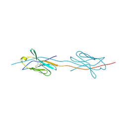
5OWN
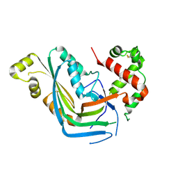 | | Structure of TgPLP1 MACPF domain | | 分子名称: | 2-acetamido-2-deoxy-beta-D-glucopyranose, Perforin-like protein 1 | | 著者 | Ni, T, Gilbert, R.J.C. | | 登録日 | 2017-09-01 | | 公開日 | 2018-04-11 | | 最終更新日 | 2024-01-17 | | 実験手法 | X-RAY DIFFRACTION (3.11 Å) | | 主引用文献 | Structures of monomeric and oligomeric forms of theToxoplasma gondiiperforin-like protein 1.
Sci Adv, 4, 2018
|
|
5OUP
 | | Structure of TgPLP1 MACPF domain | | 分子名称: | 2-acetamido-2-deoxy-beta-D-glucopyranose, Perforin-like protein 1 | | 著者 | Ni, T, Gilbert, R.J.C. | | 登録日 | 2017-08-24 | | 公開日 | 2018-04-11 | | 最終更新日 | 2020-07-29 | | 実験手法 | X-RAY DIFFRACTION (2.03 Å) | | 主引用文献 | Structures of monomeric and oligomeric forms of theToxoplasma gondiiperforin-like protein 1.
Sci Adv, 4, 2018
|
|
5OUO
 | | Structure of TgPLP1 APCbeta domain | | 分子名称: | CHLORIDE ION, MAGNESIUM ION, Perforin-like protein 1 | | 著者 | Ni, T, Gilbert, R.J.C. | | 登録日 | 2017-08-24 | | 公開日 | 2018-04-11 | | 最終更新日 | 2019-10-16 | | 実験手法 | X-RAY DIFFRACTION (1.11 Å) | | 主引用文献 | Structures of monomeric and oligomeric forms of theToxoplasma gondiiperforin-like protein 1.
Sci Adv, 4, 2018
|
|
5OUQ
 | | Structure of TgPLP1 MACPF domain | | 分子名称: | 2-acetamido-2-deoxy-beta-D-glucopyranose, Perforin-like protein 1 | | 著者 | Ni, T, Gilbert, R.J.C. | | 登録日 | 2017-08-24 | | 公開日 | 2018-04-11 | | 最終更新日 | 2024-01-17 | | 実験手法 | X-RAY DIFFRACTION (5.11 Å) | | 主引用文献 | Structures of monomeric and oligomeric forms of theToxoplasma gondiiperforin-like protein 1.
Sci Adv, 4, 2018
|
|
3ZXD
 | | wild-type lysenin | | 分子名称: | 2-(N-MORPHOLINO)-ETHANESULFONIC ACID, CHLORIDE ION, GLYCEROL, ... | | 著者 | De Colibus, L, Sonnen, A.F.P, Morris, K.J, Siebert, C.A, Abrusci, P, Plitzko, J, Hodnik, V, Leippe, M, Volpi, E, Anderluh, G, Gilbert, R.J.C. | | 登録日 | 2011-08-09 | | 公開日 | 2012-09-19 | | 最終更新日 | 2023-12-20 | | 実験手法 | X-RAY DIFFRACTION (3.3 Å) | | 主引用文献 | Structures of Lysenin Reveal a Shared Evolutionary Origin for Pore-Forming Proteins and its Mode of Sphingomyelin Recognition.
Structure, 20, 2012
|
|
2X44
 | | Structure of a strand-swapped dimeric form of CTLA-4 | | 分子名称: | CYTOTOXIC T-LYMPHOCYTE PROTEIN 4 | | 著者 | Sonnen, A.F.-P, Yu, C, Evans, E.J, Stuart, D.I, Davis, S.J, Gilbert, R.J.C. | | 登録日 | 2010-01-28 | | 公開日 | 2010-04-07 | | 最終更新日 | 2023-12-20 | | 実験手法 | X-RAY DIFFRACTION (2.6 Å) | | 主引用文献 | Domain Metastability: A Molecular Basis for Immunoglobulin Deposition?
J.Mol.Biol., 399, 2010
|
|
4BBK
 | | Structural and functional characterisation of the kindlin-1 pleckstrin homology domain | | 分子名称: | FERMITIN FAMILY HOMOLOG 1, GLYCEROL | | 著者 | Yates, L.A, Lumb, C.N, Brahme, N.N, Zalyte, R, Bird, L.E, De Colibus, L, Owens, R.J, Calderwood, D.A, Sansom, M.S.P, Gilbert, R.J.C. | | 登録日 | 2012-09-25 | | 公開日 | 2012-11-14 | | 最終更新日 | 2023-12-20 | | 実験手法 | X-RAY DIFFRACTION (2.1 Å) | | 主引用文献 | Structural and Functional Characterisation of the Kindlin-1 Pleckstrin Homology Domain
J.Biol.Chem., 287, 2012
|
|
4BQB
 | | Crystal structure of the FN5 and FN6 domains of NEO1, form 2 | | 分子名称: | 2-acetamido-2-deoxy-beta-D-glucopyranose, NEOGENIN | | 著者 | Bell, C.H, Healey, E, van Erp, S, Bishop, B, Tang, C, Gilbert, R.J.C, Aricescu, A.R, Pasterkamp, R.J, Siebold, C. | | 登録日 | 2013-05-30 | | 公開日 | 2013-06-12 | | 最終更新日 | 2020-07-29 | | 実験手法 | X-RAY DIFFRACTION (2.7 Å) | | 主引用文献 | Structure of the Repulsive Guidance Molecule (Rgm)-Neogenin Signaling Hub
Science, 341, 2013
|
|
4BQ6
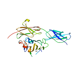 | | Crystal structure of the RGMB-NEO1 complex form 1 | | 分子名称: | 2-acetamido-2-deoxy-beta-D-glucopyranose, NEOGENIN, RGM DOMAIN FAMILY MEMBER B | | 著者 | Bell, C.H, Healey, E, van Erp, S, Bishop, B, Tang, C, Gilbert, R.J.C, Aricescu, A.R, Pasterkamp, R.J, Siebold, C. | | 登録日 | 2013-05-30 | | 公開日 | 2013-06-12 | | 最終更新日 | 2020-07-29 | | 実験手法 | X-RAY DIFFRACTION (2.3 Å) | | 主引用文献 | Structure of the Repulsive Guidance Molecule (Rgm)-Neogenin Signaling Hub
Science, 341, 2013
|
|
4BQ9
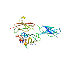
 | | Crystal structure of the FN5 and FN6 domains of NEO1, form 1 | | 分子名称: | 2-acetamido-2-deoxy-beta-D-glucopyranose, NEOGENIN | | 著者 | Bell, C.H, Healey, E, van Erp, S, Bishop, B, Tang, C, Gilbert, R.J.C, Aricescu, A.R, Pasterkamp, R.J, Siebold, C. | | 登録日 | 2013-05-30 | | 公開日 | 2013-06-12 | | 最終更新日 | 2020-07-29 | | 実験手法 | X-RAY DIFFRACTION (2.91 Å) | | 主引用文献 | Structure of the Repulsive Guidance Molecule (Rgm)-Neogenin Signaling Hub
Science, 341, 2013
|
|
4BQ7
 | | Crystal structure of the RGMB-Neo1 complex form 2 | | 分子名称: | NEOGENIN, RGM DOMAIN FAMILY MEMBER B | | 著者 | Bell, C.H, Healey, E, van Erp, S, Bishop, B, Tang, C, Gilbert, R.J.C, Aricescu, A.R, Pasterkamp, R.J, Siebold, C. | | 登録日 | 2013-05-30 | | 公開日 | 2013-06-12 | | 最終更新日 | 2019-04-03 | | 実験手法 | X-RAY DIFFRACTION (6.601 Å) | | 主引用文献 | Structure of the Repulsive Guidance Molecule (Rgm)-Neogenin Signaling Hub
Science, 341, 2013
|
|
4BQC
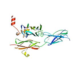
 | | Crystal structure of the FN5 and FN6 domains of NEO1 bound to SOS | | 分子名称: | 1,3,4,6-tetra-O-sulfo-beta-D-fructofuranose-(2-1)-2,3,4,6-tetra-O-sulfonato-alpha-D-glucopyranose, 2-acetamido-2-deoxy-beta-D-glucopyranose, NEOGENIN, ... | | 著者 | Bell, C.H, Healey, E, vanErp, S, Bishop, B, Tang, C, Gilbert, R.J.C, Aricescu, A.R, Pasterkamp, R.J, Siebold, C. | | 登録日 | 2013-05-30 | | 公開日 | 2013-06-12 | | 最終更新日 | 2020-07-29 | | 実験手法 | X-RAY DIFFRACTION (3.2 Å) | | 主引用文献 | Structure of the Repulsive Guidance Molecule (Rgm)-Neogenin Signaling Hub
Science, 341, 2013
|
|
4BQ8
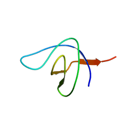
 | | Crystal structure of the RGMB-NEO1 complex form 3 | | 分子名称: | 2-acetamido-2-deoxy-beta-D-glucopyranose, NEOGENIN, RGM DOMAIN FAMILY MEMBER B | | 著者 | Bell, C.H, Healey, E, van Erp, S, Bishop, B, Tang, C, Gilbert, R.J.C, Aricescu, A.R, Pasterkamp, R.J, Siebold, C. | | 登録日 | 2013-05-30 | | 公開日 | 2013-06-12 | | 最終更新日 | 2020-07-29 | | 実験手法 | X-RAY DIFFRACTION (2.8 Å) | | 主引用文献 | Structure of the Repulsive Guidance Molecule (Rgm)-Neogenin Signaling Hub
Science, 341, 2013
|
|
2BZX
 | |
2BZY
 | |
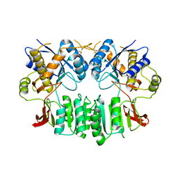
4E80
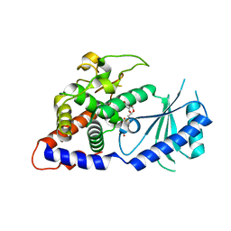 | | Structural Basis for the Activity of a Cytoplasmic RNA Terminal U-transferase | | 分子名称: | Poly(A) RNA polymerase protein cid1, URIDINE 5'-TRIPHOSPHATE | | 著者 | Yates, L.A, Fleurdepine, S, Rissland, O.S, DeColibus, L, Harlos, K, Norbury, C.J, Gilbert, R.J.C. | | 登録日 | 2012-03-19 | | 公開日 | 2012-07-04 | | 最終更新日 | 2024-02-28 | | 実験手法 | X-RAY DIFFRACTION (3.02 Å) | | 主引用文献 | Structural basis for the activity of a cytoplasmic RNA terminal uridylyl transferase.
Nat.Struct.Mol.Biol., 19, 2012
|
|
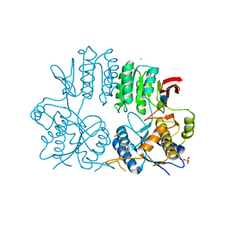
4E8F
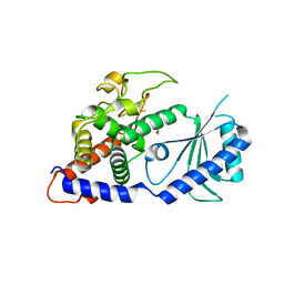 | | Structural Basis for the Activity of a Cytoplasmic RNA Terminal U-transferase | | 分子名称: | ACETATE ION, GLYCEROL, Poly(A) RNA polymerase protein cid1 | | 著者 | Yates, L.A, Fleurdepine, S, Rissland, O.S, DeColibus, L, Harlos, K, Norbury, C.J, Gilbert, R.J.C. | | 登録日 | 2012-03-20 | | 公開日 | 2012-07-04 | | 最終更新日 | 2024-02-28 | | 実験手法 | X-RAY DIFFRACTION (2.6 Å) | | 主引用文献 | Structural basis for the activity of a cytoplasmic RNA terminal uridylyl transferase.
Nat.Struct.Mol.Biol., 19, 2012
|
|
4E7X
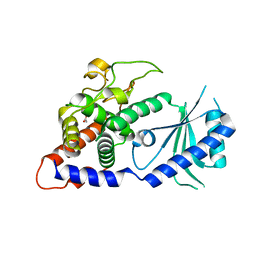 | | Structural Basis for the Activity of a Cytoplasmic RNA Terminal U-transferase | | 分子名称: | ACETATE ION, Poly(A) RNA polymerase protein cid1 | | 著者 | Yates, L.A, Fleurdepine, S, Rissland, O.S, DeColibus, L, Harlos, K, Norbury, C.J, Gilbert, R.J.C. | | 登録日 | 2012-03-19 | | 公開日 | 2012-07-04 | | 最終更新日 | 2024-02-28 | | 実験手法 | X-RAY DIFFRACTION (3.2 Å) | | 主引用文献 | Structural basis for the activity of a cytoplasmic RNA terminal uridylyl transferase.
Nat.Struct.Mol.Biol., 19, 2012
|
|
3ZX7
 | | Complex of lysenin with phosphocholine | | 分子名称: | LYSENIN, PHOSPHATE ION, PHOSPHOCHOLINE, ... | | 著者 | De Colibus, L, Sonnen, A.F.P, Morris, K.J, Siebert, C.A, Abrusci, P, Plitzko, J, Hodnik, V, Leippe, M, Volpi, E, Anderluh, G, Gilbert, R.J.C. | | 登録日 | 2011-08-08 | | 公開日 | 2012-09-19 | | 最終更新日 | 2012-10-03 | | 実験手法 | X-RAY DIFFRACTION (2.84 Å) | | 主引用文献 | Structures of Lysenin Reveal a Shared Evolutionary Origin for Pore-Forming Proteins and its Mode of Sphingomyelin Recognition.
Structure, 20, 2012
|
|
3ZXG
 | | lysenin sphingomyelin complex | | 分子名称: | LYSENIN, SULFATE ION, TRIMETHYL-[2-[[(2S,3S)-2-(OCTADECANOYLAMINO)-3-OXIDANYL-BUTOXY]-OXIDANYL-PHOSPHORYL]OXYETHYL]AZANIUM | | 著者 | De Colibus, L, Sonnen, A.F.P, Morris, K.J, Siebert, C.A, Abrusci, P, Plitzko, J, Hodnik, V, Leippe, M, Volpi, E, Anderluh, G, Gilbert, R.J.C. | | 登録日 | 2011-08-10 | | 公開日 | 2012-09-19 | | 最終更新日 | 2023-12-20 | | 実験手法 | X-RAY DIFFRACTION (3.12 Å) | | 主引用文献 | Structures of Lysenin Reveal a Shared Evolutionary Origin for Pore-Forming Proteins and its Mode of Sphingomyelin Recognition.
Structure, 20, 2012
|
|
2WJX
 | | Crystal structure of the human ionotropic glutamate receptor GluR2 ATD region at 4.1 A resolution | | 分子名称: | GLUTAMATE RECEPTOR 2 | | 著者 | Clayton, A, Siebold, C, Gilbert, R.J.C, Sutton, G.C, Harlos, K, McIlhinney, R.A.J, Jones, E.Y, Aricescu, A.R. | | 登録日 | 2009-06-01 | | 公開日 | 2009-08-18 | | 最終更新日 | 2011-07-13 | | 実験手法 | X-RAY DIFFRACTION (4.1 Å) | | 主引用文献 | Crystal Structure of the Glur2 Amino-Terminal Domain Provides Insights Into the Architecture and Assembly of Ionotropic Glutamate Receptors.
J.Mol.Biol., 392, 2009
|
|
2WJW
 | | Crystal structure of the human ionotropic glutamate receptor GluR2 ATD region at 1.8 A resolution | | 分子名称: | 2-acetamido-2-deoxy-beta-D-glucopyranose, ACETATE ION, CHLORIDE ION, ... | | 著者 | Clayton, A, Siebold, C, Gilbert, R.J.C, Sutton, G.C, Harlos, K, McIlhinney, R.A.J, Jones, E.Y, Aricescu, A.R. | | 登録日 | 2009-06-01 | | 公開日 | 2009-08-18 | | 最終更新日 | 2020-07-29 | | 実験手法 | X-RAY DIFFRACTION (1.8 Å) | | 主引用文献 | Crystal Structure of the Glur2 Amino-Terminal Domain Provides Insights Into the Architecture and Assembly of Ionotropic Glutamate Receptors.
J.Mol.Biol., 392, 2009
|
|


