2FS7
 
 | |
2G79
 | |
2G78
 | |
2G7B
 | |
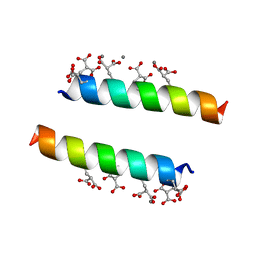
2DOH
 | | The X-ray crystallographic structure of the angiogenesis inhibitor, angiostatin, bound a to a peptide from the group A streptococcal surface protein PAM | | 分子名称: | 1,4-DIETHYLENE DIOXIDE, Angiostatin, Plasminogen-binding group A streptococcal M-like protein PAM | | 著者 | Cnudde, S.E, Prorok, M, Castellino, F.J, Geiger, J.H. | | 登録日 | 2006-04-29 | | 公開日 | 2006-12-05 | | 最終更新日 | 2023-10-25 | | 実験手法 | X-RAY DIFFRACTION (2.3 Å) | | 主引用文献 | X-ray crystallographic structure of the angiogenesis inhibitor, angiostatin, bound to a peptide from the group A streptococcal surface protein PAM
Biochemistry, 45, 2006
|
|
2DPR
 | | The crystal structures of the calcium-bound con-G and con-T(K7Glu) dimeric peptides demonstrate a novel metal-dependent helix-forming motif | | 分子名称: | CALCIUM ION, Conantokin-T | | 著者 | Cnudde, S.E, Prorok, M, Dai, Q, Castellino, F.J, Geiger, J.H. | | 登録日 | 2006-05-13 | | 公開日 | 2007-04-24 | | 最終更新日 | 2024-04-03 | | 実験手法 | X-RAY DIFFRACTION (1.7 Å) | | 主引用文献 | The crystal structures of the calcium-bound con-G and con-T[K7gamma] dimeric peptides demonstrate a metal-dependent helix-forming motif
J.Am.Chem.Soc., 129, 2007
|
|
4RLF
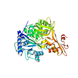
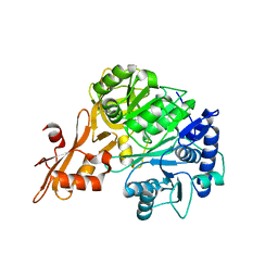
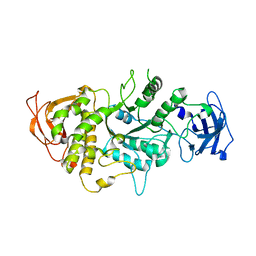
 | | Crystal structure of a benzoate coenzyme A ligase with p-Toluic acid and o-Toluic acid | | 分子名称: | 2-methylbenzoic acid, 4-METHYLBENZOIC ACID, Benzoate-coenzyme A ligase, ... | | 著者 | Strom, S, Nosrati, M, Thornburg, C, Walker, K, Geiger, J.H. | | 登録日 | 2014-10-16 | | 公開日 | 2015-09-30 | | 最終更新日 | 2023-09-20 | | 実験手法 | X-RAY DIFFRACTION (1.73 Å) | | 主引用文献 | Kinetically and Crystallographically Guided Mutations of a Benzoate CoA Ligase (BadA) Elucidate Mechanism and Expand Substrate Permissivity.
Biochemistry, 54, 2015
|
|
4RM3
 | | Crystal structure of a benzoate coenzyme A ligase with 2-Furoic acid | | 分子名称: | 2-FUROIC ACID, Benzoate-coenzyme A ligase | | 著者 | Strom, S, Nosrati, M, Thornburg, C, Walker, K.D, Geiger, J.H. | | 登録日 | 2014-10-18 | | 公開日 | 2015-09-30 | | 最終更新日 | 2023-09-20 | | 実験手法 | X-RAY DIFFRACTION (1.76 Å) | | 主引用文献 | Kinetically and Crystallographically Guided Mutations of a Benzoate CoA Ligase (BadA) Elucidate Mechanism and Expand Substrate Permissivity.
Biochemistry, 54, 2015
|
|
4RUU
 | |
4RMN
 | | Crystal structure of a benzoate coenzyme A ligase with 2-Thiophene Carboxylic acid | | 分子名称: | Benzoate-coenzyme A ligase, GLYCEROL, THIOPHENE-2-CARBOXYLIC ACID | | 著者 | Strom, S, Nosrati, M, Thornburg, C, Walker, K.D, Geiger, J.H. | | 登録日 | 2014-10-21 | | 公開日 | 2015-09-30 | | 最終更新日 | 2023-09-20 | | 実験手法 | X-RAY DIFFRACTION (1.72 Å) | | 主引用文献 | Kinetically and Crystallographically Guided Mutations of a Benzoate CoA Ligase (BadA) Elucidate Mechanism and Expand Substrate Permissivity.
Biochemistry, 54, 2015
|
|
4RM2
 | | Crystal structure of a benzoate coenzyme A ligase with 2-Fluoro benzoic acid | | 分子名称: | 2-fluorobenzoic acid, Benzoate-coenzyme A ligase | | 著者 | Strom, S, Nosrati, M, Thornburg, C, Walker, K.D, Geiger, J.H. | | 登録日 | 2014-10-18 | | 公開日 | 2015-09-30 | | 最終更新日 | 2023-09-20 | | 実験手法 | X-RAY DIFFRACTION (1.77 Å) | | 主引用文献 | Kinetically and Crystallographically Guided Mutations of a Benzoate CoA Ligase (BadA) Elucidate Mechanism and Expand Substrate Permissivity.
Biochemistry, 54, 2015
|
|
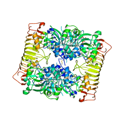
1JKF
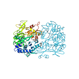
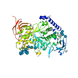
 | | Holo 1L-myo-inositol-1-phosphate Synthase | | 分子名称: | NICOTINAMIDE-ADENINE-DINUCLEOTIDE, myo-inositol-1-phosphate synthase | | 著者 | Stein, A.J, Geiger, J.H. | | 登録日 | 2001-07-12 | | 公開日 | 2002-04-10 | | 最終更新日 | 2018-01-31 | | 実験手法 | X-RAY DIFFRACTION (2.4 Å) | | 主引用文献 | The crystal structure and mechanism of 1-L-myo-inositol- 1-phosphate synthase
J.Biol.Chem., 277, 2002
|
|
1KI0
 | | The X-ray Structure of Human Angiostatin | | 分子名称: | ANGIOSTATIN, BICINE | | 著者 | Abad, M.C, Arni, R.K, Grella, D.K, Castellino, F.J, Tulinsky, A, Geiger, J.H. | | 登録日 | 2001-12-02 | | 公開日 | 2002-05-29 | | 最終更新日 | 2021-10-27 | | 実験手法 | X-RAY DIFFRACTION (1.75 Å) | | 主引用文献 | The X-ray crystallographic structure of the angiogenesis inhibitor angiostatin.
J.Mol.Biol., 318, 2002
|
|
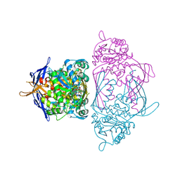
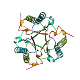
1M7X
 | | The X-ray Crystallographic Structure of Branching Enzyme | | 分子名称: | 1,4-alpha-glucan Branching Enzyme | | 著者 | Abad, M.C, Binderup, K, Rios-Steiner, J, Arni, R.K, Preiss, J, Geiger, J.H. | | 登録日 | 2002-07-23 | | 公開日 | 2002-09-18 | | 最終更新日 | 2024-05-22 | | 実験手法 | X-RAY DIFFRACTION (2.3 Å) | | 主引用文献 | The X-ray crystallographic structure of Escherichia coli branching enzyme
J.Biol.Chem., 277, 2002
|
|
7ML5
 | | Structure of the Starch Branching Enzyme I (BEI) complexed with maltododecaose from Oryza sativa L | | 分子名称: | Isoform 2 of 1,4-alpha-glucan-branching enzyme, chloroplastic/amyloplastic, alpha-D-glucopyranose-(1-4)-alpha-D-glucopyranose-(1-4)-alpha-D-glucopyranose-(1-4)-alpha-D-glucopyranose, ... | | 著者 | Nayebi Gavgani, H, Fawaz, R, Geiger, J.H. | | 登録日 | 2021-04-27 | | 公開日 | 2021-11-17 | | 最終更新日 | 2023-10-18 | | 実験手法 | X-RAY DIFFRACTION (2.35 Å) | | 主引用文献 | A structural explanation for the mechanism and specificity of plant branching enzymes I and IIb.
J.Biol.Chem., 298, 2021
|
|
7MS9
 | |
8D6H
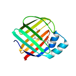
 | | Q108K:K40L:T51C:T53A:R58L:Q38F:Q4F mutant of hCRBPII bound to synthetic fluorophore CM1V after UV irradiation | | 分子名称: | (2E)-3-[7-(diethylamino)-2-oxo-2H-1-benzopyran-3-yl]prop-2-enal, bound form, ACETATE ION, ... | | 著者 | Bingham, C.R, Geiger, J.H, Borhan, B. | | 登録日 | 2022-06-06 | | 公開日 | 2023-02-01 | | 最終更新日 | 2023-10-25 | | 実験手法 | X-RAY DIFFRACTION (1.6 Å) | | 主引用文献 | Light controlled reversible Michael addition of cysteine: a new tool for dynamic site-specific labeling of proteins.
Analyst, 148, 2023
|
|
8D6N
 | | Q108K:K40L:T53A:R58L:Q38F:Q4F mutant of hCRBPII bound to synthetic fluorophore CM1V | | 分子名称: | (2E)-3-[7-(diethylamino)-2-oxo-2H-1-benzopyran-3-yl]prop-2-enal, bound form, ACETATE ION, ... | | 著者 | Bingham, C.R, Geiger, J.H, Borhan, B. | | 登録日 | 2022-06-06 | | 公開日 | 2023-02-01 | | 最終更新日 | 2023-10-25 | | 実験手法 | X-RAY DIFFRACTION (1.42 Å) | | 主引用文献 | Light controlled reversible Michael addition of cysteine: a new tool for dynamic site-specific labeling of proteins.
Analyst, 148, 2023
|
|
8DB2
 | | Q108K:K40L:T51C:T53A:R58L:Q38F mutant of hCRBPII bound to synthetic fluorophore CM1V | | 分子名称: | (2E)-3-[7-(diethylamino)-2-oxo-2H-1-benzopyran-3-yl]prop-2-enal, bound form, Retinol-binding protein 2 | | 著者 | Bingham, C.R, Geiger, J.H, Borhan, B, Staples, R. | | 登録日 | 2022-06-14 | | 公開日 | 2023-02-01 | | 最終更新日 | 2023-10-25 | | 実験手法 | X-RAY DIFFRACTION (1.5 Å) | | 主引用文献 | Light controlled reversible Michael addition of cysteine: a new tool for dynamic site-specific labeling of proteins.
Analyst, 148, 2023
|
|
8D6L
 | | Q108K:K40L:T51C:T53A:R58L:Q38F:Q4F mutant of hCRBPII bound to synthetic fluorophore CM1V | | 分子名称: | (2E)-3-[7-(diethylamino)-2-oxo-2H-1-benzopyran-3-yl]prop-2-enal, bound form, GLYCEROL, ... | | 著者 | Bingham, C.R, Geiger, J.H, Borhan, B. | | 登録日 | 2022-06-06 | | 公開日 | 2023-02-15 | | 最終更新日 | 2023-10-25 | | 実験手法 | X-RAY DIFFRACTION (1.69 Å) | | 主引用文献 | Light controlled reversible Michael addition of cysteine: a new tool for dynamic site-specific labeling of proteins.
Analyst, 148, 2023
|
|
1IG7
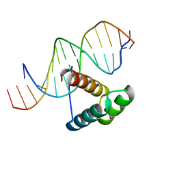 | | Msx-1 Homeodomain/DNA Complex Structure | | 分子名称: | 5'-D(*CP*AP*CP*TP*AP*AP*TP*TP*GP*AP*AP*GP*G)-3', 5'-D(P*TP*CP*CP*TP*TP*CP*AP*AP*TP*TP*AP*GP*TP*GP*AP*C)-3', Homeotic protein Msx-1 | | 著者 | Hovde, S, Abate-Shen, C, Geiger, J.H. | | 登録日 | 2001-04-17 | | 公開日 | 2001-04-23 | | 最終更新日 | 2024-04-03 | | 実験手法 | X-RAY DIFFRACTION (2.2 Å) | | 主引用文献 | Crystal structure of the Msx-1 homeodomain/DNA complex
Biochemistry, 40, 2001
|
|
8DN1
 | | Q108K:K40L:T51C:T53A:R58L:Q38F:Q4F mutant of hCRBPII bound to synthetic fluorophore CM1V at pH 7.2 | | 分子名称: | (2E)-3-[7-(diethylamino)-2-oxo-2H-1-benzopyran-3-yl]prop-2-enal, bound form, GLYCEROL, ... | | 著者 | Bingham, C.R, Borhan, B, Geiger, J.H. | | 登録日 | 2022-07-10 | | 公開日 | 2023-02-01 | | 最終更新日 | 2023-10-25 | | 実験手法 | X-RAY DIFFRACTION (1.32 Å) | | 主引用文献 | Light controlled reversible Michael addition of cysteine: a new tool for dynamic site-specific labeling of proteins.
Analyst, 148, 2023
|
|
1YTB
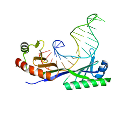 | | CRYSTAL STRUCTURE OF A YEAST TBP/TATA-BOX COMPLEX | | 分子名称: | DNA (29MER), PROTEIN (TATA BINDING PROTEIN (TBP)) | | 著者 | Kim, Y, Geiger, J.H, Hahn, S, Sigler, P.B. | | 登録日 | 1994-09-28 | | 公開日 | 1995-01-26 | | 最終更新日 | 2024-02-14 | | 実験手法 | X-RAY DIFFRACTION (1.8 Å) | | 主引用文献 | Crystal structure of a yeast TBP/TATA-box complex.
Nature, 365, 1993
|
|
1YP3
 | | Crystal structure of potato tuber ADP-glucose pyrophosphorylase in complex with ATP | | 分子名称: | ADENOSINE-5'-TRIPHOSPHATE, Glucose-1-phosphate adenylyltransferase small subunit, SULFATE ION | | 著者 | Jin, X, Ballicora, M.A, Preiss, J, Geiger, J.H. | | 登録日 | 2005-01-29 | | 公開日 | 2005-03-15 | | 最終更新日 | 2011-07-13 | | 実験手法 | X-RAY DIFFRACTION (2.6 Å) | | 主引用文献 | Crystal structure of potato tuber ADP-glucose pyrophosphorylase.
Embo J., 24, 2005
|
|
1YP2
 | | Crystal structure of potato tuber ADP-glucose pyrophosphorylase | | 分子名称: | Glucose-1-phosphate adenylyltransferase small subunit, PARA-MERCURY-BENZENESULFONIC ACID, SULFATE ION | | 著者 | Jin, X, Ballicora, M.A, Preiss, J, Geiger, J.H. | | 登録日 | 2005-01-29 | | 公開日 | 2005-03-15 | | 最終更新日 | 2011-07-13 | | 実験手法 | X-RAY DIFFRACTION (2.11 Å) | | 主引用文献 | Crystal structure of potato tuber ADP-glucose pyrophosphorylase.
Embo J., 24, 2005
|
|


