Loading
PDBj
MenuPDBj@FacebookPDBj@TwitterPDBj@YouTubewwPDB FoundationwwPDB
RCSB PDBPDBeBMRBAdv. SearchSearch help
PDB: 66 results

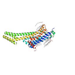
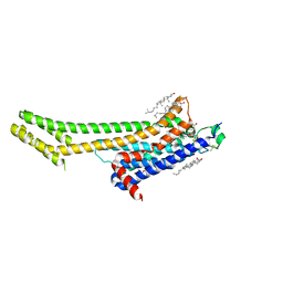
5UVI
DownloadVisualize
BU of 5uvi by Molmil
Serial Millisecond Crystallography of Membrane and Soluble Protein Micro-crystals using Synchrotron Radiation
Descriptor: (2R)-2,3-dihydroxypropyl (9Z)-octadec-9-enoate, 4-{2-[(7-amino-2-furan-2-yl[1,2,4]triazolo[1,5-a][1,3,5]triazin-5-yl)amino]ethyl}phenol, Adenosine receptor A2a,Soluble cytochrome b562,Adenosine receptor A2a, ...
Authors:Martin-Garcia, J.M, Conrad, C.E, Nelson, G, Stander, N, Zatsepin, N.A, Zook, J, Zhu, L, Geiger, J, Chun, E, Kissick, D, Hilgart, M.C, Ogata, C, Ishchenko, A, Nagaratnam, N, Roy-Chowdhury, S, Coe, J, Subramanian, G, Schaffer, A, James, D, Ketawala, G, Venugopalan, N, Xu, S, Corcoran, S, Ferguson, D, Weierstall, U, Spence, J.C.H, Cherezov, V, Fromme, P, Fischetti, R.F, Liu, W.
Deposit date:2017-02-20
Release date:2017-05-24
Last modified:2023-10-04
Method:X-RAY DIFFRACTION (3.2 Å)
Cite:Serial millisecond crystallography of membrane and soluble protein microcrystals using synchrotron radiation.
IUCrJ, 4, 2017
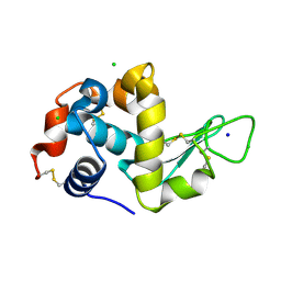
5UVJ
DownloadVisualize
BU of 5uvj by Molmil
Serial Millisecond Crystallography of Membrane and Soluble Protein Micro-crystals using Synchrotron Radiation
Descriptor: CHLORIDE ION, Lysozyme C, SODIUM ION
Authors:Martin-Garcia, J.M, Conrad, C.E, Nelson, G, Stander, N, Zatsepin, N.A, Zook, J, Zhu, L, Geiger, J, Chun, E, Kissick, D, Hilgart, M.C, Ogata, C, Ishchenko, A, Nagaratnam, N, Roy-Chowdhury, S, Coe, J, Subramanian, G, Schaffer, A, James, D, Ketawala, G, Venugopalan, N, Xu, S, Corcoran, S, Ferguson, D, Weierstall, U, Spence, J.C.H, Cherezov, V, Fromme, P, Fischetti, R.F, Liu, W.
Deposit date:2017-02-20
Release date:2017-05-24
Last modified:2023-10-04
Method:X-RAY DIFFRACTION (2.05 Å)
Cite:Serial millisecond crystallography of membrane and soluble protein microcrystals using synchrotron radiation.
IUCrJ, 4, 2017
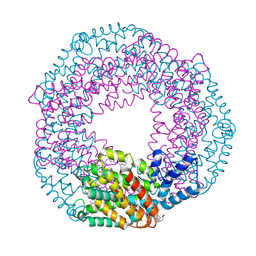
5UVK
DownloadVisualize
BU of 5uvk by Molmil
Serial Millisecond Crystallography of Membrane and Soluble Protein Micro-crystals using Synchrotron Radiation
Descriptor: C-phycocyanin alpha chain, C-phycocyanin beta chain, PHYCOCYANOBILIN
Authors:Martin-Garcia, J.M, Conrad, C.E, Nelson, G, Stander, N, Zatsepin, N.A, Zook, J, Zhu, L, Geiger, J, Chun, E, Kissick, D, Hilgart, M.C, Ogata, C, Ishchenko, A, Nagaratnam, N, Roy-Chowdhury, S, Coe, J, Subramanian, G, Schaffer, A, James, D, Ketawala, G, Venugopalan, N, Xu, S, Corcoran, S, Ferguson, D, Weierstall, U, Spence, J.C.H, Cherezov, V, Fromme, P, Fischetti, R.F, Liu, W.
Deposit date:2017-02-20
Release date:2017-05-24
Last modified:2023-10-04
Method:X-RAY DIFFRACTION (3.1 Å)
Cite:Serial millisecond crystallography of membrane and soluble protein microcrystals using synchrotron radiation.
Iucrj, 4, 2017
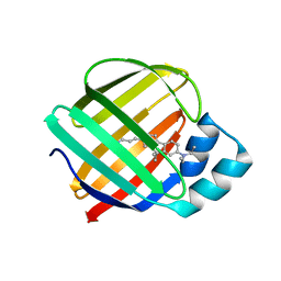
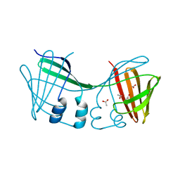
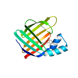
7MFX
DownloadVisualize
BU of 7mfx by Molmil
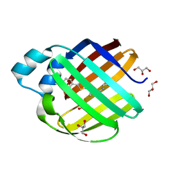
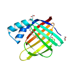
The Crystal Structure of Q108K:K40H:T53A:R58L:Q38F:Q4F Mutant of HCRBPII Bound with FR1 Chromophore Showing Excited State Intermolecular Proton Transfer
Descriptor: (4aP)-N,N-diethyl-9,9-dimethyl-7-[(1E)-prop-1-en-1-yl]-9H-fluoren-2-amine, GLYCEROL, Retinol-binding protein 2
Authors:Ghanbarpour, A, Geiger, J.
Deposit date:2021-04-11
Release date:2022-01-12
Last modified:2023-10-18
Method:X-RAY DIFFRACTION (1.59 Å)
Cite:Design of Large Stokes Shift Fluorescent Proteins Based on Excited State Proton Transfer of an Engineered Photobase.
J.Am.Chem.Soc., 143, 2021
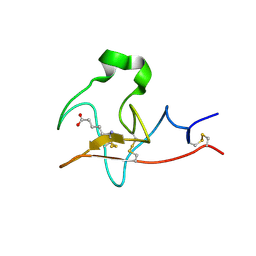
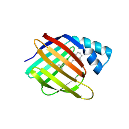
6DCM
DownloadVisualize
BU of 6dcm by Molmil
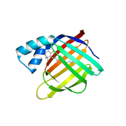
The atomic resolution crystal structure of Kringle 2 variant bound with EACA
Descriptor: 6-AMINOHEXANOIC ACID, Plasminogen
Authors:Ghanbarpour, A, Geiger, J.
Deposit date:2018-05-07
Release date:2019-05-15
Last modified:2024-10-16
Method:X-RAY DIFFRACTION (1.031 Å)
Cite:The atomic resolution crystal structure of Kringle 2 variant bound with EACA
To Be Published
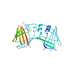
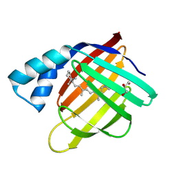
6E5E
DownloadVisualize
BU of 6e5e by Molmil
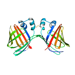
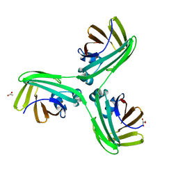
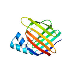
Crystal structure of the apo domain-swapped dimer Q108K:T51D mutant of human cellular retinol binding protein II
Descriptor: ACETATE ION, Retinol-binding protein 2
Authors:Ghanbarpour, A, Geiger, J.
Deposit date:2018-07-20
Release date:2019-10-16
Last modified:2023-10-11
Method:X-RAY DIFFRACTION (1.696 Å)
Cite:Engineering the hCRBPII Domain-Swapped Dimer into a New Class of Protein Switches.
J.Am.Chem.Soc., 141, 2019
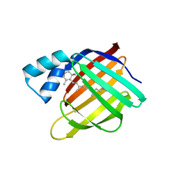
6E5Q
DownloadVisualize
BU of 6e5q by Molmil
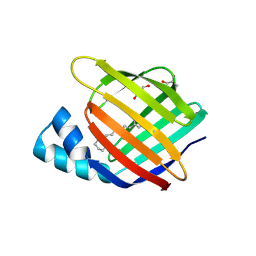
Crystal structure of the apo domain-swapped dimer Q108K:T51D:A28H mutant of human Cellular Retinol Binding Protein II
Descriptor: ACETATE ION, Retinol-binding protein 2
Authors:Ghanbarpour, A, Geiger, J.
Deposit date:2018-07-21
Release date:2019-10-16
Last modified:2023-10-11
Method:X-RAY DIFFRACTION (1.99 Å)
Cite:Engineering the hCRBPII Domain-Swapped Dimer into a New Class of Protein Switches.
J.Am.Chem.Soc., 141, 2019
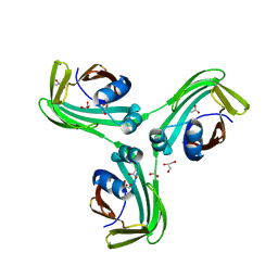
6VID
DownloadVisualize
BU of 6vid by Molmil
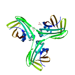
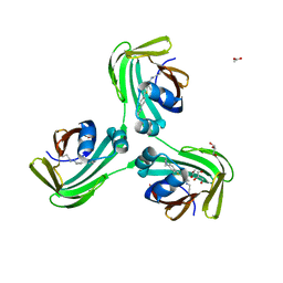
The Crystal Structure of Aps Domain-Swapped Trimer Q108K:K40D:T53A:R58L:Q38F:Q4F Variant of HCRBPII
Descriptor: ACETATE ION, GLYCEROL, Retinol-binding protein 2
Authors:Ghanbarpour, A, Geiger, J.
Deposit date:2020-01-12
Release date:2020-08-19
Last modified:2023-10-11
Method:X-RAY DIFFRACTION (2.89 Å)
Cite:The Crystal Structure of Apo Domain Swapped Trimer Q108K:K40D:T53A:R58L:Q38F:Q4F variant of HCRBPII
To Be Published
6WQA
DownloadVisualize
BU of 6wqa by Molmil
2.0A angstrom A2a adenosine receptor structure using XFEL data collected in helium atmosphere.
Descriptor: (2R)-2,3-dihydroxypropyl (9Z)-octadec-9-enoate, (2S)-2,3-dihydroxypropyl (9Z)-octadec-9-enoate, 4-{2-[(7-amino-2-furan-2-yl[1,2,4]triazolo[1,5-a][1,3,5]triazin-5-yl)amino]ethyl}phenol, ...
Authors:Lee, M.-Y, Geiger, J, Ishchenko, A, Han, G.W, Barty, A, White, T.A, Gati, C, Batyuk, A, Hunter, M.S, Aquila, A, Boutet, S, Weierstall, U, Cherezov, V, Liu, W.
Deposit date:2020-04-28
Release date:2020-11-18
Last modified:2023-10-18
Method:X-RAY DIFFRACTION (2 Å)
Cite:Harnessing the power of an X-ray laser for serial crystallography of membrane proteins crystallized in lipidic cubic phase
Iucrj, 7, 2020
6VIT
DownloadVisualize
BU of 6vit by Molmil
The Crystal Structure of Apo Domain-Swapped dimer Q108K:T51D:I32C Variant of HCRBPII with an Engineered Disulfide Bond
Descriptor: Retinol-binding protein 2
Authors:Ghanbarpour, A, Geiger, J.
Deposit date:2020-01-13
Release date:2020-08-19
Last modified:2024-10-16
Method:X-RAY DIFFRACTION (3.2 Å)
Cite:Human Cellular Retinol Binding Protein II Forms a Domain-Swapped Trimer Representing a Novel Fold and a New Template for Protein Engineering.
Chembiochem, 21, 2020
6VIS
DownloadVisualize
BU of 6vis by Molmil
The Crystal Structure of Domain-Swapped Trimer Q108K:K40D:T53A:R58L:Q38F:Q4F:V62E Variant of HCRBPII
Descriptor: GLYCEROL, Retinol-binding protein 2
Authors:Ghanbarpour, A, Geiger, J.
Deposit date:2020-01-13
Release date:2020-09-09
Last modified:2023-10-11
Method:X-RAY DIFFRACTION (2.79 Å)
Cite:Human Cellular Retinol Binding Protein II Forms a Domain-Swapped Trimer Representing a Novel Fold and a New Template for Protein Engineering.
Chembiochem, 21, 2020
6WNF
DownloadVisualize
BU of 6wnf by Molmil
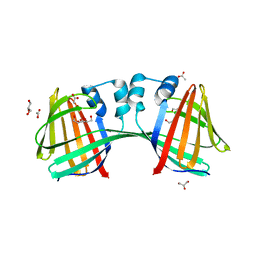
The Crystal Structure of Apo Domain-Swapped Dimer Q108K:K40D:T53A:R58L:Q38F:Q4F:F57H Variant of HCRBPII
Descriptor: ACETATE ION, GLYCEROL, Retinol-binding protein 2
Authors:Ghanbarpour, A, Geiger, J.
Deposit date:2020-04-22
Release date:2020-08-19
Last modified:2023-10-18
Method:X-RAY DIFFRACTION (1.67 Å)
Cite:Human Cellular Retinol Binding Protein II Forms a Domain-Swapped Trimer Representing a Novel Fold and a New Template for Protein Engineering.
Chembiochem, 21, 2020
6WNJ
DownloadVisualize
BU of 6wnj by Molmil
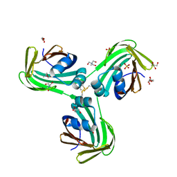
The Crystal Structure of Apo Domain-Swapped Trimer Q108K:T51D:A28C:I32C of HCRBPII
Descriptor: GLYCEROL, Retinol-binding protein 2, SULFATE ION, ...
Authors:Ghanbarpour, A, Geiger, J.
Deposit date:2020-04-22
Release date:2020-08-19
Last modified:2023-10-18
Method:X-RAY DIFFRACTION (2.1 Å)
Cite:Human Cellular Retinol Binding Protein II Forms a Domain-Swapped Trimer Representing a Novel Fold and a New Template for Protein Engineering.
Chembiochem, 21, 2020
6WP1
DownloadVisualize
BU of 6wp1 by Molmil
The Crystal Structure of Apo Domain-Swapped Trimer Q108K:K40L:T51K Variant of HCRBPII
Descriptor: ACETATE ION, GLYCEROL, Retinol-binding protein 2
Authors:Ghanbarpour, A, Geiger, J.
Deposit date:2020-04-26
Release date:2020-08-19
Last modified:2023-10-18
Method:X-RAY DIFFRACTION (2.99 Å)
Cite:Human Cellular Retinol Binding Protein II Forms a Domain-Swapped Trimer Representing a Novel Fold and a New Template for Protein Engineering.
Chembiochem, 21, 2020
7MFY
DownloadVisualize
BU of 7mfy by Molmil
The Crystal Structure of Q108K:K40L:T51V:T53S:R58W:Y19W:A33W:L117E Mutant of HCRBPII Bound with LizFluor
Descriptor: 4-{5-[(2E)-but-2-en-2-yl]thiophen-2-yl}-N,N-dimethylaniline, ACETATE ION, GLYCEROL, ...
Authors:Ghanbarpour, A, Geiger, J.
Deposit date:2021-04-12
Release date:2022-01-12
Last modified:2023-10-18
Method:X-RAY DIFFRACTION (1.26 Å)
Cite:Design of Large Stokes Shift Fluorescent Proteins Based on Excited State Proton Transfer of an Engineered Photobase.
J.Am.Chem.Soc., 143, 2021
7MFZ
DownloadVisualize
BU of 7mfz by Molmil
The Crystal Structure of Domain-Swapped Trimer Q108K:K40D:T53A:R58L:Q38F:Q4F Mutant of HCRBPII Bound with LizFluor3 Chromophore Showing Excited State Intermolecular Proton Transfer
Descriptor: 1-(4-{5-[(2E)-but-2-en-2-yl]thiophen-2-yl}phenyl)azetidine, ACETATE ION, GLYCEROL, ...
Authors:Ghanbarpour, A, Geiger, J.
Deposit date:2021-04-12
Release date:2022-01-12
Last modified:2024-10-09
Method:X-RAY DIFFRACTION (2.49 Å)
Cite:Design of Large Stokes Shift Fluorescent Proteins Based on Excited State Proton Transfer of an Engineered Photobase.
J.Am.Chem.Soc., 143, 2021
6MQJ
DownloadVisualize
BU of 6mqj by Molmil
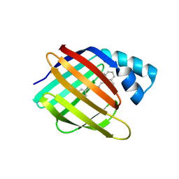
Crystal Structure of the All-Trans Retinal bound R111K:Y134F:T54V:R132Q:P39Y:R59Y:L121Q mutant of Human Cellular Retinoic Acid Binding Protein II Irradiated with 532 nm Laser for 10 minutes at 2.1 Angstrom Resolution
Descriptor: Cellular retinoic acid-binding protein 2, RETINAL
Authors:Ghanbarpour, A, Geiger, J.
Deposit date:2018-10-09
Release date:2019-01-09
Last modified:2024-10-30
Method:X-RAY DIFFRACTION (2.125 Å)
Cite:Mimicking Microbial Rhodopsin Isomerization in a Single Crystal.
J. Am. Chem. Soc., 141, 2019
6MOQ
DownloadVisualize
BU of 6moq by Molmil
Crystal Structure of the All-trans Retinal-Bound R111K:Y134F:T54V:R132Q:P39Y:R59Y:L121W Mutant of Human Cellular Retinoic Acid Binding Protein II in the Dark at 2.2 Angstrom Resolution
Descriptor: Cellular retinoic acid-binding protein 2, RETINAL
Authors:Ghanbarpour, A, Geiger, J.
Deposit date:2018-10-04
Release date:2019-01-09
Last modified:2023-10-11
Method:X-RAY DIFFRACTION (2.198 Å)
Cite:Mimicking Microbial Rhodopsin Isomerization in a Single Crystal.
J. Am. Chem. Soc., 141, 2019
6MOP
DownloadVisualize
BU of 6mop by Molmil
Crystal Structure of the All-trans Retinal-Bound R111K:Y134F:T54V:R132Q:P39Y:R59Y:L121E Mutant of Human Cellular Retinoic Acid Binding Protein II in the Dark at 1.9 Angstrom Resolution
Descriptor: Cellular retinoic acid-binding protein 2, RETINAL
Authors:Ghanbarpour, A, Geiger, J.
Deposit date:2018-10-04
Release date:2019-01-09
Last modified:2024-10-16
Method:X-RAY DIFFRACTION (1.9 Å)
Cite:Mimicking Microbial Rhodopsin Isomerization in a Single Crystal.
J. Am. Chem. Soc., 141, 2019
6MPK
DownloadVisualize
BU of 6mpk by Molmil
Crystal Structure of the 13-cis Retinal-Bound R111K:Y134F:T54V:R132Q:P39Y:R59Y:L121E:A32Y Mutant of Human Cellular Retinoic Acid Binding Protein II in the Dark at 1.58 Angstrom Resolution
Descriptor: Cellular retinoic acid-binding protein 2, GLYCEROL, RETINAL
Authors:Ghanbarpour, A, Geiger, J.
Deposit date:2018-10-07
Release date:2019-01-09
Last modified:2024-10-23
Method:X-RAY DIFFRACTION (1.58 Å)
Cite:Mimicking Microbial Rhodopsin Isomerization in a Single Crystal.
J. Am. Chem. Soc., 141, 2019
6MOV
DownloadVisualize
BU of 6mov by Molmil
Crystal Structure of the All-trans Retinal bound R111K:Y134F:T54V:R132Q:P39Y:R59Y:L121Q Human Cellular Retinoic Acid Binding Protein II in the Dark at 1.75 Angstrom Resolution
Descriptor: Cellular retinoic acid-binding protein 2, RETINAL
Authors:Ghanbarpour, A, Geiger, J.
Deposit date:2018-10-04
Release date:2019-01-09
Last modified:2023-10-11
Method:X-RAY DIFFRACTION (1.752 Å)
Cite:Mimicking Microbial Rhodopsin Isomerization in a Single Crystal.
J. Am. Chem. Soc., 141, 2019
6MQY
DownloadVisualize
BU of 6mqy by Molmil
Crystal Structure of the 13-cis Product of All-trans Retinal-Bound R111K:Y134F:T54V:R132Q:P39Y:R59Y:L121E Mutant of Human Cellular Retinoic Acid Binding Protein II Irradiated with 400 nm Laser (30 seconds) at 1.9 Angstrom
Descriptor: Cellular retinoic acid-binding protein 2, GLYCEROL, RETINAL
Authors:Ghanbarpour, A, Geiger, J.
Deposit date:2018-10-11
Release date:2019-01-09
Last modified:2024-10-23
Method:X-RAY DIFFRACTION (1.9 Å)
Cite:Mimicking Microbial Rhodopsin Isomerization in a Single Crystal.
J. Am. Chem. Soc., 141, 2019
6MOX
DownloadVisualize
BU of 6mox by Molmil
Crystal Structure of the All-trans Retinal-Bound R111K:Y134F:T54V:R132Q:P39Q:R59Y:L121E Human Cellular Retinoic Acid Binding Protein II Mutant in the Dark at 2.18 Angstrom Resolution
Descriptor: Cellular retinoic acid-binding protein 2, RETINAL
Authors:Ghanbarpour, A, Geiger, J.
Deposit date:2018-10-04
Release date:2019-01-09
Last modified:2024-10-16
Method:X-RAY DIFFRACTION (2.18 Å)
Cite:Mimicking Microbial Rhodopsin Isomerization in a Single Crystal.
J. Am. Chem. Soc., 141, 2019
6MR0
DownloadVisualize
BU of 6mr0 by Molmil
Crystal Structure of the All-trans Retinal Bound R111K:Y134F:T54V:R132Q:P39Q:R59Y:L121E Human Cellular Retinoic Acid Binding Protein II Mutant After 5 Minutes UV irradiation at 2.6 Angstrom Resolution
Descriptor: Cellular retinoic acid-binding protein 2, RETINAL
Authors:Ghanbarpour, A, Geiger, J.
Deposit date:2018-10-11
Release date:2019-01-09
Last modified:2024-10-16
Method:X-RAY DIFFRACTION (2.649 Å)
Cite:Mimicking Microbial Rhodopsin Isomerization in a Single Crystal.
J. Am. Chem. Soc., 141, 2019
6MQX
DownloadVisualize
BU of 6mqx by Molmil
Crystal Structure of All-trans Retinal-Bound R111K:Y134F:T54V:R132Q:P39Y:R59Y:L121E Human Cellular Retinoic Acid Binding Protein II Irradiated with 400 nm Laser (30 seconds) and Subsequently Dark Adapted (25 minutes) at 2.0 Angstrom Resolution
Descriptor: Cellular retinoic acid-binding protein 2, GLYCEROL, RETINAL
Authors:Ghanbarpour, A, Geiger, J.
Deposit date:2018-10-11
Release date:2019-01-09
Last modified:2023-10-11
Method:X-RAY DIFFRACTION (2.01 Å)
Cite:Mimicking Microbial Rhodopsin Isomerization in a Single Crystal.
J. Am. Chem. Soc., 141, 2019

226707

PDB entries from 2024-10-30

PDB statisticsPDBj update infoContact PDBjnumon