Loading
PDBj
MenuPDBj@FacebookPDBj@TwitterPDBj@YouTubewwPDB FoundationwwPDB
RCSB PDBPDBeBMRBAdv. SearchSearch help
PDB: 187 results

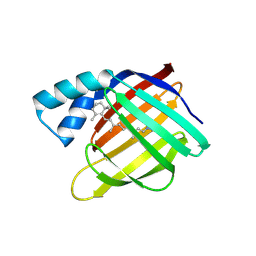
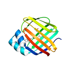
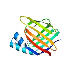
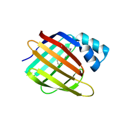
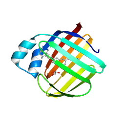
6MQZ
DownloadVisualize
BU of 6mqz by Molmil
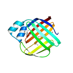
Crystal Structure of the 13-cis Product of the All-trans Retinal-Bound R111K:Y134F:T54V:R132Q:P39Y:R59Y:L121E Mutant of Human Cellular Retinoic Acid Binding Protein II Irradiated with 400 nm Laser (5 minutes) at 2.07 Angstrom
Descriptor: Cellular retinoic acid-binding protein 2, GLYCEROL, RETINAL
Authors:Ghanbarpour, A, Geiger, J.
Deposit date:2018-10-11
Release date:2019-01-09
Last modified:2023-10-11
Method:X-RAY DIFFRACTION (2.07 Å)
Cite:Mimicking Microbial Rhodopsin Isomerization in a Single Crystal.
J. Am. Chem. Soc., 141, 2019
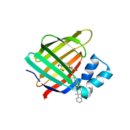
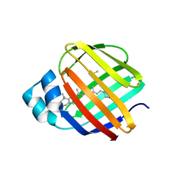
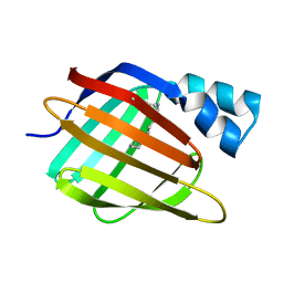
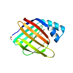
6MOR
DownloadVisualize
BU of 6mor by Molmil
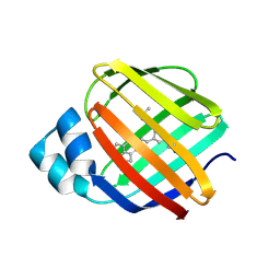
Crystal Structure of the All-Trans Retinal-Bound R111K:Y134F:T54V:R132Q:P39Y:R59Y:L121Y Mutant of Human Cellular Retinoic Acid Binding Protein II in the Dark at 1.79 Angstrom Resolution
Descriptor: Cellular retinoic acid-binding protein 2, RETINAL
Authors:Ghanbarpour, A, Geiger, J.
Deposit date:2018-10-04
Release date:2019-01-09
Last modified:2023-10-11
Method:X-RAY DIFFRACTION (1.79 Å)
Cite:Mimicking Microbial Rhodopsin Isomerization in a Single Crystal.
J. Am. Chem. Soc., 141, 2019
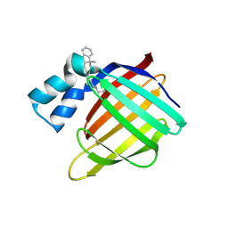
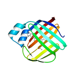
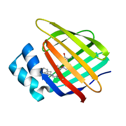
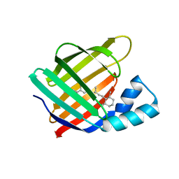
6MQW
DownloadVisualize
BU of 6mqw by Molmil
Crystal Structure of All-trans Retinal-Bound R111K:Y134F:T54V:R132Q:P39Y:R59Y:L121E Human Cellular Retinoic Acid Binding Protein II Irradiated with 400 nm laser (30 seconds) and subsequently dark adapted (10 minutes) at 2.1 Angstrom Resolution
Descriptor: Cellular retinoic acid-binding protein 2, RETINAL
Authors:Ghanbarpour, A, Geiger, J.
Deposit date:2018-10-11
Release date:2019-01-09
Last modified:2023-10-11
Method:X-RAY DIFFRACTION (2.09 Å)
Cite:Mimicking Microbial Rhodopsin Isomerization in a Single Crystal.
J. Am. Chem. Soc., 141, 2019
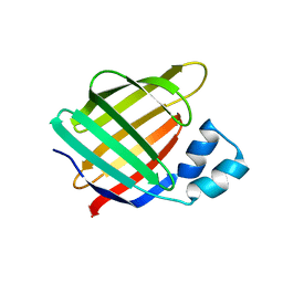
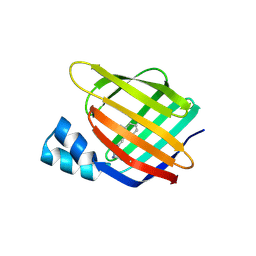
4QGW
DownloadVisualize
BU of 4qgw by Molmil
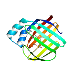
Crystal sturcture of the R132K:R111L:L121D mutant of Cellular Retinoic Acid Binding ProteinII complexed with a synthetic ligand (Merocyanine) at 1.77 angstrom resolution
Descriptor: (2E,4E,6E)-3-methyl-6-(1,3,3-trimethyl-1,3-dihydro-2H-indol-2-ylidene)hexa-2,4-dienal, 2-[3-(2-HYDROXY-1,1-DIHYDROXYMETHYL-ETHYLAMINO)-PROPYLAMINO]-2-HYDROXYMETHYL-PROPANE-1,3-DIOL, Cellular retinoic acid-binding protein 2
Authors:Nosrati, M, Yapici, I, Geiger, J.H.
Deposit date:2014-05-26
Release date:2015-01-28
Last modified:2015-02-11
Method:X-RAY DIFFRACTION (1.77 Å)
Cite:"Turn-on" protein fluorescence: in situ formation of cyanine dyes.
J.Am.Chem.Soc., 137, 2015
4QGX
DownloadVisualize
BU of 4qgx by Molmil
Crystal structure of the R132K:R111L:L121E mutant of Cellular Retinoic Acid Binding ProteinII complexed with a synthetic ligand (Merocyanine) at 1.47 angstrom resolution
Descriptor: (2E,4E,6E)-3-methyl-6-(1,3,3-trimethyl-1,3-dihydro-2H-indol-2-ylidene)hexa-2,4-dienal, Cellular retinoic acid-binding protein 2
Authors:Nosrati, M, Yapici, I, Geiger, J.H.
Deposit date:2014-05-26
Release date:2015-01-28
Last modified:2023-09-20
Method:X-RAY DIFFRACTION (1.471 Å)
Cite:"Turn-on" protein fluorescence: in situ formation of cyanine dyes.
J.Am.Chem.Soc., 137, 2015
4QGV
DownloadVisualize
BU of 4qgv by Molmil
Crystal structure of the R132K:R111L mutant of Cellular Retinoic Acid Binding ProteinII complexed with a synthetic ligand (Merocyanine) at 1.73 angstrom resolution.
Descriptor: (2E,4E,6E)-3-methyl-6-(1,3,3-trimethyl-1,3-dihydro-2H-indol-2-ylidene)hexa-2,4-dienal, Cellular retinoic acid-binding protein 2
Authors:Nosrati, M, Yapici, I, Geiger, J.H.
Deposit date:2014-05-25
Release date:2015-01-28
Last modified:2023-09-20
Method:X-RAY DIFFRACTION (1.73 Å)
Cite:"Turn-on" protein fluorescence: in situ formation of cyanine dyes.
J.Am.Chem.Soc., 137, 2015
6ON5
DownloadVisualize
BU of 6on5 by Molmil
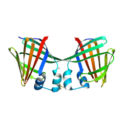
Crystal Structure of the Zn-bound Domain-Swapped Dimer Q108K:T51D:A28C:L36C:F57H Mutant of Human Cellular Retinol Binding Protein II
Descriptor: Retinol-binding protein 2, ZINC ION
Authors:Ghanbarpour, A, Geiger, J.
Deposit date:2019-04-20
Release date:2019-10-16
Last modified:2023-10-11
Method:X-RAY DIFFRACTION (1.638 Å)
Cite:Engineering the hCRBPII Domain-Swapped Dimer into a New Class of Protein Switches.
J.Am.Chem.Soc., 141, 2019
6ON8
DownloadVisualize
BU of 6on8 by Molmil
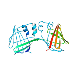
Crystal Structure of the Reduced Form of Apo Domain-Swapped Dimer Q108K:T51D:A28C:L36C:F57H Mutant of Human Cellular Retinol Binding Protein II
Descriptor: Retinol-binding protein 2
Authors:Ghanbarpour, A, Geiger, J.
Deposit date:2019-04-20
Release date:2019-10-16
Last modified:2023-10-11
Method:X-RAY DIFFRACTION (2.395 Å)
Cite:Engineering the hCRBPII Domain-Swapped Dimer into a New Class of Protein Switches.
J.Am.Chem.Soc., 141, 2019
5F58
DownloadVisualize
BU of 5f58 by Molmil
Crystal structure of the Q108K:K40L:T51V:R58F mutant of human Cellular Retinol Binding Protein II in complex with All-trans-Retinal after 24 hours of incubation at 1.54 Angstrom Resolution
Descriptor: ACETATE ION, RETINAL, Retinol-binding protein 2
Authors:Nosrati, M, Nossoni, Z, Geiger, J.H.
Deposit date:2015-12-04
Release date:2016-12-07
Last modified:2023-09-27
Method:X-RAY DIFFRACTION (1.54 Å)
Cite:Crystal structure of the Q108K:K40L:T51V:R58F mutant of human Cellular Retinol Binding Protein II in complex with All-trans-Retinal after 24 hours of incubation at 1.54 Angstrom Resolution
To Be Published
5F6B
DownloadVisualize
BU of 5f6b by Molmil
Crystal structure of the Q108K:K40L:T51V:R58Y:Y19W mutant of human Cellular Retinol Binding Protein II in complex with All-trans-Retinal at 1.3 Angstrom Resolution
Descriptor: ACETATE ION, RETINAL, Retinol-binding protein 2
Authors:Nosrati, M, Nossoni, Z, Geiger, J.H.
Deposit date:2015-12-05
Release date:2016-12-07
Last modified:2023-09-27
Method:X-RAY DIFFRACTION (1.31 Å)
Cite:Crystal structure of the Q108K:K40L:T51V:R58Y:Y19W mutant of human Cellular Retinol Binding ProteinII in complex with All-trans-Retinal at 1.31 Angstrom Resolution
To Be Published
5FAZ
DownloadVisualize
BU of 5faz by Molmil
Crystal structure of the Q108K:K40L:T51V mutant of human Cellular Retinol Binding Protein II in complex with All-trans-Retinal after 24 hours of incubation at 1.4 Angstrom Resolution
Descriptor: ACETATE ION, RETINAL, Retinol-binding protein 2
Authors:Nosrati, M, Nossoni, Z, Geiger, J.H.
Deposit date:2015-12-13
Release date:2016-12-14
Last modified:2023-09-27
Method:X-RAY DIFFRACTION (1.4 Å)
Cite:Crystal structure of the Q108K:K40L:T51V mutant of human Cellular Retinol Binding Protein II in complex with All-trans-Retinal after 24 hours of incubation at 1.54 Angstrom Resolution
To Be Published
6C7Z
DownloadVisualize
BU of 6c7z by Molmil
Crystal structure of the Q108K:K40L:T51V:R58F mutant of human Cellular Retinol Binding Protein II in complex with synthetic Ligand Julolidine
Descriptor: (2E,4E)-3-methyl-5-(2,3,6,7-tetrahydro-1H,5H-pyrido[3,2,1-ij]quinolin-9-yl)penta-2,4-dienal, ACETATE ION, Retinol-binding protein 2
Authors:Nosrati, M, Geiger, J.H.
Deposit date:2018-01-23
Release date:2018-04-25
Last modified:2023-10-04
Method:X-RAY DIFFRACTION (1.42 Å)
Cite:A Genetically Encoded Ratiometric pH Probe: Wavelength Regulation-Inspired Design of pH Indicators.
Chembiochem, 19, 2018
5FEN
DownloadVisualize
BU of 5fen by Molmil
Crystal structure of the Q108K:K40L:T53C mutant of human Cellular Retinol Binding Protein II in complex with All-trans-Retinal after 24 hours of incubation at 1.55 Angstrom Resolution
Descriptor: ACETATE ION, RETINAL, Retinol-binding protein 2
Authors:Nossoni, Z, Nosrati, M, Geiger, J.H.
Deposit date:2015-12-17
Release date:2016-12-21
Last modified:2023-09-27
Method:X-RAY DIFFRACTION (1.55 Å)
Cite:Crystal structure of the Q108K:K40L:T53C mutant of human Cellular Retinol Binding Protein II in complex with All-trans-Retinal after 24 hours of incubation at 1.55 Angstrom Resolution
To Be Published
5FFH
DownloadVisualize
BU of 5ffh by Molmil
Crystal structure of the Q108K:K40L:T51V:R58W:Y19W mutant of human Cellular Retinol Binding Protein II in complex with All-trans-Retinal at 1.68 Angstrom Resolution
Descriptor: ACETATE ION, RETINAL, Retinol-binding protein 2
Authors:Nossoni, Z, Nosrati, M, Geiger, J.H.
Deposit date:2015-12-18
Release date:2016-12-21
Last modified:2023-09-27
Method:X-RAY DIFFRACTION (1.68 Å)
Cite:Crystal structure of the Q108K:K40L:T51V:R58W:Y19W mutant of human Cellular Retinol Binding ProteinII in complex with All-trans-Retinal at 1.68 Angstrom Resolution
To Be Published
5F7G
DownloadVisualize
BU of 5f7g by Molmil
Crystal structure of the Q108K:K40L:T51V:R58Y:Y19W:Q38L mutant of human Cellular Retinol Binding Protein II in complex with All-trans-Retinal at 1.48 Angstrom Resolution
Descriptor: ACETATE ION, RETINAL, Retinol-binding protein 2
Authors:Nosrati, M, Nossoni, Z, Geiger, J.H.
Deposit date:2015-12-08
Release date:2016-12-14
Last modified:2023-09-27
Method:X-RAY DIFFRACTION (1.48 Å)
Cite:Crystal structure of the Q108K:K40L:T51V:R58F mutant of human Cellular Retinol Binding Protein II in complex with All-trans-Retinal after 24 hours of incubation at 1.54 Angstrom Resolution
To Be Published
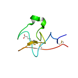
6DCM
DownloadVisualize
BU of 6dcm by Molmil
The atomic resolution crystal structure of Kringle 2 variant bound with EACA
Descriptor: 6-AMINOHEXANOIC ACID, Plasminogen
Authors:Ghanbarpour, A, Geiger, J.
Deposit date:2018-05-07
Release date:2019-05-15
Last modified:2023-11-15
Method:X-RAY DIFFRACTION (1.031 Å)
Cite:The atomic resolution crystal structure of Kringle 2 variant bound with EACA
To Be Published
4YKO
DownloadVisualize
BU of 4yko by Molmil
Crystal structure of the R111K:Y134F:T54V:R132Q:P39Q:R59Y:A32Y:F3Q mutant of human Cellular Retinoic Acid Binding Protein II with Retinal at 1.58 angstrom resolution
Descriptor: Cellular retinoic acid-binding protein 2, RETINAL
Authors:Nosrati, M, Geiger, J.
Deposit date:2015-03-04
Release date:2016-03-09
Last modified:2023-09-27
Method:X-RAY DIFFRACTION (1.57 Å)
Cite:A Photoisomerizing Rhodopsin Mimic Observed at Atomic Resolution.
J.Am.Chem.Soc., 138, 2016
3CR6
DownloadVisualize
BU of 3cr6 by Molmil
Crystal Structure of the R132K:R111L:A32E Mutant of Cellular Retinoic Acid Binding Protein Type II Complexed with C15-aldehyde (a retinal analog) at 1.22 Angstrom resolution.
Descriptor: 1,3,3-trimethyl-2-[(1E,3E)-3-methylpenta-1,3-dien-1-yl]cyclohexene, Cellular retinoic acid-binding protein 2
Authors:Jia, X, Geiger, J.H.
Deposit date:2008-04-04
Release date:2009-03-17
Last modified:2023-08-30
Method:X-RAY DIFFRACTION (1.22 Å)
Cite:Two distinctive orientations of binding determined by a single mutation in the CRABPII mutant-C15-aldehyde complexes
To be Published
3F8A
DownloadVisualize
BU of 3f8a by Molmil
Crystal Structure of the R132K:R111L:L121E:R59W Mutant of Cellular Retinoic Acid-Binding Protein Type II Complexed with C15-aldehyde (a retinal analog) at 1.95 Angstrom resolution.
Descriptor: 1,3,3-trimethyl-2-[(1E,3E)-3-methylpenta-1,3-dien-1-yl]cyclohexene, 2-[3-(2-HYDROXY-1,1-DIHYDROXYMETHYL-ETHYLAMINO)-PROPYLAMINO]-2-HYDROXYMETHYL-PROPANE-1,3-DIOL, Cellular retinoic acid-binding protein 2
Authors:Jia, X, Geiger, J.H.
Deposit date:2008-11-12
Release date:2009-11-10
Last modified:2023-09-06
Method:X-RAY DIFFRACTION (1.95 Å)
Cite:Probing Wavelength Regulation with an Engineered Rhodopsin Mimic and a C15-Retinal Analogue
Chempluschem, 77, 2012
3FA6
DownloadVisualize
BU of 3fa6 by Molmil
Crystal structure of the R132K:Y134F:R111L:L121D:T54V mutant of cellular retinoic acid-binding protein II complexed with C15-aldehyde (a retinal analog) at 1.54 angstrom resolution
Descriptor: 1,3,3-trimethyl-2-[(1E,3E)-3-methylpenta-1,3-dien-1-yl]cyclohexene, Cellular retinoic acid-binding protein 2
Authors:Jia, X, Geiger, J.H.
Deposit date:2008-11-15
Release date:2009-10-27
Last modified:2023-12-27
Method:X-RAY DIFFRACTION (1.54 Å)
Cite:Two distinctive orientations of binding determined by a single mutation in the CRABPII mutant-C15-aldehyde complexes
To be Published
3F9D
DownloadVisualize
BU of 3f9d by Molmil
Crystal structure of the R132K:R111L:T54E mutant of cellular retinoic acid-binding protein II complexed with C15-aldehyde (a retinal analog) at 2.00 angstrom resolution
Descriptor: 1,3,3-trimethyl-2-[(1E,3E)-3-methylpenta-1,3-dien-1-yl]cyclohexene, Cellular retinoic acid-binding protein 2
Authors:Jia, X, Geiger, J.H.
Deposit date:2008-11-13
Release date:2009-10-27
Last modified:2023-09-06
Method:X-RAY DIFFRACTION (2 Å)
Cite:Two distinctive orientations of binding determined by a single mutation in the CRABPII mutant-C15-aldehyde complexes
To be Published
3D1J
DownloadVisualize
BU of 3d1j by Molmil
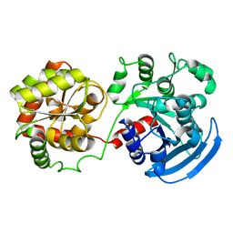
Crystal Structure of E.coli GS mutant dmGS(C7S;C408S)
Descriptor: Glycogen synthase
Authors:Sheng, F, Geiger, J.H.
Deposit date:2008-05-06
Release date:2009-03-24
Last modified:2023-08-30
Method:X-RAY DIFFRACTION (3 Å)
Cite:The Crystal Structures of the Open and Catalytically Competent Closed Conformation of Escherichia coli Glycogen Synthase.
J.Biol.Chem., 284, 2009
3COP
DownloadVisualize
BU of 3cop by Molmil
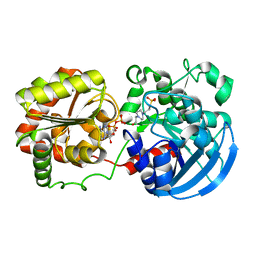
Crystal Structure of E.coli GS mutant E377A in complex with ADP and acceptor analogue HEPPSO
Descriptor: (2R)-2-hydroxy-3-[4-(2-hydroxyethyl)piperazin-1-yl]propane-1-sulfonic acid, ADENOSINE-5'-DIPHOSPHATE, Glycogen synthase, ...
Authors:Sheng, F, Geiger, J.H.
Deposit date:2008-03-28
Release date:2009-03-24
Last modified:2023-08-30
Method:X-RAY DIFFRACTION (2.3 Å)
Cite:The Crystal Structures of the Open and Catalytically Competent Closed Conformation of Escherichia coli Glycogen Synthase.
J.Biol.Chem., 284, 2009
7LHJ
DownloadVisualize
BU of 7lhj by Molmil
Crystal structure of Q108K:K40L:T51V:T53C:R58W:T29L:Y19W:Q4A mutant of cellular retinol binding protein II complex with 15-cis-retinal
Descriptor: ACETATE ION, GLYCEROL, RETINAL, ...
Authors:Ehyaei, N, Geiger, J.H, Borhan, B.
Deposit date:2021-01-25
Release date:2021-02-10
Last modified:2023-10-18
Method:X-RAY DIFFRACTION (1.26 Å)
Cite:Crystal structure of Q108K:K40L:T51V:T53C:R58W:T29L:Y19W:Q4A mutant of cellular retinol binding protein II complex with 15cis- retinal
To Be Published
7LHN
DownloadVisualize
BU of 7lhn by Molmil
Crystal structure of Q108K:K40L:T51V:T53C:R58W:T29L:Y19W:Q4A mutant of cellular retinol binding protein II complex with all-trans-retinal after exposure to visible light
Descriptor: GLYCEROL, RETINAL, Retinol-binding protein 2
Authors:Ehyaei, N, Geiger, J.H, Borhan, B.
Deposit date:2021-01-25
Release date:2021-04-14
Last modified:2023-10-18
Method:X-RAY DIFFRACTION (2.11 Å)
Cite:Crystal structure of Q108K:K40L:T51V:T53C:R58W:T29L:Y19W:Q4A mutant of cellular retinol binding protein II complex with all-trans-retinal after exposure to visible light
To Be Published

220472

数据于2024-05-29公开中

PDB statisticsPDBj update infoContact PDBjnumon