4M6S
 
 | |
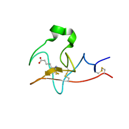
1IG7
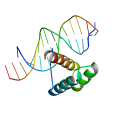 | | Msx-1 Homeodomain/DNA Complex Structure | | 分子名称: | 5'-D(*CP*AP*CP*TP*AP*AP*TP*TP*GP*AP*AP*GP*G)-3', 5'-D(P*TP*CP*CP*TP*TP*CP*AP*AP*TP*TP*AP*GP*TP*GP*AP*C)-3', Homeotic protein Msx-1 | | 著者 | Hovde, S, Abate-Shen, C, Geiger, J.H. | | 登録日 | 2001-04-17 | | 公開日 | 2001-04-23 | | 最終更新日 | 2024-04-03 | | 実験手法 | X-RAY DIFFRACTION (2.2 Å) | | 主引用文献 | Crystal structure of the Msx-1 homeodomain/DNA complex
Biochemistry, 40, 2001
|
|
4LQ1
 | |
4QZT
 | |
1P1K
 | |
1P1H
 | |
4GKC
 | |
4EDE
 | |
4EEJ
 | |
1P1J
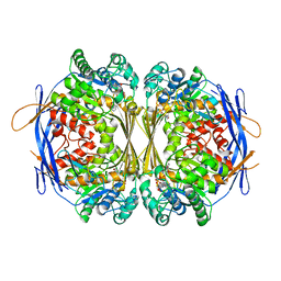
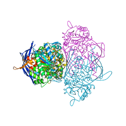 | | Crystal structure of the 1L-myo-inositol 1-phosphate synthase complexed with NADH | | 分子名称: | 1,4-DIHYDRONICOTINAMIDE ADENINE DINUCLEOTIDE, GLYCEROL, Inositol-3-phosphate synthase, ... | | 著者 | Jin, X, Geiger, J.H. | | 登録日 | 2003-04-12 | | 公開日 | 2003-07-08 | | 最終更新日 | 2024-02-14 | | 実験手法 | X-RAY DIFFRACTION (1.7 Å) | | 主引用文献 | Structures of NAD(+)- and NADH-bound 1-l-myo-inositol 1-phosphate synthase.
Acta Crystallogr.,Sect.D, 59, 2003
|
|
7LHM
 | |
1P1F
 | |
1P1I
 | |
6VID
 | |
6WQA
 | | 2.0A angstrom A2a adenosine receptor structure using XFEL data collected in helium atmosphere. | | 分子名称: | (2R)-2,3-dihydroxypropyl (9Z)-octadec-9-enoate, (2S)-2,3-dihydroxypropyl (9Z)-octadec-9-enoate, 4-{2-[(7-amino-2-furan-2-yl[1,2,4]triazolo[1,5-a][1,3,5]triazin-5-yl)amino]ethyl}phenol, ... | | 著者 | Lee, M.-Y, Geiger, J, Ishchenko, A, Han, G.W, Barty, A, White, T.A, Gati, C, Batyuk, A, Hunter, M.S, Aquila, A, Boutet, S, Weierstall, U, Cherezov, V, Liu, W. | | 登録日 | 2020-04-28 | | 公開日 | 2020-11-18 | | 最終更新日 | 2024-11-13 | | 実験手法 | X-RAY DIFFRACTION (2 Å) | | 主引用文献 | Harnessing the power of an X-ray laser for serial crystallography of membrane proteins crystallized in lipidic cubic phase
Iucrj, 7, 2020
|
|
7LHJ
 | |
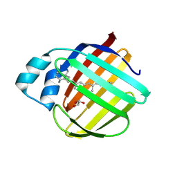
4ZJ0
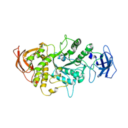
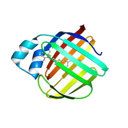
 | | The crystal structure of monomer Q108K:K40L:Y60W CRBPII bound to all-trans-retinal | | 分子名称: | ACETATE ION, RETINAL, Retinol-binding protein 2 | | 著者 | Nossoni, Z, Assar, Z, Wang, W, Vasileiou, C, Borhan, B, Geiger, J.H. | | 登録日 | 2015-04-28 | | 公開日 | 2016-05-11 | | 最終更新日 | 2024-11-20 | | 実験手法 | X-RAY DIFFRACTION (1.5 Å) | | 主引用文献 | Domain-Swapped Dimers of Intracellular Lipid-Binding Proteins: Evidence for Ordered Folding Intermediates.
Structure, 24, 2016
|
|
7LHO
 | |
7LHN
 | |
6DCM
 | |
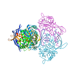
5E70
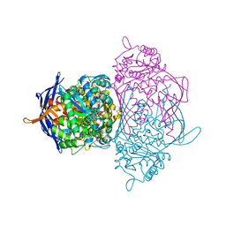
 | | Crystal structure of Ecoli Branching Enzyme with gamma cyclodextrin | | 分子名称: | 1,4-alpha-glucan branching enzyme GlgB, Cyclooctakis-(1-4)-(alpha-D-glucopyranose), GLYCEROL | | 著者 | Feng, L, Nosrati, M, Geiger, J.H. | | 登録日 | 2015-10-11 | | 公開日 | 2015-12-16 | | 最終更新日 | 2023-09-27 | | 実験手法 | X-RAY DIFFRACTION (2.33 Å) | | 主引用文献 | Crystal structures of Escherichia coli branching enzyme in complex with cyclodextrins.
Acta Crystallogr D Struct Biol, 72, 2016
|
|
4QGW
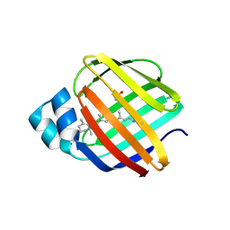
 | | Crystal sturcture of the R132K:R111L:L121D mutant of Cellular Retinoic Acid Binding ProteinII complexed with a synthetic ligand (Merocyanine) at 1.77 angstrom resolution | | 分子名称: | (2E,4E,6E)-3-methyl-6-(1,3,3-trimethyl-1,3-dihydro-2H-indol-2-ylidene)hexa-2,4-dienal, 2-[3-(2-HYDROXY-1,1-DIHYDROXYMETHYL-ETHYLAMINO)-PROPYLAMINO]-2-HYDROXYMETHYL-PROPANE-1,3-DIOL, Cellular retinoic acid-binding protein 2 | | 著者 | Nosrati, M, Yapici, I, Geiger, J.H. | | 登録日 | 2014-05-26 | | 公開日 | 2015-01-28 | | 最終更新日 | 2015-02-11 | | 実験手法 | X-RAY DIFFRACTION (1.77 Å) | | 主引用文献 | "Turn-on" protein fluorescence: in situ formation of cyanine dyes.
J.Am.Chem.Soc., 137, 2015
|
|
4QGX
 | | Crystal structure of the R132K:R111L:L121E mutant of Cellular Retinoic Acid Binding ProteinII complexed with a synthetic ligand (Merocyanine) at 1.47 angstrom resolution | | 分子名称: | (2E,4E,6E)-3-methyl-6-(1,3,3-trimethyl-1,3-dihydro-2H-indol-2-ylidene)hexa-2,4-dienal, Cellular retinoic acid-binding protein 2 | | 著者 | Nosrati, M, Yapici, I, Geiger, J.H. | | 登録日 | 2014-05-26 | | 公開日 | 2015-01-28 | | 最終更新日 | 2024-10-09 | | 実験手法 | X-RAY DIFFRACTION (1.471 Å) | | 主引用文献 | "Turn-on" protein fluorescence: in situ formation of cyanine dyes.
J.Am.Chem.Soc., 137, 2015
|
|
6C7Z
 | | Crystal structure of the Q108K:K40L:T51V:R58F mutant of human Cellular Retinol Binding Protein II in complex with synthetic Ligand Julolidine | | 分子名称: | (2E,4E)-3-methyl-5-(2,3,6,7-tetrahydro-1H,5H-pyrido[3,2,1-ij]quinolin-9-yl)penta-2,4-dienal, ACETATE ION, Retinol-binding protein 2 | | 著者 | Nosrati, M, Geiger, J.H. | | 登録日 | 2018-01-23 | | 公開日 | 2018-04-25 | | 最終更新日 | 2024-11-13 | | 実験手法 | X-RAY DIFFRACTION (1.42 Å) | | 主引用文献 | A Genetically Encoded Ratiometric pH Probe: Wavelength Regulation-Inspired Design of pH Indicators.
Chembiochem, 19, 2018
|
|
4QGV
 | | Crystal structure of the R132K:R111L mutant of Cellular Retinoic Acid Binding ProteinII complexed with a synthetic ligand (Merocyanine) at 1.73 angstrom resolution. | | 分子名称: | (2E,4E,6E)-3-methyl-6-(1,3,3-trimethyl-1,3-dihydro-2H-indol-2-ylidene)hexa-2,4-dienal, Cellular retinoic acid-binding protein 2 | | 著者 | Nosrati, M, Yapici, I, Geiger, J.H. | | 登録日 | 2014-05-25 | | 公開日 | 2015-01-28 | | 最終更新日 | 2023-09-20 | | 実験手法 | X-RAY DIFFRACTION (1.73 Å) | | 主引用文献 | "Turn-on" protein fluorescence: in situ formation of cyanine dyes.
J.Am.Chem.Soc., 137, 2015
|
|


