6CCD
 
 | |
2H34
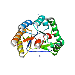
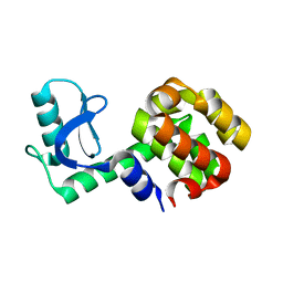
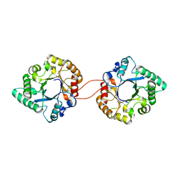
 | | Apoenzyme crystal structure of the tuberculosis serine/threonine kinase, PknE | | 分子名称: | BROMIDE ION, SODIUM ION, Serine/threonine-protein kinase pknE | | 著者 | Gay, L.M, Ng, H.L, Alber, T. | | 登録日 | 2006-05-22 | | 公開日 | 2006-07-18 | | 最終更新日 | 2017-10-18 | | 実験手法 | X-RAY DIFFRACTION (2.8 Å) | | 主引用文献 | A Conserved Dimer and Global Conformational Changes in the Structure of apo-PknE Ser/Thr Protein Kinase from Mycobacterium tuberculosis.
J.Mol.Biol., 360, 2006
|
|
1T6O
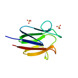
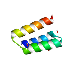
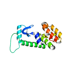
 | | Nucleocapsid-binding domain of the measles virus P protein (amino acids 457-507) in complex with amino acids 486-505 of the measles virus N protein | | 分子名称: | linker, phosphoprotein | | 著者 | Kingston, R.L, Hamel, D.J, Gay, L.S, Dahlquist, F.W, Matthews, B.W. | | 登録日 | 2004-05-06 | | 公開日 | 2004-08-03 | | 最終更新日 | 2023-08-23 | | 実験手法 | X-RAY DIFFRACTION (2 Å) | | 主引用文献 | Structural basis for the attachment of a paramyxoviral polymerase to its template.
Proc.Natl.Acad.Sci.USA, 101, 2004
|
|
2Y8U
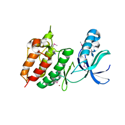
 | | A. nidulans chitin deacetylase | | 分子名称: | CHITIN DEACETYLASE, CHLORIDE ION, COBALT (II) ION, ... | | 著者 | Penman, G, Gay, L.M, van Aalten, D.M.F. | | 登録日 | 2011-02-10 | | 公開日 | 2012-02-22 | | 最終更新日 | 2024-05-08 | | 実験手法 | X-RAY DIFFRACTION (1.99 Å) | | 主引用文献 | Structure and function of a broad-specificity chitin deacetylase from Aspergillus nidulans FGSC A4.
Sci Rep, 7, 2017
|
|
3B2X
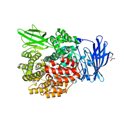
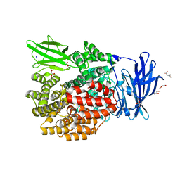 | | Crystal Structure of E. coli Aminopeptidase N in complex with Lysine | | 分子名称: | Aminopeptidase N, GLYCEROL, LYSINE, ... | | 著者 | Addlagatta, A, Gay, L, Matthews, B.W. | | 登録日 | 2007-10-19 | | 公開日 | 2008-05-06 | | 最終更新日 | 2023-08-30 | | 実験手法 | X-RAY DIFFRACTION (1.5 Å) | | 主引用文献 | Structural basis for the unusual specificity of Escherichia coli aminopeptidase N.
Biochemistry, 47, 2008
|
|
3BBZ
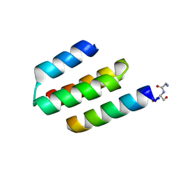
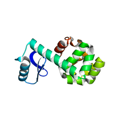
 | | Structure of the nucleocapsid-binding domain from the mumps virus phosphoprotein | | 分子名称: | BROMIDE ION, FORMIC ACID, P protein | | 著者 | Kingston, R.L, Gay, L.S, Baase, W.S, Matthews, B.W. | | 登録日 | 2007-11-11 | | 公開日 | 2008-05-27 | | 最終更新日 | 2024-05-29 | | 実験手法 | X-RAY DIFFRACTION (2.1 Å) | | 主引用文献 | Structure of the nucleocapsid-binding domain from the mumps virus polymerase; an example of protein folding induced by crystallization
J.Mol.Biol., 379, 2008
|
|
1P56
 | |
1P5C
 | |
3EOJ
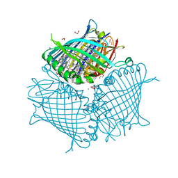 | | Fmo protein from Prosthecochloris Aestuarii 2K AT 1.3A Resolution | | 分子名称: | 1,2-ETHANEDIOL, AMMONIUM ION, BACTERIOCHLOROPHYLL A, ... | | 著者 | Tronrud, D.E, Wen, J, Gay, L, Blankenship, R.E. | | 登録日 | 2008-09-27 | | 公開日 | 2009-05-12 | | 最終更新日 | 2023-09-06 | | 実験手法 | X-RAY DIFFRACTION (1.3 Å) | | 主引用文献 | The structural basis for the difference in absorbance spectra for the FMO antenna protein from various green sulfur bacteria.
Photosynth.Res., 100, 2009
|
|
2HPT
 | | Crystal Structure of E. coli PepN (Aminopeptidase N)in complex with Bestatin | | 分子名称: | 2-(3-AMINO-2-HYDROXY-4-PHENYL-BUTYRYLAMINO)-4-METHYL-PENTANOIC ACID, Aminopeptidase N, GLYCEROL, ... | | 著者 | Addlagatta, A, Matthews, B.W, Gay, L. | | 登録日 | 2006-07-17 | | 公開日 | 2006-08-15 | | 最終更新日 | 2023-08-30 | | 実験手法 | X-RAY DIFFRACTION (2.3 Å) | | 主引用文献 | Structure of aminopeptidase N from Escherichia coli suggests a compartmentalized, gated active site.
Proc.Natl.Acad.Sci.Usa, 103, 2006
|
|
2HPO
 | | Structure of Aminopeptidase N from E. coli Suggests a Compartmentalized, Gated Active Site | | 分子名称: | Aminopeptidase N, GLYCEROL, ZINC ION | | 著者 | Addlagatta, A, Matthews, B.W, Gay, L. | | 登録日 | 2006-07-17 | | 公開日 | 2006-08-15 | | 最終更新日 | 2024-02-14 | | 実験手法 | X-RAY DIFFRACTION (1.65 Å) | | 主引用文献 | Structure of aminopeptidase N from Escherichia coli suggests a compartmentalized, gated active site.
Proc.Natl.Acad.Sci.Usa, 103, 2006
|
|
1OYU
 | |
2Y8V
 | | Structure of chitinase, ChiC, from Aspergillus fumigatus. | | 分子名称: | CLASS III CHITINASE, PUTATIVE, SODIUM ION | | 著者 | Rush, C.L, Schuettelkopf, A.W, Gay, L.M, van Aalten, D.M.F. | | 登録日 | 2011-02-10 | | 公開日 | 2012-02-22 | | 最終更新日 | 2024-05-08 | | 実験手法 | X-RAY DIFFRACTION (1.99 Å) | | 主引用文献 | Structure of Chitinase, Chic, from Aspergillus Fumigatus.
To be Published
|
|


