Loading
PDBj
MenuPDBj@FacebookPDBj@TwitterPDBj@YouTubewwPDB FoundationwwPDB
RCSB PDBPDBeBMRBAdv. SearchSearch help
PDB: 39 results

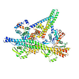
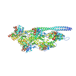
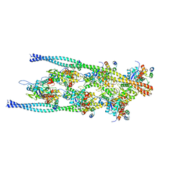
8UWY
DownloadVisualize
BU of 8uwy by Molmil
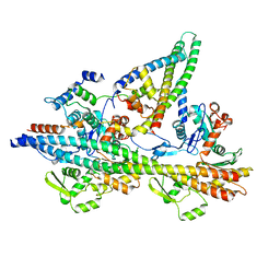
The structure of the native cardiac thin filament troponin core in Ca2+-free rotated state from the upper strand
Descriptor: Actin, alpha cardiac muscle 1, Tropomyosin alpha-1 chain, ...
Authors:Galkin, V.E, Risi, C.M.
Deposit date:2023-11-08
Release date:2024-03-06
Last modified:2024-03-13
Method:ELECTRON MICROSCOPY (5.5 Å)
Cite:Troponin Structural Dynamics in the Native Cardiac Thin Filament Revealed by Cryo Electron Microscopy.
J.Mol.Biol., 436, 2024
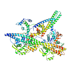
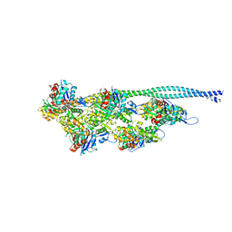
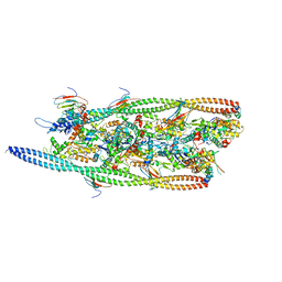
8UZX
DownloadVisualize
BU of 8uzx by Molmil
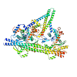
The structure of the native cardiac thin filament troponin core in Ca2+-bound fully activated state from the upper strand
Descriptor: ADENOSINE-5'-DIPHOSPHATE, Actin, alpha cardiac muscle 1, ...
Authors:Galkin, V.E, Risi, C.M.
Deposit date:2023-11-16
Release date:2024-03-06
Last modified:2024-03-13
Method:ELECTRON MICROSCOPY (5.4 Å)
Cite:Troponin Structural Dynamics in the Native Cardiac Thin Filament Revealed by Cryo Electron Microscopy.
J.Mol.Biol., 436, 2024
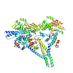
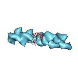
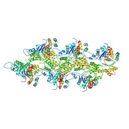
8UZY
DownloadVisualize
BU of 8uzy by Molmil
The structure of the native cardiac thin filament troponin core in Ca2+-bound partially activated state from the upper strand
Descriptor: ADENOSINE-5'-DIPHOSPHATE, Actin, alpha cardiac muscle 1, ...
Authors:Galkin, V.E, Risi, C.M.
Deposit date:2023-11-16
Release date:2024-03-06
Last modified:2024-03-13
Method:ELECTRON MICROSCOPY (5.3 Å)
Cite:Troponin Structural Dynamics in the Native Cardiac Thin Filament Revealed by Cryo Electron Microscopy.
J.Mol.Biol., 436, 2024
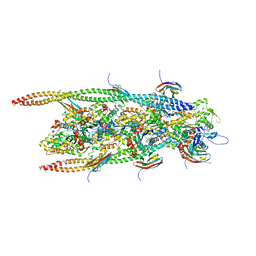
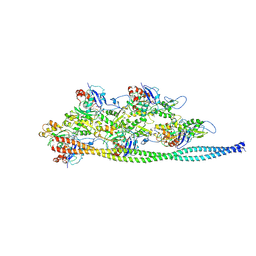
8UWW
DownloadVisualize
BU of 8uww by Molmil
The structure of the native cardiac thin filament troponin core in Ca2+-free state from the upper strand
Descriptor: Actin, alpha cardiac muscle 1, Tropomyosin alpha-1 chain, ...
Authors:Galkin, V.E, Risi, C.M.
Deposit date:2023-11-08
Release date:2024-03-06
Last modified:2024-03-13
Method:ELECTRON MICROSCOPY (5.1 Å)
Cite:Troponin Structural Dynamics in the Native Cardiac Thin Filament Revealed by Cryo Electron Microscopy.
J.Mol.Biol., 436, 2024
8V01
DownloadVisualize
BU of 8v01 by Molmil
The structure of the native cardiac thin filament troponin core in Ca2+-bound fully activated state 1 from the lower strand
Descriptor: ADENOSINE-5'-DIPHOSPHATE, Actin, alpha cardiac muscle 1, ...
Authors:Galkin, V.E, Risi, C.M.
Deposit date:2023-11-16
Release date:2024-03-06
Last modified:2024-03-13
Method:ELECTRON MICROSCOPY (5.5 Å)
Cite:Troponin Structural Dynamics in the Native Cardiac Thin Filament Revealed by Cryo Electron Microscopy.
J.Mol.Biol., 436, 2024
6G2T
DownloadVisualize
BU of 6g2t by Molmil
human cardiac myosin binding protein C C1 Ig-domain bound to native cardiac thin filament
Descriptor: Actin, cytoplasmic 2, Myosin-binding protein C, ...
Authors:Risi, C, Belknap, B, Forgacs, E, Harris, S.P, Schroder, G.F, White, H.D, Galkin, V.E.
Deposit date:2018-03-23
Release date:2018-10-17
Last modified:2024-05-15
Method:ELECTRON MICROSCOPY (9 Å)
Cite:N-Terminal Domains of Cardiac Myosin Binding Protein C Cooperatively Activate the Thin Filament.
Structure, 26, 2018
5NOL
DownloadVisualize
BU of 5nol by Molmil
Ca2+-induced Movement of Tropomyosin on Native Cardiac Thin Filaments - "Closed" state
Descriptor: ADENOSINE-5'-DIPHOSPHATE, Cardiac muscle alpha actin 1, MAGNESIUM ION, ...
Authors:Risi, C, Eisner, J, Belknap, B, Heeley, D.H, White, H.D, Schroeder, G.F, Galkin, V.E.
Deposit date:2017-04-12
Release date:2017-07-19
Last modified:2024-05-15
Method:ELECTRON MICROSCOPY (8 Å)
Cite:Ca(2+)-induced movement of tropomyosin on native cardiac thin filaments revealed by cryoelectron microscopy.
Proc. Natl. Acad. Sci. U.S.A., 114, 2017
5NOJ
DownloadVisualize
BU of 5noj by Molmil
Ca2+-induced Movement of Tropomyosin on Native Cardiac Thin Filaments - "OPEN" state
Descriptor: ADENOSINE-5'-DIPHOSPHATE, Actin, alpha skeletal muscle, ...
Authors:Risi, C, Eisner, J, Belknap, B, Heeley, D.H, White, H.D, Schroeder, G.F, Galkin, V.E.
Deposit date:2017-04-12
Release date:2017-08-02
Last modified:2017-08-09
Method:ELECTRON MICROSCOPY (11 Å)
Cite:Ca(2+)-induced movement of tropomyosin on native cardiac thin filaments revealed by cryoelectron microscopy.
Proc. Natl. Acad. Sci. U.S.A., 114, 2017
2QU4
DownloadVisualize
BU of 2qu4 by Molmil
Model for Bacterial ParM Filament
Descriptor: Plasmid segregation protein parM
Authors:Orlova, A, Garner, E.C, Galkin, V.E, Heuser, J, Mullins, R.D, Egelman, E.H.
Deposit date:2007-08-03
Release date:2007-09-18
Last modified:2024-02-21
Method:ELECTRON MICROSCOPY (16 Å)
Cite:The structure of bacterial ParM filaments.
Nat.Struct.Mol.Biol., 14, 2007
5NOG
DownloadVisualize
BU of 5nog by Molmil
Ca2+-induced Movement of Tropomyosin on Native Cardiac Thin Filaments - "Blocked" state
Descriptor: ADENOSINE-5'-DIPHOSPHATE, Cardiac muscle alpha actin 1, MAGNESIUM ION, ...
Authors:Risi, C, Eisner, J, Belknap, B, Heeley, D.H, White, H.D, Schroeder, G.F, Galkin, V.E.
Deposit date:2017-04-12
Release date:2017-07-19
Last modified:2017-08-30
Method:ELECTRON MICROSCOPY (11 Å)
Cite:Ca(2+)-induced movement of tropomyosin on native cardiac thin filaments revealed by cryoelectron microscopy.
Proc. Natl. Acad. Sci. U.S.A., 114, 2017
7TIT
DownloadVisualize
BU of 7tit by Molmil
Cardiac thin filament decorated with regulatory M-domain of cardiac myosin binding protein C
Descriptor: Myosin-binding protein C, cardiac-type, cardiac actin, ...
Authors:Risi, C.M, Galkin, V.E.
Deposit date:2022-01-14
Release date:2022-11-23
Last modified:2024-06-05
Method:ELECTRON MICROSCOPY (8 Å)
Cite:Cryo-Electron Microscopy Reveals Cardiac Myosin Binding Protein-C M-Domain Interactions with the Thin Filament.
J.Mol.Biol., 434, 2022
7TJ7
DownloadVisualize
BU of 7tj7 by Molmil
Cardiac thin filament decorated with C1 Ig-domain and regulatory M-domain of cardiac myosin binding protein C (cMyBP-C)
Descriptor: Myosin-binding protein C, cardiac-type, cardiac actin, ...
Authors:Risi, C.M, Galkin, V.E.
Deposit date:2022-01-14
Release date:2022-11-23
Last modified:2024-06-05
Method:ELECTRON MICROSCOPY (8 Å)
Cite:Cryo-Electron Microscopy Reveals Cardiac Myosin Binding Protein-C M-Domain Interactions with the Thin Filament.
J.Mol.Biol., 434, 2022
7TIJ
DownloadVisualize
BU of 7tij by Molmil
Cardiac F-actin decorated with regulatory M-domain of cardiac myosin binding protein C
Descriptor: Myosin-binding protein C, cardiac-type, cardiac actin
Authors:Risi, C.M, Galkin, V.E.
Deposit date:2022-01-13
Release date:2022-11-23
Last modified:2024-06-05
Method:ELECTRON MICROSCOPY (8 Å)
Cite:Cryo-Electron Microscopy Reveals Cardiac Myosin Binding Protein-C M-Domain Interactions with the Thin Filament.
J.Mol.Biol., 434, 2022
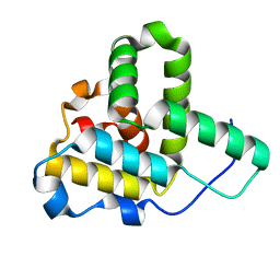
1Q5Z
DownloadVisualize
BU of 1q5z by Molmil
Crystal Structure of the C-terminal Actin Binding Domain of Salmonella Invasion Protein A (SipA)
Descriptor: SipA
Authors:Stebbins, C.E, Lilic, M, Galkin, V.E, Orlova, A, VanLoock, M.S, Egelman, E.H.
Deposit date:2003-08-11
Release date:2003-10-07
Last modified:2024-02-14
Method:X-RAY DIFFRACTION (1.8 Å)
Cite:Salmonella SipA polymerizes actin by stapling filaments with nonglobular protein arms.
Science, 301, 2003
<12


222926

건을2024-07-24부터공개중

PDB statisticsPDBj update infoContact PDBjnumon