1W4N
 
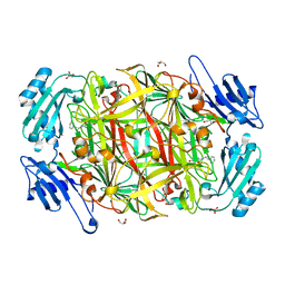 | | AGAO covalent complex with Tranylcypromine | | 分子名称: | COPPER (II) ION, GLYCEROL, PHENYLETHYLAMINE OXIDASE, ... | | 著者 | Duff, A.P, Trambaiolo, D.M, Langley, D.B, Juda, G.A, Shepard, E.M, Dooley, D.M, Freeman, H.C, Guss, J.M. | | 登録日 | 2004-07-27 | | 公開日 | 2005-12-08 | | 最終更新日 | 2023-12-13 | | 実験手法 | X-RAY DIFFRACTION (1.65 Å) | | 主引用文献 | Complexes of the Copper-Containing Amine Oxidase from Arthrobacter Globiformis with the Inhibitors Benzylhydrazine and Tranylcypromine.
Acta Crystallogr.,Sect.F, 64, 2008
|
|
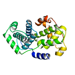
1VIN
 | |
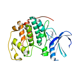
1VUB
 | | CCDB, A TOPOISOMERASE POISON FROM E. COLI | | 分子名称: | CCDB, CHLORIDE ION | | 著者 | Loris, R, Dao-Thi, M.-H, Bahasi, E.M, Van Melderen, L, Poortmans, F, Liddington, R, Couturier, M, Wyns, L. | | 登録日 | 1998-04-17 | | 公開日 | 1998-07-15 | | 最終更新日 | 2024-04-03 | | 実験手法 | X-RAY DIFFRACTION (2.6 Å) | | 主引用文献 | Crystal structure of CcdB, a topoisomerase poison from E. coli.
J.Mol.Biol., 285, 1999
|
|
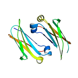
1W8C
 | |
4Q9B
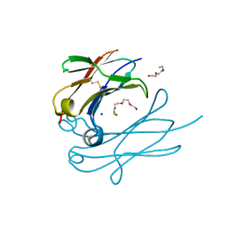
 | | IgNAR antibody domain C2 | | 分子名称: | Novel antigen receptor | | 著者 | Feige, J.M, Graewert, M.A, Marcinowski, M, Hennig, J, Behnke, J, Auslaender, D, Herold, E.M, Peschek, J, Castro, C.D, Flajnik, M.F, Hendershot, L.M, Sattler, M, Groll, M, Buchner, J. | | 登録日 | 2014-04-30 | | 公開日 | 2014-07-02 | | 最終更新日 | 2023-09-20 | | 実験手法 | X-RAY DIFFRACTION (1.5 Å) | | 主引用文献 | The structural analysis of shark IgNAR antibodies reveals evolutionary principles of immunoglobulins.
Proc.Natl.Acad.Sci.USA, 111, 2014
|
|
6E0I
 | | Crystal structure of Glucokinase in complex with compound 72 | | 分子名称: | 1-{4-[5-({3-[(2-methylpyridin-3-yl)oxy]-5-[(pyridin-2-yl)sulfanyl]pyridin-2-yl}amino)-1,2,4-thiadiazol-3-yl]piperidin-1 -yl}ethan-1-one, DIMETHYL SULFOXIDE, Glucokinase, ... | | 著者 | Hinklin, R.J, Baer, B.R, Boyd, S.A, Chicarelli, M.D, Condroski, K.R, DeWolf, W.E, Fischer, J, Frank, M, Hingorani, G.P, Lee, P.A, Neitzel, N.A, Pratt, S.A, Singh, A, Sullivan, F.X, Turner, T, Voegtli, W.C, Wallace, E.M, Williams, L, Aicher, T.D. | | 登録日 | 2018-07-06 | | 公開日 | 2019-07-10 | | 最終更新日 | 2024-03-13 | | 実験手法 | X-RAY DIFFRACTION (1.9 Å) | | 主引用文献 | Discovery and preclinical development of AR453588 as an anti-diabetic glucokinase activator.
Bioorg.Med.Chem., 28, 2020
|
|
3EEE
 | | Probing the function of heme distortion in the H-NOX family | | 分子名称: | CHLORIDE ION, Methyl-accepting chemotaxis protein, OXYGEN MOLECULE, ... | | 著者 | Olea Jr, C, Boon, E.M, Pellicena, P, Kuriyan, J, Marletta, M.A. | | 登録日 | 2008-09-04 | | 公開日 | 2008-11-25 | | 最終更新日 | 2023-08-30 | | 実験手法 | X-RAY DIFFRACTION (2.12 Å) | | 主引用文献 | Probing the function of heme distortion in the H-NOX family.
Acs Chem.Biol., 3, 2008
|
|
6E1M
 | | Structure of AtTPC1(DDE) reconstituted in saposin A | | 分子名称: | 1,2-DIACYL-GLYCEROL-3-SN-PHOSPHATE, CALCIUM ION, PALMITIC ACID, ... | | 著者 | Kintzer, A.F, Green, E.M, Cheng, Y, Stroud, R.M. | | 登録日 | 2018-07-10 | | 公開日 | 2018-09-19 | | 最終更新日 | 2019-12-18 | | 実験手法 | ELECTRON MICROSCOPY (3.3 Å) | | 主引用文献 | Structural basis for activation of voltage sensor domains in an ion channel TPC1.
Proc. Natl. Acad. Sci. U.S.A., 115, 2018
|
|
6E1P
 | | Structure of AtTPC1(DDE) in state 2 | | 分子名称: | CALCIUM ION, PALMITIC ACID, Two pore calcium channel protein 1 | | 著者 | Kintzer, A.F, Green, E.M, Cheng, Y, Stroud, R.M. | | 登録日 | 2018-07-10 | | 公開日 | 2018-09-19 | | 最終更新日 | 2019-12-18 | | 実験手法 | ELECTRON MICROSCOPY (3.7 Å) | | 主引用文献 | Structural basis for activation of voltage sensor domains in an ion channel TPC1.
Proc. Natl. Acad. Sci. U.S.A., 115, 2018
|
|
3EF2
 | | Structure of the Marasmius oreades mushroom lectin (MOA) in complex with Galalpha(1,3)[Fucalpha(1,2)]Gal and Calcium. | | 分子名称: | ACETATE ION, Agglutinin, CALCIUM ION, ... | | 著者 | Grahn, E.M, Goldstein, I.J, Krengel, U. | | 登録日 | 2008-09-08 | | 公開日 | 2009-06-30 | | 最終更新日 | 2023-11-15 | | 実験手法 | X-RAY DIFFRACTION (1.8 Å) | | 主引用文献 | Structural Characterization of a Lectin from the Mushroom Marasmius oreades in Complex with the Blood Group B Trisaccharide and Calcium.
J.Mol.Biol., 390, 2009
|
|
6E0E
 | | Crystal structure of Glucokinase in complex with compound 6 | | 分子名称: | 2-({2-[(4-methyl-1,3-thiazol-2-yl)amino]pyridin-3-yl}oxy)benzonitrile, Glucokinase, alpha-D-glucopyranose | | 著者 | Hinklin, R.J, Baer, B.R, Boyd, S.A, Chicarelli, M.D, Condroski, K.R, DeWolf, W.E, Fischer, J, Frank, M, Hingorani, G.P, Lee, P.A, Neitzel, N.A, Pratt, S.A, Singh, A, Sullivan, F.X, Turner, T, Voegtli, W.C, Wallace, E.M, Williams, L, Aicher, T.D. | | 登録日 | 2018-07-06 | | 公開日 | 2019-07-10 | | 最終更新日 | 2024-03-13 | | 実験手法 | X-RAY DIFFRACTION (2.7 Å) | | 主引用文献 | Discovery and preclinical development of AR453588 as an anti-diabetic glucokinase activator.
Bioorg.Med.Chem., 28, 2020
|
|
6E1N
 | | Structure of AtTPC1(DDE) in state 1 | | 分子名称: | 1,2-DIACYL-GLYCEROL-3-SN-PHOSPHATE, CALCIUM ION, PALMITIC ACID, ... | | 著者 | Kintzer, A.F, Green, E.M, Cheng, Y, Stroud, R.M. | | 登録日 | 2018-07-10 | | 公開日 | 2018-09-19 | | 最終更新日 | 2019-12-18 | | 実験手法 | ELECTRON MICROSCOPY (3.7 Å) | | 主引用文献 | Structural basis for activation of voltage sensor domains in an ion channel TPC1.
Proc. Natl. Acad. Sci. U.S.A., 115, 2018
|
|
6E1K
 | | Structure of AtTPC1(DDE) reconstituted in saposin A with cat06 Fab | | 分子名称: | CALCIUM ION, PALMITIC ACID, Two pore calcium channel protein 1, ... | | 著者 | Kintzer, A.F, Green, E.M, Cheng, Y, Stroud, R.M. | | 登録日 | 2018-07-10 | | 公開日 | 2018-09-19 | | 最終更新日 | 2019-12-18 | | 実験手法 | ELECTRON MICROSCOPY (3.3 Å) | | 主引用文献 | Structural basis for activation of voltage sensor domains in an ion channel TPC1.
Proc. Natl. Acad. Sci. U.S.A., 115, 2018
|
|
6E9R
 | | DHF46 filament | | 分子名称: | DHF46 filament | | 著者 | Lynch, E.M, Shen, H, Fallas, J.A, Kollman, J.M, Baker, D. | | 登録日 | 2018-08-01 | | 公開日 | 2018-11-21 | | 最終更新日 | 2024-03-13 | | 実験手法 | ELECTRON MICROSCOPY (5.9 Å) | | 主引用文献 | De novo design of self-assembling helical protein filaments.
Science, 362, 2018
|
|
4XXR
 | | Atomic Resolution X-Ray Crystal Structure of a Ruthenocene Conjugated Beta-Lactam Antibiotic in Complex with CTX-M-14 E166A Beta-Lactamase | | 分子名称: | CTX-M-14 Class A Beta-Lactamase, POTASSIUM ION, [(1,2,3,4,5-eta)-1-(4-{[(4-carboxy-5,5-dimethyl-1,3-thiazolidin-2-yl)methyl]amino}-4-oxobutanoyl)cyclopentadienyl][(1,2,3,4,5-eta)-cyclopentadienyl]ruthenium, ... | | 著者 | Lewandowski, E.M, Chen, Y. | | 登録日 | 2015-01-30 | | 公開日 | 2015-03-18 | | 最終更新日 | 2023-09-27 | | 実験手法 | X-RAY DIFFRACTION (1.18 Å) | | 主引用文献 | Antibacterial properties and atomic resolution X-ray complex crystal structure of a ruthenocene conjugated beta-lactam antibiotic.
Chem.Commun.(Camb.), 51, 2015
|
|
6E9Z
 | | DHF119 filament | | 分子名称: | DHF119 filament | | 著者 | Lynch, E.M, Shen, H, Fallas, J.A, Kollman, J.M, Baker, D. | | 登録日 | 2018-08-01 | | 公開日 | 2018-11-21 | | 最終更新日 | 2024-03-13 | | 実験手法 | ELECTRON MICROSCOPY (3.4 Å) | | 主引用文献 | De novo design of self-assembling helical protein filaments.
Science, 362, 2018
|
|
6EEY
 | |
6EIA
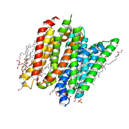 | | PepTSt in complex with HEPES (100 mM) | | 分子名称: | (2R)-2,3-DIHYDROXYPROPYL(7Z)-PENTADEC-7-ENOATE, (2S)-2,3-DIHYDROXYPROPYL(7Z)-PENTADEC-7-ENOATE, 4-(2-HYDROXYETHYL)-1-PIPERAZINE ETHANESULFONIC ACID, ... | | 著者 | Martinez Molledo, M, Quistgaard, E.M, Loew, C. | | 登録日 | 2017-09-18 | | 公開日 | 2018-02-21 | | 最終更新日 | 2024-01-17 | | 実験手法 | X-RAY DIFFRACTION (2 Å) | | 主引用文献 | Multispecific Substrate Recognition in a Proton-Dependent Oligopeptide Transporter.
Structure, 26, 2018
|
|
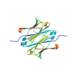
4Q9C
 | | IgNAR antibody domain C3 | | 分子名称: | CHLORIDE ION, Novel antigen receptor, SODIUM ION, ... | | 著者 | Feige, J.M, Graewert, M.A, Marcinowski, M, Hennig, J, Behnke, J, Auslaender, D, Herold, E.M, Peschek, J, Castro, C.D, Flajnik, M.F, Hendershot, L.M, Sattler, M, Groll, M, Buchner, J. | | 登録日 | 2014-04-30 | | 公開日 | 2014-07-02 | | 最終更新日 | 2023-09-20 | | 実験手法 | X-RAY DIFFRACTION (2.8 Å) | | 主引用文献 | The structural analysis of shark IgNAR antibodies reveals evolutionary principles of immunoglobulins.
Proc.Natl.Acad.Sci.USA, 111, 2014
|
|
4Q97
 | | IgNAR antibody domain C1 | | 分子名称: | Novel antigen receptor | | 著者 | Feige, J.M, Graewert, M.A, Marcinowski, M, Hennig, J, Behnke, J, Auslaender, D, Herold, E.M, Peschek, J, Castro, C.D, Flajnik, M.F, Hendershot, L.M, Sattler, M, Groll, M, Buchner, J. | | 登録日 | 2014-04-29 | | 公開日 | 2014-07-02 | | 最終更新日 | 2023-09-20 | | 実験手法 | X-RAY DIFFRACTION (2.4 Å) | | 主引用文献 | The structural analysis of shark IgNAR antibodies reveals evolutionary principles of immunoglobulins.
Proc.Natl.Acad.Sci.USA, 111, 2014
|
|
4Y75
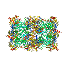 | | Yeast 20S proteasome in complex with Ac-PAF-ep | | 分子名称: | 2-(N-MORPHOLINO)-ETHANESULFONIC ACID, Ac-PAF-ep, CHLORIDE ION, ... | | 著者 | Huber, E.M, Groll, M. | | 登録日 | 2015-02-13 | | 公開日 | 2015-06-17 | | 最終更新日 | 2024-01-10 | | 実験手法 | X-RAY DIFFRACTION (2.8 Å) | | 主引用文献 | Systematic Analyses of Substrate Preferences of 20S Proteasomes Using Peptidic Epoxyketone Inhibitors.
J.Am.Chem.Soc., 137, 2015
|
|
4Y8N
 | |
4QUY
 | | yCP beta5-A49S-mutant | | 分子名称: | CHLORIDE ION, MAGNESIUM ION, Probable proteasome subunit alpha type-7, ... | | 著者 | Huber, E.M, Heinemeyer, W, Groll, M. | | 登録日 | 2014-07-14 | | 公開日 | 2015-02-04 | | 最終更新日 | 2023-09-20 | | 実験手法 | X-RAY DIFFRACTION (2.8 Å) | | 主引用文献 | Bortezomib-Resistant Mutant Proteasomes: Structural and Biochemical Evaluation with Carfilzomib and ONX 0914.
Structure, 23, 2015
|
|
4QW3
 | | yCP beta5-C63F mutant in complex with bortezomib | | 分子名称: | CHLORIDE ION, MAGNESIUM ION, N-[(1R)-1-(DIHYDROXYBORYL)-3-METHYLBUTYL]-N-(PYRAZIN-2-YLCARBONYL)-L-PHENYLALANINAMIDE, ... | | 著者 | Huber, E.M, Heinemeyer, W, Groll, M. | | 登録日 | 2014-07-16 | | 公開日 | 2015-02-04 | | 最終更新日 | 2023-09-20 | | 実験手法 | X-RAY DIFFRACTION (2.9 Å) | | 主引用文献 | Bortezomib-Resistant Mutant Proteasomes: Structural and Biochemical Evaluation with Carfilzomib and ONX 0914.
Structure, 23, 2015
|
|
4Y69
 | |


