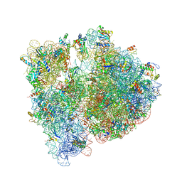
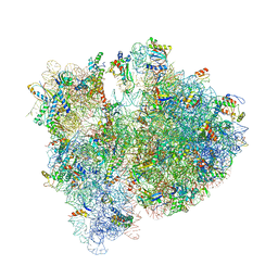
4V7S
 
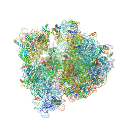 | | Crystal structure of the E. coli ribosome bound to telithromycin. | | 分子名称: | 16S rRNA, 23S rRNA, 30S ribosomal protein S10, ... | | 著者 | Dunkle, J.A, Xiong, L, Mankin, A.S, Cate, J.H.D. | | 登録日 | 2010-08-05 | | 公開日 | 2014-07-09 | | 最終更新日 | 2023-09-20 | | 実験手法 | X-RAY DIFFRACTION (3.2547 Å) | | 主引用文献 | Structures of the Escherichia coli ribosome with antibiotics bound near the peptidyl transferase center explain spectra of drug action.
Proc.Natl.Acad.Sci.USA, 107, 2010
|
|
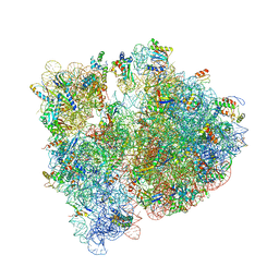
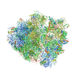
4V7U
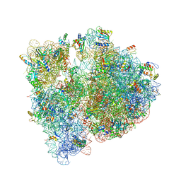 | | Crystal structure of the E. coli ribosome bound to erythromycin. | | 分子名称: | 16S rRNA, 23S rRNA, 30S ribosomal protein S10, ... | | 著者 | Dunkle, J.A, Xiong, L, Mankin, A.S, Cate, J.H.D. | | 登録日 | 2010-08-15 | | 公開日 | 2014-07-09 | | 最終更新日 | 2023-09-20 | | 実験手法 | X-RAY DIFFRACTION (3.1 Å) | | 主引用文献 | Structures of the Escherichia coli ribosome with antibiotics bound near the peptidyl transferase center explain spectra of drug action.
Proc.Natl.Acad.Sci.USA, 107, 2010
|
|
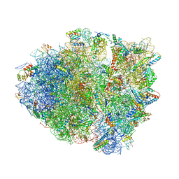
4V7V
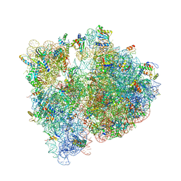 | | Crystal structure of the E. coli ribosome bound to clindamycin. | | 分子名称: | 16S rRNA, 23S rRNA, 30S ribosomal protein S10, ... | | 著者 | Dunkle, J.A, Xiong, L, Mankin, A.S, Cate, J.H.D. | | 登録日 | 2010-08-16 | | 公開日 | 2014-07-09 | | 最終更新日 | 2014-12-10 | | 実験手法 | X-RAY DIFFRACTION (3.2891 Å) | | 主引用文献 | Structures of the Escherichia coli ribosome with antibiotics bound near the peptidyl transferase center explain spectra of drug action.
Proc.Natl.Acad.Sci.USA, 107, 2010
|
|
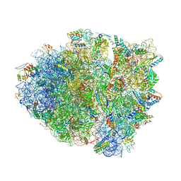
4V9D
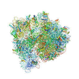 | | Structures of the bacterial ribosome in classical and hybrid states of tRNA binding | | 分子名称: | 16S rRNA, 23S rRNA, 30S ribosomal protein S10, ... | | 著者 | Dunkle, J.A, Wang, L, Feldman, M.B, Pulk, A, Chen, V.B, Kapral, G.J, Noeske, J, Richardson, J.S, Blanchard, S.C, Cate, J.H.D. | | 登録日 | 2012-07-31 | | 公開日 | 2014-07-09 | | 最終更新日 | 2023-09-20 | | 実験手法 | X-RAY DIFFRACTION (3 Å) | | 主引用文献 | Structures of the bacterial ribosome in classical and hybrid states of tRNA binding.
Science, 332, 2011
|
|
4V7T
 | | Crystal structure of the E. coli ribosome bound to chloramphenicol. | | 分子名称: | 16S rRNA, 23S rRNA, 30S ribosomal protein S10, ... | | 著者 | Dunkle, J.A, Xiong, L, Mankin, A.S, Cate, J.H.D. | | 登録日 | 2010-08-14 | | 公開日 | 2014-07-09 | | 最終更新日 | 2023-09-20 | | 実験手法 | X-RAY DIFFRACTION (3.1942 Å) | | 主引用文献 | Structures of the Escherichia coli ribosome with antibiotics bound near the peptidyl transferase center explain spectra of drug action.
Proc.Natl.Acad.Sci.USA, 107, 2010
|
|
4WWW
 | | Crystal structure of the E. coli ribosome bound to CEM-101 | | 分子名称: | (3aS,4R,7S,9R,10R,11R,13R,15R,15aR)-1-{4-[4-(3-aminophenyl)-1H-1,2,3-triazol-1-yl]butyl}-4-ethyl-7-fluoro-11-methoxy-3a ,7,9,11,13,15-hexamethyl-2,6,8,14-tetraoxotetradecahydro-2H-oxacyclotetradecino[4,3-d][1,3]oxazol-10-yl 3,4,6-trideoxy-3-(dimethylamino)-beta-D-xylo-hexopyranoside, 16S rRNA, 23S rRNA, ... | | 著者 | Dunkle, J.A, Zhang, W, Cate, J.H.D, Mankin, A.S. | | 登録日 | 2014-11-12 | | 公開日 | 2014-12-24 | | 最終更新日 | 2023-09-27 | | 実験手法 | X-RAY DIFFRACTION (3.1 Å) | | 主引用文献 | Binding and action of CEM-101, a new fluoroketolide antibiotic that inhibits protein synthesis.
Antimicrob. Agents Chemother., 54, 2010
|
|
7RRN
 | |
6UY5
 | |
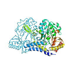
7RUJ
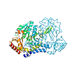 | | E. coli cysteine desulfurase SufS N99A | | 分子名称: | CHLORIDE ION, Cysteine desulfurase, PYRIDOXAL-5'-PHOSPHATE | | 著者 | Dunkle, J.A, Gogar, R, Frantom, P.A. | | 登録日 | 2021-08-17 | | 公開日 | 2023-01-25 | | 最終更新日 | 2023-10-25 | | 実験手法 | X-RAY DIFFRACTION (2.5 Å) | | 主引用文献 | The beta-latch structural element of the SufS cysteine desulfurase mediates active site accessibility and SufE transpersulfurase positioning.
J.Biol.Chem., 299, 2023
|
|
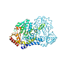
7RW3
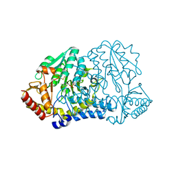 | | E. coli cysteine desulfurase SufS N99D | | 分子名称: | Cysteine desulfurase, PYRIDOXAL-5'-PHOSPHATE | | 著者 | Dunkle, J.A, Gogar, R, Frantom, P.A. | | 登録日 | 2021-08-19 | | 公開日 | 2023-01-25 | | 最終更新日 | 2023-11-15 | | 実験手法 | X-RAY DIFFRACTION (2.3 Å) | | 主引用文献 | The beta-latch structural element of the SufS cysteine desulfurase mediates active site accessibility and SufE transpersulfurase positioning.
J.Biol.Chem., 299, 2023
|
|
6MRH
 | |
6MRE
 | |
6MRI
 | |
6MR2
 | |
6MR6
 | |
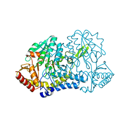
6O12
 | | E. coli cysteine desulfurase SufS H123A | | 分子名称: | CHLORIDE ION, Cysteine desulfurase, PYRIDOXAL-5'-PHOSPHATE | | 著者 | Dunkle, J.A, Frantom, P.A. | | 登録日 | 2019-02-17 | | 公開日 | 2019-06-26 | | 最終更新日 | 2023-10-11 | | 実験手法 | X-RAY DIFFRACTION (2.05 Å) | | 主引用文献 | Direct observation of intermediates in the SufS cysteine desulfurase reaction reveals functional roles of conserved active-site residues.
J.Biol.Chem., 294, 2019
|
|
6O11
 | |
6O13
 | |
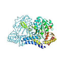
6O10
 | | E. coli cysteine desulfurase SufS | | 分子名称: | CHLORIDE ION, Cysteine desulfurase, PYRIDOXAL-5'-PHOSPHATE | | 著者 | Dunkle, J.A, Frantom, P.A. | | 登録日 | 2019-02-17 | | 公開日 | 2019-06-26 | | 最終更新日 | 2023-10-11 | | 実験手法 | X-RAY DIFFRACTION (2 Å) | | 主引用文献 | Direct observation of intermediates in the SufS cysteine desulfurase reaction reveals functional roles of conserved active-site residues.
J.Biol.Chem., 294, 2019
|
|
4OX9
 | | Crystal structure of the aminoglycoside resistance methyltransferase NpmA bound to the 30S ribosomal subunit | | 分子名称: | 16S rRNA, 16S rRNA (adenine(1408)-N(1))-methyltransferase, 30S ribosomal protein S10, ... | | 著者 | Dunkle, J.A, Conn, G.L, Dunham, C.M. | | 登録日 | 2014-02-04 | | 公開日 | 2014-04-09 | | 最終更新日 | 2023-09-27 | | 実験手法 | X-RAY DIFFRACTION (3.8035 Å) | | 主引用文献 | Molecular recognition and modification of the 30S ribosome by the aminoglycoside-resistance methyltransferase NpmA.
Proc.Natl.Acad.Sci.USA, 111, 2014
|
|
4W4G
 | | Postcleavage state of 70S bound to HigB toxin and AAA (lysine) codon | | 分子名称: | 16S rRNA, 23S rRNA, 30S ribosomal protein S10, ... | | 著者 | Schureck, M.A, Maehigashi, T, Dunkle, J.A, Dunham, C.M. | | 登録日 | 2014-08-14 | | 公開日 | 2015-10-21 | | 最終更新日 | 2023-12-27 | | 実験手法 | X-RAY DIFFRACTION (3.3 Å) | | 主引用文献 | Defining the mRNA recognition signature of a bacterial toxin protein.
Proc.Natl.Acad.Sci.USA, 112, 2015
|
|
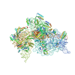
4V6D
 | | Crystal structure of the E. coli 70S ribosome in an intermediate state of ratcheting | | 分子名称: | 16S rRNA, 23S rRNA, 30S ribosomal protein S10, ... | | 著者 | Zhang, W, Dunkle, J.A, Cate, J.H.D. | | 登録日 | 2009-06-27 | | 公開日 | 2014-07-09 | | 最終更新日 | 2014-12-10 | | 実験手法 | X-RAY DIFFRACTION (3.814 Å) | | 主引用文献 | Structures of the ribosome in intermediate States of ratcheting.
Science, 325, 2009
|
|
4V8J
 | | Crystal structure of the bacterial ribosome ram mutation G347U. | | 分子名称: | 16S rRNA, 23S rRNA, 30S ribosomal protein S10, ... | | 著者 | Fagan, C.E, Dunkle, J.A, Maehigashi, T, Dunham, C.M. | | 登録日 | 2011-12-20 | | 公開日 | 2014-07-09 | | 最終更新日 | 2019-07-17 | | 実験手法 | X-RAY DIFFRACTION (3.9 Å) | | 主引用文献 | Reorganization of an intersubunit bridge induced by disparate 16S ribosomal ambiguity mutations mimics an EF-Tu-bound state.
Proc.Natl.Acad.Sci.USA, 110, 2013
|
|
4YZV
 | | Precleavage 70S structure of the P. vulgaris HigB deltaH92 toxin bound to the ACA codon | | 分子名称: | 16S rRNA, 23S rRNA, 30S ribosomal protein S10, ... | | 著者 | Schureck, M.A, Dunkle, J.A, Maehigashi, T, Dunham, C.M. | | 登録日 | 2015-03-25 | | 公開日 | 2015-10-21 | | 最終更新日 | 2023-09-27 | | 実験手法 | X-RAY DIFFRACTION (3.1 Å) | | 主引用文献 | Defining the mRNA recognition signature of a bacterial toxin protein.
Proc.Natl.Acad.Sci.USA, 112, 2015
|
|
4YPB
 | | Precleavage 70S structure of the P. vulgaris HigB DeltaH92 toxin bound to the AAA codon | | 分子名称: | 16S rRNA, 23S rRNA, 30S ribosomal protein S10, ... | | 著者 | Schureck, M.A, Dunkle, J.A, Maehigashi, T, Dunham, C.M. | | 登録日 | 2015-03-12 | | 公開日 | 2015-10-21 | | 最終更新日 | 2023-09-27 | | 実験手法 | X-RAY DIFFRACTION (3.4 Å) | | 主引用文献 | Defining the mRNA recognition signature of a bacterial toxin protein.
Proc.Natl.Acad.Sci.USA, 112, 2015
|
|


