Loading
PDBj
MenuPDBj@FacebookPDBj@TwitterPDBj@YouTubewwPDB FoundationwwPDB
RCSB PDBPDBeBMRBAdv. SearchSearch help
PDB: 104 results

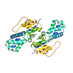
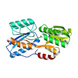
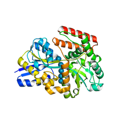
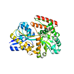
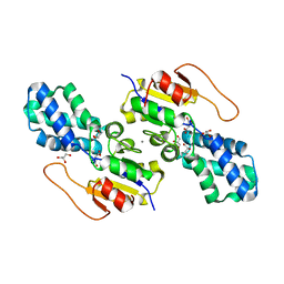
8WBT
DownloadVisualize
BU of 8wbt by Molmil
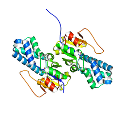
Crystal structure of cis-Epoxysuccinate Hydrolases KlCESH[L] mutant D48N complexed with L-TA
Descriptor: (S)-2-haloacid dehalogenase, CALCIUM ION, GLYCEROL, ...
Authors:Dong, S, Xuan, J.S, Feng, Y.G, Cui, Q.
Deposit date:2023-09-10
Release date:2024-01-31
Last modified:2024-02-21
Method:X-RAY DIFFRACTION (2.05 Å)
Cite:Deciphering the stereo-specific catalytic mechanisms of cis-epoxysuccinate hydrolases producing L(+)-tartaric acid.
J.Biol.Chem., 300, 2024
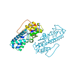
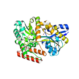
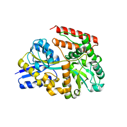
8WBS
DownloadVisualize
BU of 8wbs by Molmil
Crystal structure of cis-Epoxysuccinate Hydrolases KlCESH[L]-D48N complexed with sulfate ions
Descriptor: (S)-2-haloacid dehalogenase, CALCIUM ION, SULFATE ION
Authors:Dong, S, Xuan, J.S, Feng, Y.G, Cui, Q.
Deposit date:2023-09-10
Release date:2024-01-31
Last modified:2024-02-21
Method:X-RAY DIFFRACTION (2.03 Å)
Cite:Deciphering the stereo-specific catalytic mechanisms of cis-epoxysuccinate hydrolases producing L(+)-tartaric acid.
J.Biol.Chem., 300, 2024
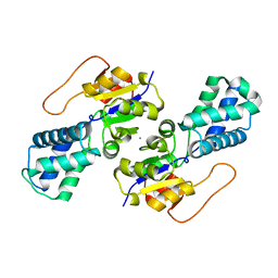
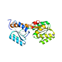
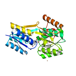
8WBO
DownloadVisualize
BU of 8wbo by Molmil
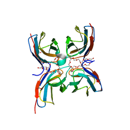
Crystal structure of cis-Epoxysuccinate Hydrolases RhCESH[L] mutant D18N complexed with sulfate ions
Descriptor: Epoxide hydrolase, SULFATE ION
Authors:Dong, S, Xuan, J.S, Feng, Y.G, Cui, Q.
Deposit date:2023-09-10
Release date:2024-01-31
Last modified:2024-02-21
Method:X-RAY DIFFRACTION (1.58 Å)
Cite:Deciphering the stereo-specific catalytic mechanisms of cis-epoxysuccinate hydrolases producing L(+)-tartaric acid.
J.Biol.Chem., 300, 2024
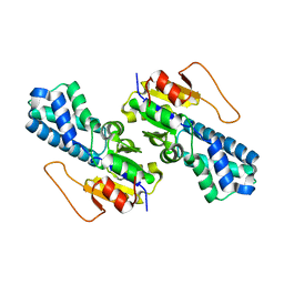
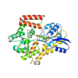
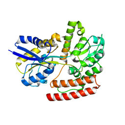
8WBR
DownloadVisualize
BU of 8wbr by Molmil
Crystal structure of cis-Epoxysuccinate Hydrolases KlCESH[L]
Descriptor: (S)-2-haloacid dehalogenase, CALCIUM ION
Authors:Dong, S, Xuan, J.S, Feng, Y.G, Cui, Q.
Deposit date:2023-09-10
Release date:2024-01-31
Last modified:2024-02-21
Method:X-RAY DIFFRACTION (2.02 Å)
Cite:Deciphering the stereo-specific catalytic mechanisms of cis-epoxysuccinate hydrolases producing L(+)-tartaric acid.
J.Biol.Chem., 300, 2024
8WBP
DownloadVisualize
BU of 8wbp by Molmil
Crystal structure of cis-Epoxysuccinate Hydrolases RhCESH[L] mutant E212Q
Descriptor: Epoxide hydrolase
Authors:Dong, S, Xuan, J.S, Feng, Y.G, Cui, Q.
Deposit date:2023-09-10
Release date:2024-01-31
Last modified:2024-10-23
Method:X-RAY DIFFRACTION (1.87 Å)
Cite:Deciphering the stereo-specific catalytic mechanisms of cis-epoxysuccinate hydrolases producing L(+)-tartaric acid.
J.Biol.Chem., 300, 2024
8WBQ
DownloadVisualize
BU of 8wbq by Molmil
Crystal structure of cis-Epoxysuccinate Hydrolases RhCESH[L] mutant E212Q complexed with L-TA.
Descriptor: Epoxide hydrolase, L(+)-TARTARIC ACID
Authors:Dong, S, Xuan, J.S, Feng, Y.G, Cui, Q.
Deposit date:2023-09-10
Release date:2024-01-31
Last modified:2024-02-21
Method:X-RAY DIFFRACTION (2.2 Å)
Cite:Deciphering the stereo-specific catalytic mechanisms of cis-epoxysuccinate hydrolases producing L(+)-tartaric acid.
J.Biol.Chem., 300, 2024
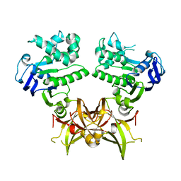
7CG5
DownloadVisualize
BU of 7cg5 by Molmil
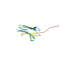
Structure of the sensor domain (long construct) of the anti-sigma factor RsgI4 in Pseudobacteroides cellulosolvens
Descriptor: Anti-sigma factor RsgI, N-terminal
Authors:Dong, S, Feng, Y.
Deposit date:2020-06-30
Release date:2021-06-30
Last modified:2023-11-29
Method:X-RAY DIFFRACTION (2.85 Å)
Cite:Structure of the sensor domain of the anti-sigma factor RsgI4 in Pseudobacteroides cellulosolvens
To Be Published
7CG8
DownloadVisualize
BU of 7cg8 by Molmil
Structure of the sensor domain (short construct) of the anti-sigma factor RsgI4 in Pseudobacteroides cellulosolvens
Descriptor: 2,5,8,11,14,17,20,23,26,29,32,35,38,41,44,47,50,53,56,59,62,65,68,71,74,77,80-HEPTACOSAOXADOOCTACONTAN-82-OL, ACETATE ION, Anti-sigma factor RsgI, ...
Authors:Dong, S, Feng, Y.
Deposit date:2020-06-30
Release date:2021-06-30
Last modified:2023-11-29
Method:X-RAY DIFFRACTION (1.5 Å)
Cite:Structure of the sensor domain of the anti-sigma factor RsgI4 in Pseudobacteroides cellulosolvens
To Be Published
7CG1
DownloadVisualize
BU of 7cg1 by Molmil
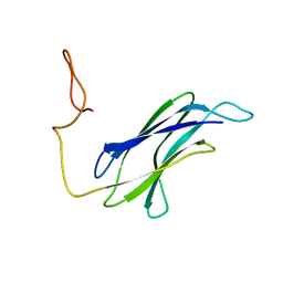
Solution structure of the sensor domain of the anti-sigma factor RsgI4 in Pseudobacteroides cellulosolvens
Descriptor: Anti-sigma factor RsgI, N-terminal
Authors:Dong, S, Feng, Y.
Deposit date:2020-06-30
Release date:2021-06-30
Last modified:2024-05-15
Method:SOLUTION NMR
Cite:Structure of the sensor domain of the anti-sigma factor RsgI4 in Pseudobacteroides cellulosolvens
To Be Published
7DMK
DownloadVisualize
BU of 7dmk by Molmil
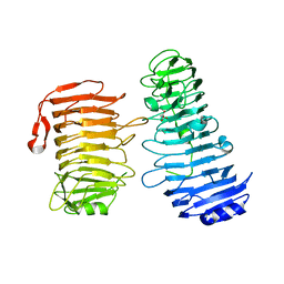
PL6 alginate lyase BcAlyPL6
Descriptor: BcAlyPL6, CALCIUM ION, MALONATE ION
Authors:Dong, S, Wang, B, Ma, X.Q, Li, F.L, Feng, Y.G.
Deposit date:2020-12-04
Release date:2021-10-06
Last modified:2023-11-29
Method:X-RAY DIFFRACTION (2.213 Å)
Cite:Structural basis for the exolytic activity of polysaccharide lyase family 6 alginate lyase BcAlyPL6 from human gut microbe Bacteroides clarus.
Biochem.Biophys.Res.Commun., 547, 2021
8IVY
DownloadVisualize
BU of 8ivy by Molmil
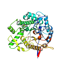
Beta-Glucosidase BglA mutant E166Q in complex with glucose
Descriptor: Beta-glucosidase, GLYCEROL, beta-D-glucopyranose
Authors:Dong, S, Xiao, Y, Feng, Y.
Deposit date:2023-03-29
Release date:2023-08-23
Method:X-RAY DIFFRACTION (1.95 Å)
Cite:Key roles of beta-glucosidase BglA for the catabolism of both laminaribiose and cellobiose in the lignocellulolytic bacterium Clostridium thermocellum.
Int.J.Biol.Macromol., 250, 2023
7X0L
DownloadVisualize
BU of 7x0l by Molmil
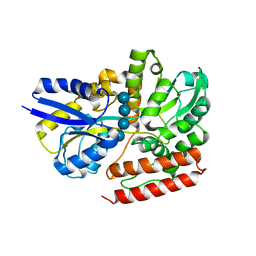
Crystal structure of sugar binding protein CbpB complexed wtih cellotetraose from Clostridium thermocellum
Descriptor: CbpB, beta-D-glucopyranose-(1-4)-beta-D-glucopyranose-(1-4)-beta-D-glucopyranose-(1-4)-beta-D-glucopyranose
Authors:Dong, S, Yao, X, Feng, Y.
Deposit date:2022-02-22
Release date:2022-09-14
Last modified:2024-05-29
Method:X-RAY DIFFRACTION (1.9 Å)
Cite:Deciphering Cellodextrin and Glucose Uptake in Clostridium thermocellum.
Mbio, 13, 2022
7X0H
DownloadVisualize
BU of 7x0h by Molmil
Crystal structure of sugar binding protein CbpA complexed wtih glucose from Clostridium thermocellum
Descriptor: CbpA, beta-D-glucopyranose
Authors:Dong, S, Yao, X, Feng, Y.
Deposit date:2022-02-22
Release date:2022-09-14
Last modified:2023-11-29
Method:X-RAY DIFFRACTION (1.85 Å)
Cite:Deciphering Cellodextrin and Glucose Uptake in Clostridium thermocellum.
Mbio, 13, 2022
7X0P
DownloadVisualize
BU of 7x0p by Molmil
Crystal structure of sugar binding protein CbpD from Clostridium thermocellum
Descriptor: CbpD
Authors:Dong, S, Yao, X, Feng, Y.
Deposit date:2022-02-22
Release date:2022-09-14
Last modified:2023-11-29
Method:X-RAY DIFFRACTION (1.5 Å)
Cite:Deciphering Cellodextrin and Glucose Uptake in Clostridium thermocellum.
Mbio, 13, 2022
7X0K
DownloadVisualize
BU of 7x0k by Molmil
Crystal structure of sugar binding protein CbpB complexed wtih cellotriose from Clostridium thermocellum
Descriptor: CpbB, beta-D-glucopyranose-(1-4)-beta-D-glucopyranose-(1-4)-beta-D-glucopyranose
Authors:Dong, S, Yao, X, Feng, Y.
Deposit date:2022-02-22
Release date:2022-09-14
Last modified:2022-11-09
Method:X-RAY DIFFRACTION (1.7 Å)
Cite:Deciphering Cellodextrin and Glucose Uptake in Clostridium thermocellum.
Mbio, 13, 2022
7X0G
DownloadVisualize
BU of 7x0g by Molmil
Crystal structure of sugar binding protein CbpA from Clostridium thermocellum
Descriptor: CbpA
Authors:Dong, S, Yao, X, Feng, Y.
Deposit date:2022-02-22
Release date:2022-09-14
Last modified:2023-11-29
Method:X-RAY DIFFRACTION (2.1 Å)
Cite:Deciphering Cellodextrin and Glucose Uptake in Clostridium thermocellum.
Mbio, 13, 2022
7X0O
DownloadVisualize
BU of 7x0o by Molmil
Crystal structure of sugar binding protein CbpC from Clostridium thermocellum
Descriptor: CbpC
Authors:Dong, S, Yao, X, Feng, Y.
Deposit date:2022-02-22
Release date:2022-09-14
Last modified:2024-05-29
Method:X-RAY DIFFRACTION (2 Å)
Cite:Deciphering Cellodextrin and Glucose Uptake in Clostridium thermocellum.
Mbio, 13, 2022
7X0M
DownloadVisualize
BU of 7x0m by Molmil
Crystal structure of sugar binding protein CbpB complexed wtih cellopentaose from Clostridium thermocellum
Descriptor: CbpB, beta-D-glucopyranose-(1-4)-beta-D-glucopyranose-(1-4)-beta-D-glucopyranose-(1-4)-beta-D-glucopyranose-(1-4)-beta-D-glucopyranose
Authors:Dong, S, Yao, X, Feng, Y.
Deposit date:2022-02-22
Release date:2022-09-14
Last modified:2024-05-29
Method:X-RAY DIFFRACTION (2 Å)
Cite:Deciphering Cellodextrin and Glucose Uptake in Clostridium thermocellum.
Mbio, 13, 2022
7X0Q
DownloadVisualize
BU of 7x0q by Molmil
Crystal structure of ATPase Clo1313_2554 from Clostridium thermocellum
Descriptor: ABC transporter related protein
Authors:Dong, S, Yao, X, Feng, Y.
Deposit date:2022-02-22
Release date:2022-09-14
Last modified:2023-11-29
Method:X-RAY DIFFRACTION (2.9 Å)
Cite:Deciphering Cellodextrin and Glucose Uptake in Clostridium thermocellum.
Mbio, 13, 2022
7X0R
DownloadVisualize
BU of 7x0r by Molmil
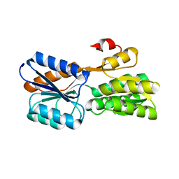
Crystal structure of substrate binding protein Lbp complexed wtih guanosine from Clostridium thermocellum
Descriptor: GUANOSINE, Lbp, ZINC ION
Authors:Dong, S, Yao, X, Feng, Y.
Deposit date:2022-02-22
Release date:2022-09-14
Last modified:2023-11-29
Method:X-RAY DIFFRACTION (1.47 Å)
Cite:Deciphering Cellodextrin and Glucose Uptake in Clostridium thermocellum.
Mbio, 13, 2022
7X0I
DownloadVisualize
BU of 7x0i by Molmil
Crystal structure of sugar binding protein CbpB from Clostridium thermocellum
Descriptor: CbpB
Authors:Dong, S, Yao, X, Feng, Y.
Deposit date:2022-02-22
Release date:2022-09-14
Last modified:2024-05-29
Method:X-RAY DIFFRACTION (1.7 Å)
Cite:Deciphering Cellodextrin and Glucose Uptake in Clostridium thermocellum.
Mbio, 13, 2022
7X0J
DownloadVisualize
BU of 7x0j by Molmil
Crystal structure of sugar binding protein CbpB complexed wtih cellobiose from Clostridium thermocellum
Descriptor: CbpB, beta-D-glucopyranose-(1-4)-beta-D-glucopyranose
Authors:Dong, S, Yao, X, Feng, Y.
Deposit date:2022-02-22
Release date:2022-09-14
Last modified:2024-05-29
Method:X-RAY DIFFRACTION (1.7 Å)
Cite:Deciphering Cellodextrin and Glucose Uptake in Clostridium thermocellum.
Mbio, 13, 2022
7X0N
DownloadVisualize
BU of 7x0n by Molmil
Crystal structure of sugar binding protein CbpB complexed wtih laminaribiose from Clostridium thermocellum
Descriptor: CbpB, beta-D-glucopyranose-(1-3)-beta-D-glucopyranose
Authors:Dong, S, Yao, X, Feng, Y.
Deposit date:2022-02-22
Release date:2022-09-14
Last modified:2024-05-29
Method:X-RAY DIFFRACTION (1.68 Å)
Cite:Deciphering Cellodextrin and Glucose Uptake in Clostridium thermocellum.
Mbio, 13, 2022
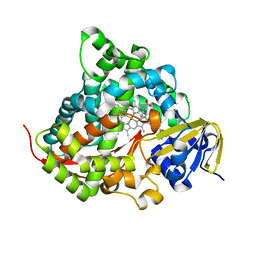
7YD9
DownloadVisualize
BU of 7yd9 by Molmil
Crystal structure of the P450 BM3 heme domain mutant F87G/T268V/A184V/A328V in complex with N-imidazolyl-hexanoyl-L-phenylalanine,methylbenzene and hydroxylamine
Descriptor: (2S)-2-(6-imidazol-1-ylhexanoylamino)-3-phenyl-propanoic acid, Bifunctional cytochrome P450/NADPH--P450 reductase, HYDROXYAMINE, ...
Authors:Dong, S, Chen, J, Jiang, Y, Cong, Z, Feng, Y.
Deposit date:2022-07-04
Release date:2023-05-24
Last modified:2023-11-29
Method:X-RAY DIFFRACTION (1.748 Å)
Cite:Regiodivergent and Enantioselective Hydroxylation of C-H bonds by Synergistic Use of Protein Engineering and Exogenous Dual-Functional Small Molecules.
Angew.Chem.Int.Ed.Engl., 62, 2023
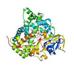
7YDA
DownloadVisualize
BU of 7yda by Molmil
Crystal structure of the P450 BM3 heme domain mutant F87V/T268V/A184V in complex with N-imidazolyl-pentanoyl-L-phenylalanine and hydroxylamine
Descriptor: (2~{S})-2-(5-imidazol-1-ylpentanoylamino)-3-phenyl-propanoic acid, Bifunctional cytochrome P450/NADPH--P450 reductase, HYDROXYAMINE, ...
Authors:Dong, S, Chen, J, Jiang, Y, Cong, Z, Feng, Y.
Deposit date:2022-07-04
Release date:2023-05-24
Last modified:2023-11-29
Method:X-RAY DIFFRACTION (1.557 Å)
Cite:Regiodivergent and Enantioselective Hydroxylation of C-H bonds by Synergistic Use of Protein Engineering and Exogenous Dual-Functional Small Molecules.
Angew.Chem.Int.Ed.Engl., 62, 2023

227111

PDB entries from 2024-11-06

PDB statisticsPDBj update infoContact PDBjnumon