Loading
PDBj
メニューPDBj@FacebookPDBj@TwitterPDBj@YouTubewwPDB FoundationwwPDB
RCSB PDBPDBeBMRBAdv. SearchSearch help
PDB: 20 件

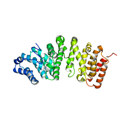
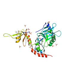
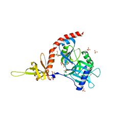
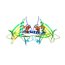
5FG1
DownloadVisualize
BU of 5fg1 by Molmil
Structure of the conserved yeast listerin (Ltn1) selenomethionine-substituted N-terminal domain, TRIGONAL FORM
分子名称: E3 ubiquitin-protein ligase listerin, POTASSIUM ION
著者Doamekpor, S.K, Lima, C.D.
登録日2015-12-19
公開日2016-07-06
最終更新日2019-11-20
実験手法X-RAY DIFFRACTION (2.55 Å)
主引用文献Structure and function of the yeast listerin (Ltn1) conserved N-terminal domain in binding to stalled 60S ribosomal subunits.
Proc.Natl.Acad.Sci.USA, 113, 2016
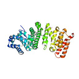
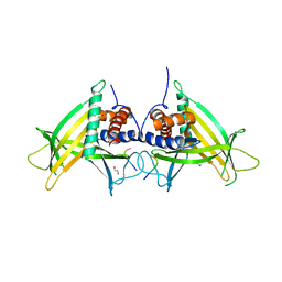
5FG0
DownloadVisualize
BU of 5fg0 by Molmil
Structure of the conserved yeast listerin (Ltn1) N-terminal domain, MONOCLINIC FORM
分子名称: 1,2-ETHANEDIOL, E3 ubiquitin-protein ligase listerin, POTASSIUM ION
著者Doamekpor, S.K, Lima, C.D.
登録日2015-12-19
公開日2016-07-06
最終更新日2024-03-06
実験手法X-RAY DIFFRACTION (2.41 Å)
主引用文献Structure and function of the yeast listerin (Ltn1) conserved N-terminal domain in binding to stalled 60S ribosomal subunits.
Proc.Natl.Acad.Sci.USA, 113, 2016
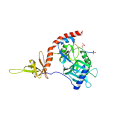
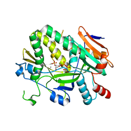
7UG9
DownloadVisualize
BU of 7ug9 by Molmil
Crystal structure of RNase AM PHP domain
分子名称: 5'-3' exoribonuclease, MANGANESE (II) ION, SULFATE ION
著者Doamekpor, S.K, Tong, L.
登録日2022-03-24
公開日2022-08-31
最終更新日2023-10-18
実験手法X-RAY DIFFRACTION (1.69 Å)
主引用文献Identification of a novel deFADding activity in human, yeast and bacterial 5' to 3' exoribonucleases.
Nucleic Acids Res., 50, 2022
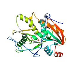
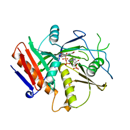
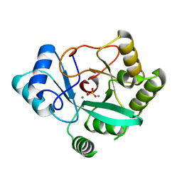
5ULJ
DownloadVisualize
BU of 5ulj by Molmil
Crystal structure of Scheffersomyces stipitis Rai1 in complex with (3'-NADP)+ and calcium ion
分子名称: CALCIUM ION, PYROPHOSPHATE, RAI1, ...
著者Doamekpor, S.K, Tong, L.
登録日2017-01-24
公開日2017-05-17
最終更新日2024-03-06
実験手法X-RAY DIFFRACTION (1.911 Å)
主引用文献5' End Nicotinamide Adenine Dinucleotide Cap in Human Cells Promotes RNA Decay through DXO-Mediated deNADding.
Cell, 168, 2017
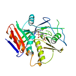
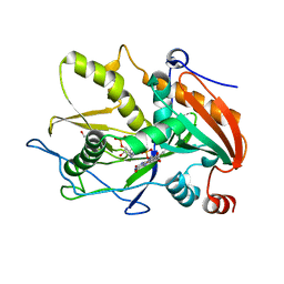
5ULI
DownloadVisualize
BU of 5uli by Molmil
Crystal Structure of mouse DXO in complex with (3'-NADP)+ and calcium ion
分子名称: CALCIUM ION, Decapping and exoribonuclease protein, [[(2R,3S,4R,5R)-5-(3-aminocarbonyl-4H-pyridin-1-yl)-3,4-bis(oxidanyl)oxolan-2-yl]methoxy-oxidanyl-phosphoryl] [(2R,3S,4R,5R)-5-(6-aminopurin-9-yl)-4-oxidanyl-3-phosphonooxy-oxolan-2-yl]methyl hydrogen phosphate
著者Doamekpor, S.K, Tong, L.
登録日2017-01-24
公開日2017-05-17
最終更新日2024-03-06
実験手法X-RAY DIFFRACTION (2.1 Å)
主引用文献5' End Nicotinamide Adenine Dinucleotide Cap in Human Cells Promotes RNA Decay through DXO-Mediated deNADding.
Cell, 168, 2017
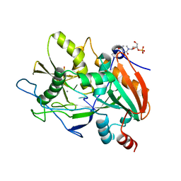
6WUI
DownloadVisualize
BU of 6wui by Molmil
Crystal Structure of mutant S. pombe Rai1 (E150S/E199Q/E239Q) in complex with 3'-FADP
分子名称: Decapping nuclease din1, [(2R,3S,4R,5R)-5-(6-amino-9H-purin-9-yl)-4-hydroxy-3-(phosphonooxy)tetrahydrofuran-2-yl]methyl (2R,3S,4S)-5-(7,8-dimethyl-2,4-dioxo-3,4-dihydrobenzo[g]pteridin-10(2H)-yl)-2,3,4-trihydroxypentyl dihydrogen diphosphate (non-preferred name)
著者Doamekpor, S.K, Tong, L.
登録日2020-05-04
公開日2020-05-20
最終更新日2023-10-18
実験手法X-RAY DIFFRACTION (1.9 Å)
主引用文献DXO/Rai1 enzymes remove 5'-end FAD and dephospho-CoA caps on RNAs.
Nucleic Acids Res., 48, 2020
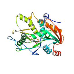
6WUF
DownloadVisualize
BU of 6wuf by Molmil
Crystal structure of mouse DXO in complex with 3'-FADP
分子名称: 1,2-ETHANEDIOL, Decapping and exoribonuclease protein, [(2R,3S,4R,5R)-5-(6-amino-9H-purin-9-yl)-4-hydroxy-3-(phosphonooxy)tetrahydrofuran-2-yl]methyl (2R,3S,4S)-5-(7,8-dimethyl-2,4-dioxo-3,4-dihydrobenzo[g]pteridin-10(2H)-yl)-2,3,4-trihydroxypentyl dihydrogen diphosphate (non-preferred name)
著者Doamekpor, S.K, Tong, L.
登録日2020-05-04
公開日2020-05-20
最終更新日2023-10-18
実験手法X-RAY DIFFRACTION (1.6 Å)
主引用文献DXO/Rai1 enzymes remove 5'-end FAD and dephospho-CoA caps on RNAs.
Nucleic Acids Res., 48, 2020
6WRE
DownloadVisualize
BU of 6wre by Molmil
Crystal structure of mouse DXO in complex with 5'-OH RNA substrate mimic and calcium ion
分子名称: 3'-URIDINEMONOPHOSPHATE, CALCIUM ION, Decapping and exoribonuclease protein, ...
著者Doamekpor, S.K, Tong, L.
登録日2020-04-29
公開日2020-05-13
最終更新日2023-10-18
実験手法X-RAY DIFFRACTION (2 Å)
主引用文献A novel 5'-hydroxyl dinucleotide hydrolase activity for the DXO/Rai1 family of enzymes.
Nucleic Acids Res., 48, 2020
6WUG
DownloadVisualize
BU of 6wug by Molmil
Crystal Structure of S. pombe Rai1 in complex with 3'-FADP
分子名称: 1,2-ETHANEDIOL, Decapping nuclease din1, MAGNESIUM ION, ...
著者Doamekpor, S.K, Tong, L.
登録日2020-05-04
公開日2020-05-20
最終更新日2023-11-15
実験手法X-RAY DIFFRACTION (1.9 Å)
主引用文献DXO/Rai1 enzymes remove 5'-end FAD and dephospho-CoA caps on RNAs.
Nucleic Acids Res., 48, 2020
6WUK
DownloadVisualize
BU of 6wuk by Molmil
Crystal structure of mouse DXO in complex with CoA
分子名称: COENZYME A, Decapping and exoribonuclease protein
著者Doamekpor, S.K, Tong, L.
登録日2020-05-04
公開日2020-05-20
最終更新日2023-10-18
実験手法X-RAY DIFFRACTION (1.599 Å)
主引用文献DXO/Rai1 enzymes remove 5'-end FAD and dephospho-CoA caps on RNAs.
Nucleic Acids Res., 48, 2020
4PZ7
DownloadVisualize
BU of 4pz7 by Molmil
PCE1 guanylyltransferase
分子名称: GLYCEROL, SULFATE ION, mRNA-capping enzyme subunit alpha
著者Doamekpor, S.K, Lima, C.D.
登録日2014-03-28
公開日2014-06-25
最終更新日2023-09-20
実験手法X-RAY DIFFRACTION (2.109 Å)
主引用文献How an mRNA capping enzyme reads distinct RNA polymerase II and Spt5 CTD phosphorylation codes.
Genes Dev., 28, 2014
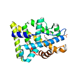
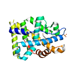
8FH2
DownloadVisualize
BU of 8fh2 by Molmil
Crystal structure of mutant Androgen Receptor ligand binding domain L702H/H875Y with DHT
分子名称: 5-ALPHA-DIHYDROTESTOSTERONE, Androgen receptor, SULFATE ION
著者Doamekpor, S.K, Tong, L.
登録日2022-12-13
公開日2023-04-12
最終更新日2024-05-22
実験手法X-RAY DIFFRACTION (1.59 Å)
主引用文献A partially open conformation of an androgen receptor ligand-binding domain with drug-resistance mutations.
Acta Crystallogr.,Sect.F, 79, 2023
4PZ8
DownloadVisualize
BU of 4pz8 by Molmil
PCE1 guanylyltransferase bound to SPT5 CTD
分子名称: SULFATE ION, Transcription elongation factor spt5, mRNA-capping enzyme subunit alpha
著者Doamekpor, S.K, Lima, C.D.
登録日2014-03-28
公開日2014-06-25
最終更新日2023-09-20
実験手法X-RAY DIFFRACTION (3.1 Å)
主引用文献How an mRNA capping enzyme reads distinct RNA polymerase II and Spt5 CTD phosphorylation codes.
Genes Dev., 28, 2014
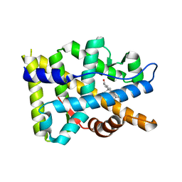
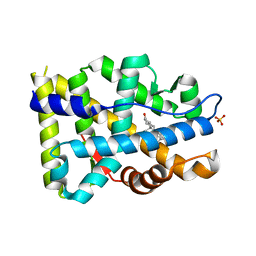
8FGZ
DownloadVisualize
BU of 8fgz by Molmil
Crystal structure of mutant Androgen Receptor ligand binding domain L702H/H875Y/F877L with DHT
分子名称: 5-ALPHA-DIHYDROTESTOSTERONE, Androgen receptor, SULFATE ION
著者Doamekpor, S.K, Tong, L.
登録日2022-12-13
公開日2023-04-12
最終更新日2024-05-22
実験手法X-RAY DIFFRACTION (1.61 Å)
主引用文献A partially open conformation of an androgen receptor ligand-binding domain with drug-resistance mutations.
Acta Crystallogr.,Sect.F, 79, 2023
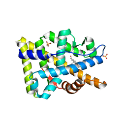
8FH1
DownloadVisualize
BU of 8fh1 by Molmil
Crystal structure of mutant Androgen Receptor ligand binding domain F877L/T878A with DHT
分子名称: 5-ALPHA-DIHYDROTESTOSTERONE, Androgen receptor, SULFATE ION
著者Doamekpor, S.K, Tong, L.
登録日2022-12-13
公開日2023-04-12
最終更新日2024-05-22
実験手法X-RAY DIFFRACTION (1.69 Å)
主引用文献A partially open conformation of an androgen receptor ligand-binding domain with drug-resistance mutations.
Acta Crystallogr.,Sect.F, 79, 2023
8FGY
DownloadVisualize
BU of 8fgy by Molmil
Crystal structure of mutant Androgen Receptor ligand binding domain L702H/H875Y/F877L/T878A with DHT
分子名称: 5-ALPHA-DIHYDROTESTOSTERONE, Androgen receptor
著者Doamekpor, S.K, Tong, L.
登録日2022-12-13
公開日2023-04-12
最終更新日2024-05-22
実験手法X-RAY DIFFRACTION (2.2 Å)
主引用文献A partially open conformation of an androgen receptor ligand-binding domain with drug-resistance mutations.
Acta Crystallogr.,Sect.F, 79, 2023
8FH0
DownloadVisualize
BU of 8fh0 by Molmil
Crystal structure of mutant Androgen Receptor ligand binding domain H875Y/F877L/T878A with DHT
分子名称: 5-ALPHA-DIHYDROTESTOSTERONE, Androgen receptor, SULFATE ION
著者Doamekpor, S.K, Tong, L.
登録日2022-12-13
公開日2023-04-12
最終更新日2024-05-22
実験手法X-RAY DIFFRACTION (1.59 Å)
主引用文献A partially open conformation of an androgen receptor ligand-binding domain with drug-resistance mutations.
Acta Crystallogr.,Sect.F, 79, 2023
4PZ6
DownloadVisualize
BU of 4pz6 by Molmil
PCE1 guanylyltransferase bound to SER2/SER5 phosphorylated RNA pol II CTD
分子名称: DNA-directed RNA polymerase II subunit rpb1, GUANOSINE, mRNA-capping enzyme subunit alpha
著者Doamekpor, S.K, Lima, C.D.
登録日2014-03-28
公開日2014-06-25
最終更新日2023-09-20
実験手法X-RAY DIFFRACTION (2.406 Å)
主引用文献How an mRNA capping enzyme reads distinct RNA polymerase II and Spt5 CTD phosphorylation codes.
Genes Dev., 28, 2014
4PN0
DownloadVisualize
BU of 4pn0 by Molmil
Structure of S. pombe Pct1 RNA triphosphatase
分子名称: mRNA-capping enzyme subunit beta
著者Lima, C.D, Doamekpor, S.K.
登録日2014-05-22
公開日2014-11-12
最終更新日2023-09-27
実験手法X-RAY DIFFRACTION (2.6 Å)
主引用文献Fission yeast RNA triphosphatase reads an Spt5 CTD code.
Rna, 21, 2015
4PN1
DownloadVisualize
BU of 4pn1 by Molmil
Structure of S. pombe Pct1 RNA triphosphatase in complex with the Spt5 CTD
分子名称: 1,2-ETHANEDIOL, FORMIC ACID, Synthetic peptide, ...
著者Lima, C.D, Doamekpor, S.K.
登録日2014-05-22
公開日2014-11-12
最終更新日2023-09-27
実験手法X-RAY DIFFRACTION (2.803 Å)
主引用文献Fission yeast RNA triphosphatase reads an Spt5 CTD code.
Rna, 21, 2015

221716

件を2024-06-26に公開中

PDB statisticsPDBj update infoContact PDBjnumon